XS-E 가 아토피피부염이 유발된 NC/Nga Mice의 피부상태에 미치는 영향
김금란*
XS-E is Induced Atopic Dermatitis NC/Nga Mice the Impact of Skin Conditions
Kum-Lan Kim*
접수: 2014년 7월 17일 / 게재승인: 2014년 8월 11일
© 2014 The Korean Society for Biotechnology and Bioengineering
Abstract: This study reports significant improvement of ato- pic dermatitis condition as a result of experiment using Xanth- ium strumarium L. extract (XS-E) at the dorsal skin of indu- ced atopic dermatitis Nc/Nga mice. Skin clinical score has decreased (2.75±0.85, *p<0.05), showing visible change of skin condition. IgE (***p<0.001) and IgG1 (2522.00±32.80,
***p<0.001) in plasma also decreased significantly. mRNA (gene expression) level increased (RQ=2.75±0.10, ***p<0.001) within skin tissue of CD4+CD25+Foxp3+ Treg cell that’s ac- tivated by XS-E dosage, thereby discovering that there is an effect of suppressing proliferation and viability of Th2 cell, eosinophils, mast cell and inflammatory cell. Upon examin- ing cells permeated with H&E and toluidine blue staining technique, thickness of epidermis and mast cell’s permeation decreased, and the result of examining the distribution of CCR 3+ eosinophils within ALN showed that it’s level fell down to that of wild type (normal group, NC/Nga-WT). By such results, it is suggested that XS-E is highly effective on atopic dermati- tis, and it is considered that continued quantitative research and case study of clinical research such as effect of cell num- ber in individual tissues or change of total cell number are necessary.
Keywords: CD4+CD25+Foxp3+ Treg, Mite extract, CCR3+, NC/Nga mice
1. INTRODUCTION
아토피피부염은 대식세포 (macrophage)와 과립세포 (granu- locyte)로 불리는 호염구 (basophil), 호산구 (eosinophil), 호중 구 (neutrophil), 비만세포 (mast cell), 그리고 T 림프구 (T lym- phocyte) 등 면역반응에 관여 세포들에 의해 발병되는 것으 로 알려져 있으며 [1], 외부 항원에 대한 혈중 IgE (immuno- globulin E)의 증가로 나타나는 복합적 질환이라고 할 수 있 다 [2-3].
IgE는 B 림프구 (B lymphocyte)에 의해 생성되는 면역 글 로블린으로 IL (interleukin)-4, IL-5, IL-10과 같은 Th (T hel- per)2형 세포의 지시를 받으며 IL-2, IFN-γ과 같은 Th1형 세 포에 의해 억제되고 [4] Th1과 Th2의 상호작용을 통해 인체 에 유의한 면역 균형을 유지한다. 아토피피부염은 초기 Th2 형 세포들에 의한 IL-4, IL-10의 과다분비로 IL-12가 불안정 하게 생산되어 IFN-γ (interferon-gamma)의 분비 저하를 초래 하고 [5], Th2 면역반응에 의해 개시되어 만성적인 Th1 면역 반응으로 전환하게 된다 [6].
Th 세포는 흉선 (thymus)에서 성숙 (maturation)된 T cell 즉, T helper 세포 (Th cells), Cytotoxic T 세포 (Tc cell), Regulatory T 세포 (treg cells), 중 하나로 APC (antigen presenting cells)의 항원 (antigen) 섭취와 표출 (presenting)에 따라 또는 영향을 미치는 사이토카인 (cytokine)의 종류에 따라 Th1, Th2, Th3, 수원여자대학교 미용예술학과
Department of Beauty and Art, Suwon Women's University, Suwon, 441-748, Korea
Tel: +82-31-290-8200, Fax: +82-31-290-8141 e-mail: [email protected]
연구논문
전사인자 Foxp3 (forkhead box p3)에 특이적으로 발현하기 때 문에 CD25와 Foxp3를 marker로 CD4+CD25+Foxp3+ Treg 세 포를 식별할 수 있다 [8].
최근 IVIG (intravenous immunoglobulin)를 매개로 한 자가 면역 치료에 관한 연구에서 CD4+CD25+regulatory T 세포의 주된 역할을 강조하였으며 공격적 면역반응, 자가면역 질환, 그리고 자기 항원에 대한 면역학적 무반응의 유지에 대한 기 능의 중요성 [9]이 입증되었으며, Foxp3가 조절 T림프구 발 현에 관여하는 필수적인 전사인자로 Foxp3+ Tregs은 활성화 된 T림프구 기능에 대한 직접적인 억제 기능과 IL-10 또는 TGF-β와 같은 항염증 (anti-inflammatory) 사이토카인을 분비 함으로써 과도한 면역반응을 제어할 수 있다 [10]고 보고되 었다.
이처럼 T 세포는 사이토카인, 면역활성 억제, 알러지, 자가 면역질환에 있어 중요한 방어 역할을 하므로 Treg 세포에 대 한 연구의 중요성은 크다고 할 수 있다 [11].
본 연구에서는 창이자 추출물에 의해 유도된 Foxp3+ Treg 세포가 NC/Nga mice의 면역글로블린, 면역조직, Foxp3+ mRNA 유전자 발현 등에 미치는 영향에 대하여 실험하였다.
창이자 (Xanthium strumarium L.)는 국화과 (compositae)에 속하는 1년 초로 비만세포 매개성 즉각형알러지반응 억제효 과 [12], 면역세포 및 혈청 IgE 감소효과 [13], 항균효과 [14], B림프구와 비만세포의 항알러지 효과 [15]가 있는 물질로 알 려져 있고 면역기능과 관련된 연구보고에 의하여 창이자의 응용성을 알 수 있으나, LPS와 IL-4로 자극된 mice에 혈청 중 IgE 생성량 및 in vitro에서 비장세포에 대한 IgE 생성량이 억 제되었음을 보고한 결과 [16] 외에 창이자의 항알러지 작용 에 대한 연구가 미비한 실정이다.
본 연구에서는 in vivo에서 아토피피부염 (atopic dermatitis like skin)을 유발시킨 NC/Nga mice의 면역글블린, 면역조직 화학분석, Foxp3+ mRNA 유전자 발현 등을 측정 및 분석한 결과 창이자 추출물에 의해 유도된 Foxp3+ Treg 세포가 아토 피피부염 억제 효과가 있었기에 이에 보고하는 바이다.
2. MATERIALS AND METHOD 2.1. 실험재료
실험에 사용된 동물은 수컷 7주령의 SPF (specific pathogen- free) NC/Nga mice (15~20 g)로 Charles River Japan (Yoko- hama, Japan)사에서 공급받았다. NC/Nga mice는 태어난 후 일반사육환경 (conventional system)에서 노출되면 사람의 아 토피피부염과 유사한 병변의 형태 즉, 딱지가 앉은 상처 및 작은 결정의 병변이 관찰되는 동물로서 아토피피부염 실험
pan) [18]. IgE, IgG1 ELISA kit는 SHIBAYAGI사 (Shibukawa, Japan)제품, γ-PGA는 바이웰㈜에서 구입하였으며 평균 분자 량은 300 kDa이다. CD4+ T isolation kit는 MACS (Mill. Bio- tec, USA)에서, GolgiStop kit는 BD bioscience (CA, USA)에 서 각각 구매하였다. 그 외에 사용된 시약은 따로 언급이 없 는 한 시판중인 특급시약을 사용하였다. 창이자는 Xanthium strumarium L.의 열매를 말하며 허브포유에서 구입한 후 정 선하여 사용하였다.
2.2. 창이자 추출물 (Xanthium strumarium L. extract; XS- E)의 제조
창이자 600 g 분량에 증류수 (distilled water) 2,000 mL를 가하 여 열탕 추출기에서 3시간 추출하여 얻은 액을 흡입 여과한 후 이를 감압 증류장치 (rotary evaporator, BUCHI B-480, Switzerland)로 농축하였다. 농축액을 다시 동결건조기 (freeze dryer, EYELA FDU-540, Japan)를 이용하여 완전 건조시킨 15.8 g (Xanthium strumarium L. extract; 이하 XS-E)을 냉동 보 관(-80oC)하면서 사용하였다.
2.3. NC/Nga mice에 Mite extract를 이용한 아토피피부발 진 유발 및 임상평가
실험동물 NC/Nga mice (15~20 g)는 실험 당일까지 고형사료 (항생제 무첨가, 삼양사료 Co.), 충분한 물, 온도 22±2oC, 습 도 55±15%, 12시간 (light-dark cycle)의 환경에 1주간 적응시 킨 후 미리 아토피피부염이 발진된 18주령의 NC/Nga mice 와 같은 공간에서 2주간 동시 사육하여 항원감작을 하였다.
2주 후 capillary 관을 이용하여 NC/Nga mice의 눈에서 100 µL의 혈액을 채혈한 후 마취제인 chloral hydrate (10%)로 마 취하고 귀와 등 쪽 목 부위를 깨끗하게 제모한 후 피부의 미 세 상처가 치유되도록 24시간 방치하였다. Mite extract는 1 주 2회 3주간 등 부위 및 목 부분에 고르게 도포하였고 도포 2~3시간 전에 4% SDS 용액을 분무하여 피부층을 파괴하였 다 (Fig. 1). 2주 후 등 부분에 피부염이 충분히 유발되면서 긁 는 행동이 심화되면 육안 평가를 실시하였다. NC/Nga mice 의 임상적 육안 평가는 아토피성 피부염에서 일반적으로 사 용되는 Yamamoto [18]의 방법을 약간 변형하여 사용하였다.
육안평가 항목은 소양행동을 동반한 홍반 (erythema/hemor- rhage), 인설 및 가피 (dryness/scarring), 부종 (edema), 표피박 탈/미란 (excoriation/erosion), 태선화 (lichenification) 5가지 항목으로 하고, 육안평가 결과는 각각 평가한 점수의 총합으 로 나타내었다. 각각의 항목은 없음 (none, 0), 약함 (mild, 1), 중증도 (moderate, 2), 심함 (severe, 3)으로 채점하였으며 최 소 0점에서, 최고 15점 사이의 점수를 측정하였다 [19].
2.4. 혈액채혈 및 IgE 와 IgG1 측정
8·12·15주에 NC/Nga mice의 눈에 capillary관을 이용하여 약 100µL의 혈액을 채혈한 후 원심분리기 6,500 rpm에서 20분 간 원심분리한 후 30 µL의 혈청을 분리하였다. 이렇게 취한 혈청은 -70oC에서 냉동 보관하였다. NC/Nga mice의 혈청 내 IgE와 IgG1 농도 측정은 Enzyme-linked immuno-sorbent assay kit (Shibayagi, Japan)로 측정하였다. 각 well에 채혈한 혈청 5 µL (1/10 dilution)와 dilution buffer 45 µL를 혼합하여 분주하고, 25oC 실온에서 2시간 방치한 후 washing 완충용액 으로 2회 세척한 다음 antibody biotin-IgE conjugated를 넣고 2시간 방치하였다. 다시 2회 수세한 후 완충용액으로 세척하 여 antibody Avidin-HRP conjugated 100 µL를 처리하고 1시 간 실온에서 방치한 후 다시 세척하였다. TMB 기질을 100 µL씩 분주하고 암소에서 30분간 방치한 후 100 µL의 stop 용 액을 처리한 후 ELISA leader 450 nm에서 각각 IgE와 IgG1 에 대한 흡광도를 측정하였다 [17].
2.5. 피부조직에서 foxp3+ mRNA 유전자 발현 측정 2.5.1. 등 피부조직과 ALN (axillary lymph node)에서 RNA 분리
아토피피부염이 유발된 NC/Nga mice의 등 피부조직과 ALN 를 적출하여 각각에 RNAzolB 500µL를 넣고 homogenizer로 용해될 때까지 분쇄하였다. 이 분쇄된 조직에 혼합 부유액 CHCl3 (chloroform) 50µL를 첨가하였고 15초간 다시 혼합한 다음 15분간 얼음에 방치한 후 13,000 rpm에서 원심분리하 였다. 원심분리기에서 회수된 상층액 중 약 200 µL에 2- propanol 200µL를 동량 혼합한 후 천천히 흔들어 섞은 후 얼 음에서 15분간 방치하였다. 이 용액을 다시 13,000 rpm에서 원심분리한 후 80% EtOH로 수세하였고 vaccum pump에서 3분간 건조하여 RNA를 추출하였다. 추출한 RNA는 DEPC (diethyl pyrocarbonate)를 처리한 20 µL의 증류수에 녹여 heating block 75oC에서 불활성화시킨 후 first strand cDNA합 성에 사용하였다 [20].
2.5.2. 역전사 (reverse transcription)중합효소 연쇄반응 역전사중합효소 연쇄반응은 준비된 total RNA 2 µg을 DNase I (10 U/mL) 2 U/tube를 37oC heating block에서 30분 동안 반 응한 후 75oC에서 10분 동안 변성시킨 후에 2.5 mL 10 mM dNTPs mix, 1 mL random sequence hexanucleotides (25 pmole/
25 mL), RNA inhibitor로서 1 mL RNase inhibitor (20 U/mL), 1 mL 100 mM DTT, 4.5 mL 5 × RT buffer (250 mM Tris-HCl
(pH 8.3), 375 mM KCl, 15 mM MgCl2를 가한 후, 1 mL의 M- mLV RT (200 U/mL)를 다시 가하고 DEPC 처리된 증류수로 서 최종 부피가 20 mL가 되도록 하였다. 이 20 mL의 반응 혼 합액을 잘 섞은 뒤 2,000 rpm에서 5초 동안 원심 침강하여 37oC heating block에서 60분 동안 반응시켜 first-strand cDNA 를 합성한 다음, 95oC에서 5분 동안 방치하여 M-mLV RT를 불활성화 시킨 후 합성이 완료된 cDNA를 polymerase chain reaction (PCR)에 사용하였다.
2.5.3. Real time quantitative PCR (RT-PCR)
RT-PCR은 Applied Biosystems 7500 Real-Time PCR system (Applied Biosystems, USA)를 이용하여 수행하였다 [21]. 사 용된 Foxp3 probe는 GCCACTCCAGCTCCCGGCAACTTCT (FAM)을 사용하였으며 probe는 Applied Biosystems사에서 제공되는 제품 (Sper-Taqman PCR Master mix, ABI)을 사용 하였으며 primer의 최종 농도가 200 nM이 되게 반응시켰다.
RT-PCR의 조건으로는 pre-denaturation은 50oC에서 2분, 94oC 에서 10분, 그리고 40 cycles을 95oC에서 0.15분, 60oC에서 1 분 수행하였다. 양성대조군 (Mite-FK506)과 실험군 (Mite- XS-E)의 internal standard로 G3PDH를 사용하여 target group 의 Quantitative PCR y = x(1+e)n, x=starting quantity, y=yield, n=number of cycles, e=efficiency로 계산하여 RQ (relative quantitative)를 측정하였다.
2.6. 조직검사 분석
NC/Nga mice의 등 피부조직을 생검하여 10% paraform-alde- hyde에서 24시간 동안 포르말린에 고정한 후 파라핀으로 포 맷하였고, 5 µM 두께로 block을 만들었다. 이렇게 분리한 조 직부분은 염증을 일으키는 표피 (epidermis), 진피 (dermis), 각질형성세포 (keratinocytes), 호중구 (neutrophils), 호산구 (eo- sinophil) 그 외 다른 세포와 부종을 식별하는 hematoxyline/
eosin (H&E) 염색과 비만세포를 염색하는 toluidine blue 염색 으로 비만세포의 침윤을 광학현미경 (Nikon, Japan, ×200)으 로 관찰하였다 [22].
2.7. 면역화학조직염색 (immune-histochemical staining) 본 실험에 사용한 모든 NC/Nga mice는 15주령에서 면역화 학조직염색을 하였다. 등 피부를 생검하여 10% 포르말린 용 액에 고정한 다음 파라핀 블록을 만든 후 rat anti-mice CCR3 mAb와 rat anti-mice CD4 mAb를 사용하였다. 조직절편을 4 µm 두께로 세절한 후 probe-on plus slide에 부착시켜 건조시 Fig. 1. Experimental design for the induction of atopic dermatitis like skin lesions in NC/Nga mice.
을 차단시켰다. 단백질을 차단시킨 후 일차 단일항체에 1시 간 동안 부착시킨 다음 완충액으로 수세하였다. LSAB kit를 이용하여 PE-conjugated goat anti-rat IgG에 30분간 반응시켰 다. 3회 Tris-buffered saline with 0.1% Tween 20 (TBST)용액 으로 수세한 후 잘 건조하여 형광위상차현미경 (×400 배율) 을 사용하여 관찰하였다 [23].
2.8. 통계처리
다양한 실험으로부터 얻은 결과는 mean±standard error로 기 록하였고, 유의성 검증은 Student's t-test분석방법을 이용하 여 결정하였다 [24].
3. RESULTS AND DISCUSSION
3.1. NC/Nga mice의 아토피피부염 억제 효과
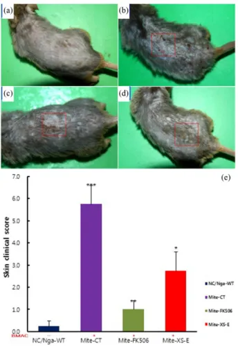
NC/Nga mice 12주령의 등 부위를 4% SDS용액으로 분무하 고 Mite extract를 주 2회 2주간 도포하여 아토피피부염을 유 발하였다. Fig. 2(b)는 NC/Nga mice에 Mite extract를 3주간 도포하고, 약물 도포 약 2주간 (총 4회)으로 400 mg 정도 도 포한 결과이다. 등 피부 부위에 소양증을 동반한 홍반, 구진, 인설과 가피 등 사람의 아토피피부염과 같은 병변이 나타났 다. 이 NC/Nga mice에 Mite extract를 1주간 추가 도포하여 3 주간 (총 6회)으로 600 mg 정도 도포하였다. Fig. 2(c)는 Mite extract 도포 종료 후 2주째 사진으로 양성대조군으로 FK506 을 3주간 도포한 결과, 양성대조군 (Mite-FK506, Fig. 2(c))과 실험군 (Mite-XS-E, Fig. 2(d))의 등 피부 부위에 홍반, 구진, 인설과 가피 등이 대조군에 비하여 현저하게 감소하였다.
Fig. 2(e)는 Mite extract를 NC/Nga mice에 3주간 도포하고 5주 후 등 피부의 skin clinical score를 측정한 결과이다. 대조 군 (Mite-CT)은 5.75±0.85로 정상군 (NC/Nga-WT) 0.30±0.30 에 비하여 15배 이상 증가하였고, 양성대조군 (Mite-FK506) 은 1.00±0.41로 대조군에 비하여 5배 이상 (***p<0.001), 실 험군 (Mite-XS-E)은 2.75±0.85로 2배 이상 유의성 있게 감소 하였다 (*p<0.05). Jun Hiroi (1998) 등의 아토피피부염이 유 도된 NC/Nga mice에 FK506을 도포하여 CD4, 비만세포, eosinophils, IL-4, IL-5, IgE 등의 감소와 피부의 alopecia, atrophy가 현저하게 완화되었다는 결과[25]와 유사한 결과로 XS-E이 홍반, 부종, 인설, 가피, 태선화 등에 효과적일 것이 라고 사료된다.
3.2. 혈청 중 immuno-globulin 측정
아토피피부염과 같은 알레르기 질환은 그 균형이 Th2 쪽으 로 치우친 결과로 유발된 면역질환 중의 하나이다. 이렇듯
알레르기 발생과 조절에 있어 가장 중요한 세포는 Th2 세포 로, 항체의 isotype을 IgM 또는 IgG에서 IgE로 바꾸어주는 Th2 세포가 활성화되면서 시작되며, Th2 세포가 생산하는 Th type 2 cytokine (IL-4, IL-10 etc)에 의해서 B 세포의 IgE 생 성, 호산구의 활성화, 염증반응 등을 나타낸다 [26].
Fig. 3은 혈청 내 IgE (Fig. 3(a))의 양과 혈장 내 IgG1 (Fig.
3(b))의 양을 측정한 결과이다.
Fig. 3(a)에 나타난 바와 같이 IgE의 양은 정상군 (normal;
NC/Nga-WT)이 103.3에서 283.2로 자연적으로 증가되고 대 조군 (control; Mite-CT)은 95.9에서 293.5로 15주에서 정상군 과 비슷하게 증가하였다. 그리고 3주간 0.3%의 FK506 도포 한 양성대조군 (Mite extract-FK506, Mite-FK506)은 99.1에서 201.3으로 유의하게 감소하였고 (***p<0.001), 실험군 (Mite- XS-E) 또한 135.4에서 281.2로 증가하다가 12주 이후 감소하 여 15주에는 162.0으로 혈장 내 IgE 수준이 유의성 있는 감소 Fig. 2. Topical application of XS-E and FK506 treatment of atopic dermatitis-like NC/Nga mice induced by Mite extract for 3 weeks.
Atopic dermatitis NC/Nga mice was induced by Mite extract treat- ment in the dorsal skin, before the treatment of Mite extract (nor- mal; NC/Nga-WT, (a)), Mite extract treatment for 3 weeks (control;
Mite-CT, (b)), Mite extract treatment for 3 weeks with FK506 (0.3%)- ointment (Mite-FK506, (c)), Mite extract treatment for 3 weeks with skin apply of XS-E (220 mg/kg) orally administration (Mite-XS-E, (d)), and Mite extract treatment for 3 weeks.
를 나타내었다 (***p<0.001). Fig. 3(b)는 혈장 내 IgG1의 수 준은 B세포가 IL-4의 반응으로 분화되어 IgE가 분비될 때 같 이 증가되는 Ig이다. 본 연구결과에서 혈장 내 IgG1의 수준 은 정상군이 1456.50±146.20, 양성대조군이 929.50±51.50 대 조군 3249.50±124.70의 3배 감소하였고 (***p<0.001), 실험 군은 2522.00±32.80로 대조군에 비하여 통계학적으로 유의 성 있는 감소를 나타내었다(***p<0.001). 이는 Park (2007) 등 의 다래 (Actinidiaarguta)로부터 분리한 PG102가 NC/Nga mice에서 SATA-6와 전사인자 인산화, GATA-3의 억제작용 으로 인해 Th2 사이토카인 및 IgE 분비 및 발현이 감소된 연 구와 유사한 결과이다 [27].
3.3. 피부 조직에서 Foxp3 mRNA 유전자 발현 분석 Miyara (2007) 등을 비롯한 최근 실험과 임상 연구들은 유전 적으로 혹은 고의적으로 일으킨 Tregs 결핍이 자가면역 질환 과 염증을 일으키는 고도한 면역 반응으로 이어진다고 보고 했다 [28]. Tregs은 thymus에서 나타나는 natural Tregs (nTregs) 와 naive CD4+ T 세포로부터 바깥쪽에서 발달된 induced Tregs (iTregs) 두 가지로 구분된다.
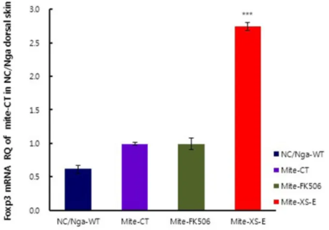
본 연구는 XS-E 투여로 활성화된 CD4+CD25+Foxp3+ Treg 세포가 Mite extract로 유도된 NC/Nga mice의 피부발진에 미 치는 영향을 알아보기 위하여, 등 피부를 biopsy하여 real- time PCR로 Foxp3 mRNA유전자 발현량을 측정하였다 (Fig.
4.). 그 결과 대조군에 대한 Foxp3 mRNA의 유전자발현의 상 대정량 값 (RQ)은 BMAC를 도포하지 않은 정상군 (normal;
NC/Nga-WT) 0.612±0.064에 비하여 대조군 (control; Mite- CT) 0.992±0.008, FK506 0.3% 도포군 0.991±0.034로 유의한 차이가 나타나지 않았으나, 실험군 (Mite-XS-E)의 Foxp3 mRNA 유전자발현의 RQ값이 2.745±0.100로 대조군에 비하 여 약 3배 이상 유의성 있게 증가하였다 (***p<0.001). 이 결 과는 XS-E에 의하여 유도된 Treg 세포가 Th2 세포, 호산구세 Fig. 3. Serum IgE and IgG1 elevation and development of atopic
dermatitis skin lesions-induced in NC/Nga mice by Mite extract.
(a): Atopic dermatitis NC/Nga mice was induced by Mite extract treat- ment in the dorsal skin, before the treatment of Mite extract (normal;
NC/Nga-WT), Mite extract treatment for 3 weeks (control; Mite-CT), Mite extract treatment for 3 weeks with FK506 (0.3%)-ointment (Mite- FK506), Mite extract treatment for 3 weeks with skin apply of XS-E orally administration (Mite-XS-E) for 3 weeks. (b): Blood was collec- ted from the retro-orbital plexus under ether an esthesia. Serum sam- ples were obtained by centrifugation and stored at -20oC until use. Total IgE and IgG1 levels were measured by a sandwich ELISA using an ELISA kit (Shibayagi, Japan). Each point represents the mean±SE of six mices. Statistically significant value compared with Mite extract- Control group data by T test (**p<0.01, ***p<0.001).
Fig. 4. Effects of XS-E treatment on Foxp3+ mRNA expression in dorsal skin tissue in atopic dermatitis-like NC/Nga mice induced by Mite extract.
Atopic dermatitis NC/Nga mice was induced by Mite extract treat- ment in the dorsal skin, before the treatment of Mite extract (nor- mal; NC/Nga-WT), Mite extract treatment for 3 weeks (contro; Mite- CT), Mite extract treatment for 3 weeks with FK506 (0.3%)-oint- ment (Mite-FK506), Mite extract treatment for 3 weeks with XS-E (2 mg/day/mice) orally administration (Mite-XS-E), Mite extract treatment for 3 weeks with XS-E (2 mg/day/mice) for 3 weeks. Fox p3+mRNA synthesized by real-time PCR was analyzed. The amount of Taqman probe was measured at the end of each cycle. The cycle number at which the emission intensity of the sample rises above the baseline is referred as to the RQ (relative quantitative) and is proportional to the target concentration. Real time PCR was perfor- med in duplicate and analyzed by a Applied Biopsy stems 7500 Real-Time PCR system. Each point represents the mean±SE of six mice. Statistically significant value compared with Mite-CT group data by T test (***p<0.001).
포, 비만세포, 그리고 염증세포의 증식 및 활성을 억제하여 피부발진이 감소한 결과라고 사료되며, De Groot과 그의 연 구진들이 보고한 Treg 활성 부위 (Tregitopes)에 의해 PBMC 에서의 iTregs 확산과 이에 따른 CD25+, CTLA-4, CTLA-4, GITR같은 세포 표면 marker의 발현 증가의 결과 [29]와 일치 하였다. 그러나 XS-E이 Treg 세포를 활성화시키는데 있어서 CD25+, CTLA-4, GITR 간의 상호작용에 의한 기전인지에 대 한 추후 연구가 필요하다고 생각된다.
3.4. 피부조직 분석
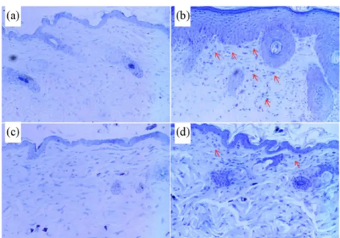
실험 종료 후 SPF와 conventional 조건에 사육한 NC/Nga mice의 등 피부조직을 생검하여 H & E과 toluidine blue 염색 을 실시하여 표피에 침윤된 비만세포를 관찰한 결과는 Fig. 5 와 Fig. 6과 같다.
Fig. 5(a)와 Fig. 6(a)는 SPF 조건에서 사육한 15주령의 정상 군 NC/Nga mice의 피부조직으로 표피가 얇게 분포하고 비 만세포는 거의 관찰되지 않았으나, 대조군의 표피 두께 (Fig.
5(b), 6(b))는 비후 (hyperplasia)하게 확장 (long red arrow)되 어 주변 피부상태 (hyperkeratosis, acanthosis, hypergranulosis, parakeratosis)와 비만세포 (Fig. 6(b), red arrow)의 침윤 등이 정상군에 비하여 현저하게 증가된 것을 관찰할 수 있었다.
양성대조군 (Fig. 5(c), 6(c))의 일부는 대조군에 비하여 정상 군에 가깝게 표피의 두께가 줄어들었고, 나머지 일부의 두께 (red arrow)와 주변 피부상태와 비만세포 (Fig. 6(c), red arrow)
침윤 등이 현저하게 감소하였다. XS-E를 도포한 실험군의 표 피 두께 (Fig. 5(d))와 (Fig. 6(d), red arrow)는 대조군에 비하여 현저하게 줄어들었고 나머지 일부의 표피 두께 (red arrow), 그리고 그 주변의 피부상태와 비만세포 (Fig. 6(d), red arrow) 침윤이 감소하였다.
이러한 결과는 Kim (2007) 등의 NC/Nga mice의 ALN와 아 토피피부염 부위에 CpG-ODN을 투여하여 피부발진, 표피의 두께 및 염증면역세포 수가 현저하게 감소한 결과 [30]와 일 치하였다.
3.5. ALN (axillary lymph node)에서 면역화학조직염색 아토피피부염은 기저막 (basement membrane)두께, 호산구 수, 비만세포수, Th2형의 사이토카인 (IL-4, IL-5)을 발현하 는 T림프구와 CCR3, CCR5, CCR6, 그리고 CXCR3과 같은 chemokine 수용체의 발현이 증가한다. 이러한 C-C유형의 케 모카인은 호산구 및 호염기구에서도 발현될 뿐만 아니라 Th2 세포 등 염증세포 활성화를 유도하여 만성적 피부질환 으로 악화시킨다.
본 실험에서는 면역조직화학염색 (LSAB2 HRP, Rabbit/
mice (DAB) kit)을 통하여 biopsy한 NC/Nga mice의 ALN을 적출하여 CCR3+호산구 (eosinophils)(Fig. 7(a)~(d), strong brown color, red arrow)를 광학현미경으로 관찰하였다.
CCR3+호산구의 분포는 Fig. 7(a)~(d)와 같다. 대조군 (Fig.
7(b))은 정상군 ((Fig. 7(a))에 비하여 ALN 조직 사이의 침윤이 Fig. 5. Histological features of dorsal skin group in atopic derma-
titis-like NC/Nga mice induced by Mite extract.
Atopic dermatitis NC/Nga mice was induced by Mite extract treat- ment in the dorsal skin, (a); before the treatment of Mite extract (normal; NC/Nga-WT), (b); Mite extract treatment for 3 weeks (control; Mite-CT), (c); Mite extract treatment for 3 weeks with FK506 (0.3%)-ointment (Mite-FK506), (d); Mite extract treatment for 3 weeks with XS-E (220 mg/kg) orally administration (Mite- XS-E) for 3 weeks. NC/Nga mice skin biopsy were stained with hematoxylin and eosin (H & E) and shows the thickening of the epidermis (red arrow) by bright microscope (×100). Data represent individual values and the average value of four individual mice in each group.
Fig. 6. Histological status of the skin stained with toluidine blue of dorsal skin group in atopic dermatitis-like NC/Nga mice induced by Mite extract.
Atopic dermatitis NC/Nga mice was induced by Mite extract treat- ment in the dorsal skin, (a); before the treatment of Mite extract (normal; NC/Nga-WT), (b); Mite extract treatment for 3 weeks (control; Mite-CT), (c); Mite extract treatment for 3 weeks with FK 506 (0.3%)-ointment (Mite-FK506), (d); Mite extract treatment for 3 weeks with XS-E (220 mg/kg) orally administration (Mite-XS-E) for 3 weeks. NC/Nga mice skin biopsy were stained with toluidine blue staining and shows the degranulated mast cells in the dermis (red arrow) by bright microscope (×100). Data represent individual values and the average value of four individual mice in each group.
현저하게 증가된 것으로 나타났으며, 양성대조군 (Fig. 7(c)) 과 실험군 (Fig. 7(d))의 CCR3+호산구의 분포는 대조군 (Fig.
7(b))에 비하여 정상군에 가깝게 감소된 것을 관찰할 수 있었 다.
이러한 결과는 XS-E이 투여된 NC/Nga mice의 CD4+CD 25+Foxp3+ Treg 세포의 발현으로 Th2형의 사이토카인 (IL-4, IL-5)이 억제되고 CCR3+세포의 증식과 활성화가 감소됨으로 서 아토피피부염의 완화를 가져왔다고 사료된다. Lee (2009) 가 반하 (半半) 추출물로 NC/Nga 생쥐의 피부발진 부위에 적 용한 실험한 결과 CD4+CD25+ 조절 T 세포상승 및 CD3+ CCR3+ Th2 세포 침윤 억제 효과가 나타난 연구 [31]와 동일 한 결과가 나타났다.
4. CONCLUSION
아토피피부염은 소양증을 동반한 만성/재발성의 피부염으로 유·소아기에 높은 발병률을 보임에도 불구하고 정확한 치료 제가 없이 스테로이드제 등을 통한 대증 요법이 사용되어 왔 으며, 스테로이드제 오남용에 따른 부작용은 잘 알려진 바이 다.
본 연구는 아토피피부염에 치료효과는 높으면서 부작용이 적은 천연물질의 계발을 위해 기초 자료검색을 통해 가능성 이 있다고 사료되는 창이자를 선별하여 Mite cream으로 아
토피피부염이 발생된 NC/Nga mice에 창이자 추출물 (XS-E) 을 적용한 결과 아토피 피부부위의 상태가 현저하게 호전되 었으며 IgE, IgG 및 IL-4, IL-10과 같은 염증유발 사이토카인 생산이 유의성 있게 감소하였다. 또한 Foxp3 mRNA유전자 발현과 H & E 그리고 toluidine blue 조직염색으로 CD4+ CD 25+ Foxp3+ Treg 세포 활성화에 따른 CCR3+ 호산구의 활성 억제 작용이 있는 것으로 관찰되었다. 이러한 결과로 XS-E 는 아토피피부염에 매우 효과적이라고 판단되며, 향후 각 조 직별 세포수 또는 총세포수의 변화와 같은 정량평가와 임상 연구와 같은 실증적인 연구가 지속적으로 필요하다고 사료 된다.
REFERENCES
1. Sicherer, S. H. and H. A. Sampson (1999) Food hypersensitivity and atopic dermatitis: Pathophysiology, epidemiology, diagnosis, and management. Allergy. Clinical Immunol. 104: 114-122.
2. Rousset, F., J. Robert, M. Andary, J. P. Bonnin, G. Souillet, I. Chre- tien, F. Briere, J. Pene, and J. E. Vries (1991) Shifts in interleukin-4 and interferon-gamma production by T cells of patients with ele- vated serum IgE levels and the modulatory effects of these lym- phokines on spontaneous IgE synthesis. Allergy Clin Immunol. 87:
58-69.
3. Kleij, H. P., A. D. Kraneveld, A. H. Houwelingen, M. Kool, A. C.
Weitenberg, F. A. Redegeld, and F. P. Nijkamp (2004) Murine mo- del for non-IgE-mediated Asthma. Inflammation. 28: 115-125.
4. LEE, M. Y (2002) Hydroquinone, a metabolite of benzene, en- hances interleukin-4 production in CD4+ T cells and increases IgE levels in antigen-primed mice. Ph. D. Pharmacy. Graduate School of Chunnam National University, Korea.
5. Matsumoto, M., R. C. Kawamoto, K. Sato, H. Itakura, A. Sawada, J. Ushio, H. Suto, H. Mitsuishi, K. Hikasa, and H. Matsuda (1999) IgE hyperproduction through enhanced tyrosine phosphorylation of Janus kinase 3 in NC/Nga mice, a model for human atopic der- matitis. Immunol. 162: 1056-1063.
6. Miyamoto, K., S. Miyake, and T. Yamamura (2001) A synthetic glycolipid prevents autoimmune encephalomyelitis by inducing TH2 bias of natural killer T cells. Nature. 413: 531-534.
7. Hori, S., T. Nomura, and S. Sakaguchi (2003) Control of regula- tory T cell development by the transcription factor Foxp3. Science 14: 1030-1031.
8. Shih, I. L. and Y. T. Van (2001) The production of poly- (gamma- glutamic acid) from microorganisms and its various applications.
Bioresour Technol. 79: 207-225.
9. Bayry, J., S. Siberil, F. Triebel, D. F. Tough, and S. V. Kaveri (2007) Rescuing CD4+CD25+ regulatory T-cell functions in rheumatoid arthritis by cytokine-targeted monoclonal antibody therapy. Drug Discovery Today. 12: 548-552.
10. Wan, Y.Y (2010) Regulatory T cells: immune suppression and beyond. Cell Mol. Immunol. 7: 204-210.
11. Coffman, R. L., J. Ohara, M. W. Bond, J. Carty, A. Zlotnik, and W.
E. Paul (1986) B cell stimulatory factor enhances the IgE response Fig. 7. Immuno-histochemical staining of the tissue stained with
CCR3+ cells of ALN in atopic dermatitis-like NC/Nga mice.
Atopic dermatitis NC/Nga mice was induced by Mite extract treat- ment in the dorsal skin, (a); before the treatment of Mite extract (normal, SPF, NC/Nga-WT), (b); Mite extract treatment for 3 weeks (control, Mite-CT), (c); Mite extract treatment for 3 weeks with FK 506 (0.3%)-ointment (Mite-FK506), (d); Mite extract treatment for 3 weeks with XS-E (220 mg/kg) orally administration (Mite-XS-E) for 3 weeks. Following 3 weeks, mice ALN biopsy stained with anti-mice CD4 mAb respectively. ALN biopsy were stained with anti-mice CCR3 mAb, used LSAB2 HRP. Rabbit/mice (DAB) kit and shows the CCR3+cells in the dermis (red arrow) by bright mic- roscope (×100).
Asthma Model. Oriental preventive medical society. 5: 93-105.
14. Kim, H. S. and J. O. Shin (1997) Isolation and Antimicrobial of Xanthium strumarium L. Extract. Microbiol. Biotechnol. 25: 183- 188.
15. Seo, Y. B., Y. B. Choi, Y. K. Lee, and Y. C. Lee (2002) Study on the Effects of Xanthii Fructus about the anti-allergic Action on IC- 2 Cell. Herbology. 17: 39-54.
16. Kim, S. H., S. H. Kim, H. Y. Lee, S. H. Park, M .S. Zheong, M. Y.
Lee, Y, H. Shim, S. H. Moon, M. J. Jo, S. H. Lee, J. B. Na, D. H.
Kang, S. H. Shin, J. M. Lee, G. J. Ji, and C. B. Park (2006) 90 days Oral Repeated Dose Toxicity and Genotoxicity study of a Xanth- ium strumarium L. pp. 1-4. The Annual Report of KFDA, vol. 10, KOR.
17. Matsuda, H., N. Watanabe, G. P. Geba, M. Sperl, J. Tsudzuki, and J. Hiroi (1997) Development of atopic dermatitis like skin lesion with IgE hyperproduction in NC/Nga mice. Immunol. 9: 461-466.
18. Yamamoto, M., T. Haruna, K. Yasui, H. Takahashi, M. Iduhara, S.
Takaki, M. Deguchi, and A. Arimura (2007) A novel atopic der- matitis model induced by topical application with dermatopha- goides farinae extract in NC/Nga mice. Allergol. 56: 139-48.
19. Dogru, M., C. Katakamami, N. Nakagawa, K. Tetsumoto, and M.
Yamamoto (1998) Impression cytology in atopic dermatitis. Oph- thalmology. 105: 1478-1484.
20. Montagne, A., O. Grepinet, M. Peloille, F. Lantier, and A. C. Lal- manach (2001) Quantification of ovine cytokine gene expression by a competitive RT-PCR method. Immunol. Methods. 253: 83-93.
21. Galli, S. J (2006) Allergy. Biol. 10: 93-95.
22. Hashimoto, Y., I. Arai, Y. Nakanishi, T. Sakurai, and A. Nakamu- ra (2004) Scratching of their skin by NC/Nga mice leads to devel-
lisher, USA.
25. Jun, H., S. Takanori, M. Kyoko, K. Shinichi, S. Sato, T. Ogawa, M. Tsudzuki, H. Matsuda, A. Wada, and K. Esaki (1998) Effect of Tacrolimus Hydrate (FK506) Ointment on Spontaneous Dermati- tis in NC/Nga Mice. Pharmacol. 76: 175-183.
26. Ohmen, J. D., J. M. Hanifin, and B. J. Nickoloff (1995) Overex- pression of IL-10 in atopic dermatitis. Contrasting cytokine pat- terns with delayed-type hypersensitivity reactions. Immunol. 154:
1956-1963.
27. Park, E. J., K. C. Park, H. K. Eo, J. K. Seo, M. W. Son, K. H. Kim, Y. S. Chang, S. H. Cho, K. U. Min, M. R. Jin, and S. Y. Kim (2007) Suppression of Spontaneous Dermatitis in NC/Nga Murine Model by PG102 Isolated from Actinidia arguta. Investigative Dermatol. 127: 1154–1160.
28. Miyara, M. and S. Sakaguchi (2007) Natural regulatory T cells:
Mechanisms of suppression. Trends in Molecular Medicine 13:
108-116.
29. De Groot, A. S., L. Moise1, J. A. McMurry, E. Wambre, L. V.
Overtvelt, P. Moingeon, D. W. Scott, and W. Martin (2008) Acti- vation of natural regulatory T cells by IgG Fc-derived peptide “Tre- gitopes”. Blood. 8: 112-120.
30. Kim, Y. S., y. h. Kim, K. J. Lee, H. J. Kwon, D. S. Kim, and T. Y.
Kim (2007) Improvement of Atopic Dermatitis in NC/Nga Mice by Topical Application of CpG Phosphodiester-ODN. Allergy Im- munol. 144: 315-324.
31. Lee, Y. C. (2009) A Therapeutic Effect of Pinellia Ternata via the Increase of CD4+CD25+ Regulatory T Cells and the Suppression of CD3+CCR3+ Cellular Infiltration During Allergic Airway Infla- mmation. Herbol. 24: 73-78.