PKC 활성 조절을 통한 두시 하태독법의 항염증작용이 Mite 항원 유도 아토피유사피부염 발병 조절에 미치는 효과
안상현1․김기봉2,3
1세명대학교 한의과대학 해부학교실, 2부산대학교 한의학전문대학원 소아과학교실, 3부산대학교한방병원 한방소아과
Received: November 1, 2016 ∙ Revised: November 13, 2016 ∙ Accepted: November 14, 2016 Corresponding Author: Kim Kibong, KMD, Ph.D.
Department of Pediatrics, Korean Medicine Hospital, Pusan National University 20, Geumo-ro, Mulgeum-eup, Yangsan-si, Gyeongsangnam-do, 50612, Republic of Korea Tel: +82-55-360-5952, Fax: +82-55-360-5952
E-mail: [email protected]
ⓒ The Association of Pediatrics of Korean Medicine. All rights reserved. This is an open-access article distributed under the tenus of the Creative Commons Attribution Non-Commercial License (http://creativecommons.org/licenses/by-nc/3.0/), which permits unrestricted non-commercial use, distribution, and reproduction in any medium, provided the original work is properly cited.
Abstract
Anti-inflammatory Effects of Hataedock with Douchi in Atopic Dermatitis-like Skin Lesions in House Dust Mite-Induced NC/Nga Mice
Ahn Sang Hyun1
․Kim Ki Bong
2,31
Department of Anatomy, College of Korean Medicine, Semyung University
2
Department of Pediatrics, School of Korean Medicine, Pusan National University
3
Department of Pediatrics, Korean Medicine Hospital, Pusan National University
Objectives
Hataedock (HTD) is an oral Korean herbal medical oral treatment that removes fetal toxic heat and meconium from new born babies. The purpose of this study is to evaluate whether Hataedock treatment of Duchi extracts has anti-inflammation effects in atopic dermatitis-like skin lesions in House Dust Mite-Induced NC/Nga Mice.
Methods
The mice were divided into 3 groups (n=10 per group) as follows: the control group (Ctrl group), AD-induced group (AE group), AD-induced with HTD treatment group (DT group). 3-week-old NC/Nga mice were introduced to Hataedock treatment, made of Duchi extract. After 4 weeks, House Dust Mite-Induced application was used six times per week for 3 weeks to induce the first atopic dermatitis, and second AD in 7 weeks after. To examine skin injuries and anti-inflammatory effect, PKC, MMP-9, iNOS immunohistochemistry were used.
Results
The alleviate effect of the skin damage and angiogenesis was observed in DT group. The damage of stratum corneum, hyperplasia, edema, infiltration of lymphocytes and distribution of capillary were decreased in DT group.
Also, the study results suggested that Hataedock treatment made of Duchi extracts in DT group remarkably decreased skin damages by 51% (p < 0.001), as well as PKC by 91%, MMP-9 by 48% (p < 0.001), iNOS by 51% (p < 0.001).
Conclusions
Based on the study results, we observed that Hataedock treatment of Duchi extracts alleviates AD by diminishing various inflammatory cytokines, initial steps of AD development, in the skin lesions. Potential applications for prevention and treatment of atopic dermatitis are expected.
Key words: Hataedock, Duchi, Atopic dermatitis, PKC, MMP-9, iNOS
ISSN 1226-8038(Print), 2287-9463(Online), http://dx.doi.org/10.7778/jpkm.2016.30.4.077
78 Anti-inflammatory Effects of Hataedock with Douchi in Atopic Dermatitis-like Skin Lesions in House Dust Mite-Induced NC/Nga Mice
Ⅰ. Introduction
아토피 피부염 (Atopic Dermatitis, 이하 AD)은 염증 성 만성 피부질환으로 소아에서 흔히 발생하며, 유전, 환경, 약물성, 심리적, 면역학적, 피부 장벽 요인 등의 다양한 요인들의 복잡한 상호관계로 인해 야기되는 난 치성 질환이다
1-2). 한국의 경우 AD 발병의 48%가 9세 미만의 소아에 집중되어 있으며
3), 이는 출산시 Th2 skewed condition상태의 영·유아가 아직 정상 Th1/Th2 balance로 도달하지 못한 상태에서 AD 관련 병인에 노 출된 결과로 볼 수 있다
4).
한의학에서는 이러한 AD의 원인을 Th2 skewed con- dition으로 인해 발생하는 열성 증상인 태열 (胎熱)로 보았다. AD는 태열이 제대로 제거되지 못한 상황에서 발생하는 질환이며, AD의 원인을 稟性不耐과 濕熱內 蘊한 상태에서 다시 風濕熱邪에 감작되는 내인과 외인 의 상호작용으로 발생하는 것으로 이해하였다
5).
태열은 영유아에서 발병하는 다양한 질환들의 원인 으로 인식하고, 이를 제거하기 위해 출생 직후 하태독 법 (下胎毒法)을 시행하였다
6). 하태독법은 두시법 (豆 豉法), 감초법 (甘草法), 황련법 (黃連法), 주밀법 (朱蜜 法) 등이 있으며, 한약재를 달인 약물을 부드러운 천이 나 비단에 묻혀 입안의 더러운 것들을 닦아주면서 소 량 먹이는 방법이다
7). 이 중 두시는 신량해표약 (辛凉 解表藥)으로 체표 (體表)의 열을 발산시키는 효능이 있 으며, 최근 연구에서 염증 증상들을 완화시킨다는 보 고들이 있다
8-10). 선행연구에 의하면 발효콩이 AD 증상 완화와 소양감 억제 효과가 있음을 보고하고 있으며
11), Th2 response를 통한 Th1과 Th2 조절 효과
12)및 eosino- phil airway inflammation억제 효과를 보고하고 있다
9,13). 이러한 내용은 두시의 면역학적 변화 야기 가능성을 보여준다.
AD의 발병은 각질 내 Ceramide 결핍에 따른 sphin- gosine생성 감소와 house dust mite, Dermatophagoides pteronissinus의 노출이 protein kinase C (PKC)의 활성을 유도함으로써 시작된다
14). 이는 Th2 skewed condition induced cytokine인 IL-4, IL-5, IL-13의 생성 증가에 의 해 야기된다
15). 특히, IL-4 생성은 B cell의 IgE 분비를 유도하여 비만세포의 탈과립화 (degranulate)를 일으키 며, 이는 부종을 유도하는 MMP-9, 혈관 확장에 관여하 는 염증효소인 i-NOS, 그리고 동통과 소양증 관련 인 자인 substance P 등을 생성하여 피부손상을 가속화한
다
10). AD치료제로 사이토카인 생산을 억제하는 스테 로이드제가 많이 사용되고 있으나, 장기투여 시 피부 위축, 골유실로 인한 골절, 성장지연 등의 부작용을 야 기할 수 있어 대안적 치료법에 대한 요구가 높다
16).
본 연구는 두시 추출물의 AD 발병 조절 효과를 검 증하기 위해서 Nc/Nga 생쥐 피부에 sodium dodeecyl sulfate로 각질층의 lipid lamella를 제거한 후 D. pter- onissinus 항원에 노출시켜 PKC 활성을 일으킨 후, 두 시 처리 생쥐 피부 내 PKC, MMP-9, iNOS에 대한 면역 조직적 변화관찰을 통해 두시의 항염증 효과를 확인하 고 AD 치료제로의 가용성을 조사하였다.
Ⅱ. Materials and Methods
1. 두시추출물의 제조와 성분 분석
두시 (duochi, fermameted Glycine max Merr.; 남영제약 영농조합, 무주, 한국)는 서리태 (Glycine max Merr.)를 청호 (Artemisia Apiacea Herba)와 상엽 (Mori Folium)의 전 탕액 (1:1)에 삶은 후, 37~38 ℃에서 5일간 발효시킨 다음 열풍 건조시켜 제조하였다. 두시 열수추출물을 얻기 위해 두시 100 g을 파쇄한 후 증류수 1000 ㎖에 넣고 3시간동안 전탕한 후 여과하였다. 그 여액을 ro- tary evaporator에서 50 ㎖으로 농축한 후 동결 건조하여 추출물 15 g (수득률 15%) 얻었다.
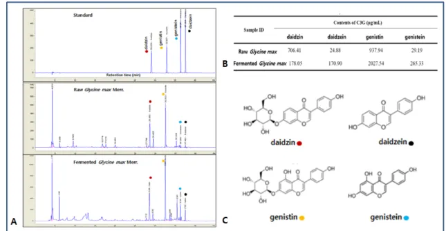
청호와 상엽을 이용한 발효에 따른 두시의 isoflavone 의 성분 변화를 조화하기 위해 HPLC을 실시하였다
10). 두시에 포함된 isoflavone은 genistin, genistiein, daidzine, daidzein으로 발효에 의해 genistin, genistein, daidzein는 증가하였고, daidzin은 감소하였다 (Fig. 1).
2. 피부염 실험모델
중앙실험동물 (한국)에서 분양 받은 태령 3주된
Nc/Nga 수컷 생쥐를 사용하였다. 대조군 (Ctrl군), 아토
피 피부염 유발군 (AE군), 두시 하태독법 시행 후 아토
피 피부염 유발군 (DT군)으로 나누었으며, 각 군에 각
10마리씩 배정하였다. DT군에 두시추출물 (20 ㎎/㎏)
를 음용 투여하는 하태독법을 실시한 후 4주 경과 후
생쥐 등 쪽 부위 피부를 면도한 다음 계면활성제
(surfactant)인 5% sodium dodeecyl sulfate (SDS: Sigma,
USA) 1 ㎖을 면봉으로 20회 문질러서 각질층의 lipid
lamella를 제거한 후 D. farinae crude extract striper (100
㎎, Biostir, Japan)을 3주동안 주 6회씩 도포하여 아토 피피부염을 1차 유발하였다. 1차유발 후 1주일 경과 후 동일한 방법으로 2차 유발하였다. 2차유발 72시간 후 sodium pentobarbital 용액으로 마취한 후 처지하였다.
얻어진 등쪽 피부를 10% NBF에 실온에서 24시간동안 고정한 후 통상적인 방법으로 paraffin에 포매하고 5 ㎛ 두께로 연속절편을 만들었다 (Fig. 2).
3. PKC 활성 조절 관찰
피부 ceramide 장벽 손상에 따른 protein kinase C (PKC) 활성 변화를 조사하기 위해 항 PKC 항체를 이 용한 면역조직화학적 염색을 실시하였다. 우선 피부절
편을 proteinase K (20 ㎍/㎖)에 5분 동안 proteolysis 과 정을 거친 후 blocking serum인 10% normal goat serum 에서 2시간 동안 반응시켰다. 그리고 1차 항체인 goat anti-PKC (1:200, Santa Cruz Biotec, USA)에 4 ℃ hu- midified chamber에서 72시간 동안 반응시켰다. 그런 다음 2차 항체인 biotinylated rabbit anti-goat IgG (1:100, Santa Cruz Biotec)에 실온에서 24시간 link 하였 고, 그런 다음 avidin biotin complex kit (Vector Lab, USA)에 1시간동안 실온에서 반응시켰다. 0.05%
3,3'-diaminobenzidine과 0.01% HCl이 포함된 0.05 M tris-HCl 완충용액 (pH 7.4)에서 발색시킨 후, hematox- ylin으로 대조염색하였다.
Fig. 1. The compositional changes in douchi (Artemisia Apiacea Herba and Mori Folium fermaneted Glycine max Merr.). A. The representative chromatograms of isoflavones in the standard solution, Glycine max Merr., and douchi extracts.
Each compound was detected with AegisPak-L C18 (4.6 × 150 mm ID, 3 micro pore size) at UV 260 nm. Peak marker: ●, daidzin; ●, daidzein; ●, genistin; ●, genistein
Fig. 2. Protocol of HTD for D. farinae induced atopy like dermatitis
80 Anti-inflammatory Effects of Hataedock with Douchi in Atopic Dermatitis-like Skin Lesions in House Dust Mite-Induced NC/Nga Mice
4. 부종 조절 관찰
1) 진피내 아교섬유 분포 변화 관찰
아교섬유 (collagen fiber)의 변화를 관찰하기 위해 Masson trichrome 염색을 실시하였다. 우선 50 - 60 ℃ Bouin 용액에서 1시간동안 매염 처리한 다음 70% 에 탄올에서 picric acid를 제거하였다. Weigert iron hema- toxylin에서 10분 동안 반응시켜 핵 염색하고, Biebrich scalet-acid fuchsin와 phosphomolybdic-phosphotungstic acid에서 각각 15분간, aniline blue에 5분간 처리하여 아교섬유 (청색)을 염색한 후 관찰하였다.
2) 아교섬유분해효소 변화 관찰
아교섬유분해효소인 matrix metalloproteinases (MMP)-9 분포변화를 관찰하기 위해 goat anti mouse MMP-9 (1:100, Santa Cruz Biotec) 항체를 이용한 면역조직화학 적 염색을 실시하였다.
5. 혈관 관찰
1) 신생혈관생성의 영상분석 변화 관찰
피부를 절개하여 젖힌 후 나타난 혈관을 × 4배율로 촬영하고, Image pro Plus (Media Cybernetic, USA)에서 먼저 image 기능의 sharpen low - filter를 사용하여 혈관 을 명확하게 하였다. 그런 다음 binary morphology에서 invert 기능을 선택하여 모세혈관을 intensity 180-200 으로 전환, 부각시킨 후 관찰하였다.
2) iNOS 변화 관찰
NO 생성을 통한 혈관 확장에 관여하는 염증 효소인 inducible nitric oxide synthase (iNOS)의 조직내 분포를 조사하기 위해 rabbit anti-mouse iNOS (1:250, Santa Cruz Biotec)를 이용한 면역조직화학적 염색을 실시하였다.
6. 영상 분석
면역조직화학의 결과는 image Pro Plus (Media cy- bernetics, USA)를 이용한 영상분석을 통해 수치화 (means ± standard error) 했다. 각 군의 표본에서 임의로 선정된 피부를 x 400배율에서 촬영한 다음 positive pix- els/20,000,000 pixels로 영상분석 하였다.
7. 통계
피부손상점수와 면역조직화학 결과의 통계는 SPSS software (SPSS 20, SPSS Inc., USA)를 이루어졌으며,
one-way ANOVA 시행을 통해 유의성 (P<0.05)을 검증 하고 Duncan’s multiple range test로 사후 검증하였다.
8. 윤리
본 연구과정은 부산대학교 동물실험윤리위원회 승 인을 받아 시행되었으며 (IACUC number: PNU-2015- 0924), 실험실 동물의 관리와 사용에 대해서는 NIH 가 이드라인에 따라 시행되었다.
Ⅲ. Results
1. 외부피부 손상 완화 효과
AE군의 피부 대부분에서 각질층이 손상된 습진 (eczema)이 나타났으며, 일부 가장자리지역에서는 탈락 과정에 있는 혈병 (blood clot)과 표피 잔재도 관찰되었 다. 이에 반해 DT군은 일부 지역을 제외하고는 정상적 인 외부형태로 관찰되었으며, 피부손상점수는 AE군에 비해 51% 감소하였다 (Fig. 3).
2. PKC 활성 조절 효과
아토피 피부염에서의 각질내 ceramide결핍에 의해 AE군에서는 PKC 활성이 일어났다. 항 PKC항체를 이 용한 면역조직화학적 염색 결과, PKC 양성반응은 손 상된 각질세포의 세포사이공간 (intercellular space)과 기저막 주변부에서 증가되었다. DT군에서는 PKC 양 성반응은 감소하였는데, 영상분석 결과 AE군에 비해 91% 감소한 것으로 관찰되었다 (Fig. 4).
3. 부종 조절 효과
콜라겐 분해효소인 MMP-9의 생성은 세포외기질과 기저막을 구성하는 단백질 성분을 분해함으로써 조직파 괴를 유도하여 염증관련 세포의 이주를 용이하게 하는 부종을 야기하는데, AE군에서는 MMP-9 생성이 증가가 관찰되었다. DT군에서는 MMP-9 생성이 적었는데, AE 군에 비해 48% 감소한 것으로 관찰되었다 (Fig. 5).
Masson trichrome method를 이용한 부종 변화를 관
찰한 결과, AE군에서는 부종이 증가했으며 과립백혈
구 및 림프구의 기저층 침윤 증가, 모세혈관 분포증가,
진피내 아교섬유 분포 감소가 일어났다. DT군에서의
부종 생성은 AE군에 비해 적었다 (Fig. 5).
Fig. 3. The alleviation of external skin damages by Hataedock. The items of skin score are (1) erythema/hemorrhage (2) scarring/dryness (3) edema (4) excoriation/erosion was scored as 0 (none), 1 (mild), 2 (moderate), or 3 (severe); The sum of the individual scores was defined as the atopy skin score17). Abbreviation: Ctrl, no-AE elicited group. AE, Atopy dermatitis (AD) elicitated group; DT, Douchi as fermented Glycine Semen Preparata extract treated group after AD elicitation. *, p<0.005 compared with AE group
Fig. 4. The regulation of PKC activation by Hataedock (PKC immunohistochemistry). Abbreviation: arrow, PKC positive reaction; EP, epithelim; DE, dermis; SC, stratum corneum; Bar size, 100 ㎛
82 Anti-inflammatory Effects of Hataedock with Douchi in Atopic Dermatitis-like Skin Lesions in House Dust Mite-Induced NC/Nga Mice
4. 혈관확장 효과
NO 생성을 통한 혈관 확장으로 염증을 주도하는 iNOS는 AE군에서는 증가한 것으로 관찰되었다. 항 iNOS항체를 이용한 면역조직화학적 염색 결과, iNOS 양성반응은 진피내 대식세포에서 확인되었다. 반면 DT군은 iNOS 양성반응이 감소했는데, 영상분석 결 과는 AE군에 비해 51% 감소한 것으로 관찰되었다 (Fig. 6).
혈관 변화는 피부를 절개하여 젖힌 후 혈관을 x 4배 율로 촬영한 다음 반전 (divert)시켜 관찰하였다. 대조 군에 비해 AE군에서는 혈관의 가지수가 증가한 반면, DT군에서는 AE군에 비해 감소를 보였다 (Fig. 6).
Ⅳ. Discussion
본 연구에서는 AD-induced Nc/Nga 생쥐에 두시 추 출물을 투여하여 AD에 대한 항염증 효과를 확인하고 자 하였다. 그 결과, DT군에서 AE군에 비해 PKC 활성 조절로 인한 i-NOS, MMP-9와 같은 염증 인자들 (inflammatory factors)의 감소가 야기되었고 이는 AD의 주 증상인 소양감 및 부종, 진피 내 신생혈관의 수가 감소되는 결과를 보였다.
AD의 주요 원인 중 하나는 피부각질층 내의 ceram- ide 손실로 발생하는 피부장벽 손상이 대표적이다
18). 피부장벽의 손상은 외인성 알레르겐에 대해 장벽으로
Fig. 5. The mitigative effects of edema by Hataedock (MMP-9 immunohistochemistry & Masson trichrome method).Abbreviation: arrow, MMP-9 positive reaction; Bar size, 100 ㎛
작용하는 이러한 각질층의 disruption으로 야기된다
19). 각질층의 손상은 sphingosine을 생성시키며, 생성된 sphingosine이 protein kinase C (PKC)의 활성을 증가시 킨다
20). 활성화된 PKC는 피부 상피 기저층에서 IL-4와 같은 Th2 사이토카인의 활성을 야기한다고 보고된바 있다
21). 이와 같은 Th2 cytokine의 활성화는 항원특이 적인 IgE를 생성으로 이어져, 비만세포에 결합하며, 활 성화된 비만세포는 탈과립화하여 히스타민과 같은 염 증매개물질을 유리함을 통하여 혈관확장 및 소양감을 야기한다
22).
본 연구에서는 Nc/Nga 생쥐에 sodium dodeecyl sul- fate을 적용하여 ceramide 층을 붕괴해 피부장벽기능의 손상을 야기하여 AD가 유발되기 쉬운 조건을 제공하였
다. 이후 D. pteronissinus를 노출시켜 AD-like skin lesion 을 발생시켰다. 항원의 도포는 Th1/Th2 균형 상태를 Th2 우세형 상태로 옮겼다. 이는 한의학적 관점에서 피 부에 표열 (피부 표층의 열) 상태를 유발된 것으로 볼 수 있다. 또한 3주령된 어린 Nc/Nga mice를 사용하였는 데, Nc/Nga mice는 conventional한 조건에서 자발적으로 심각한 피부염을 유발하는 동물모델로서
23), AD이 성인 보다 소아에서 높은 발병률을 보이는 질환으로 볼 때
24), 3주령된 Nc/Nga생쥐는 적합한 동물모델로 사료된다.
AD는 Th 면역 불균형 상태와 외인성 알레르겐의 반응 으로 발생하게 되며, 이는 한의학에서 AD를 내부에 쌓 인 열 (태독)과 외부의 사기 (external manifestation)의 접 목으로 바라보는 관점과도 일치한다.
Fig. 6. The amelioration of vasodilatation in dermis by Hataedock (iNOS immunohistochemistry & angio-exposure).
Abbreviation: arrow, iNOS positive reaction; Bar size, 100 ㎛; Angio-exposure, x4
84 Anti-inflammatory Effects of Hataedock with Douchi in Atopic Dermatitis-like Skin Lesions in House Dust Mite-Induced NC/Nga Mice
연구 결과, AE군에서 Ctrl군에 비해 PKC의 활성이 증가하였고 (Fig. 4), 비만세포의 활성화는 각종 호산구 및 대식세포로부터 각종 proinflammatory cytokine의 분 비를 증가시켜 부종을 야기하는 MMP-9의 분비를 증 가시켰다 (Fig. 5). 또한 염증반응의 전사인자인 i-NOS 를 활성화시켜 혈관 투과성 증가, 부종 등의 염증반응 을 촉진하는 결과를 보여주었다 (Fig. 6). 이는 피부 내 축적된 열이 그 원인으로 볼 수 있으며, 각종 면역학적 변화는 정상적 면역체계가 열 또는 화에 의해 교란되 어 발생했다고 해석할 수 있다. 실험결과에서 보여진 다양한 염증세포의 교란 및 이로 인해 야기된 염증증 상 및 부종은 한의학에서 말하는 ‘열 (熱)’이 혈류의 흐 름 증가 및 피부 내 염증세포들의 이주를 촉진시킴으 로서 발생된 것으로 사료된다.
본 연구에서 우리는 한의학에서 차가운 성질을 가 진 두시를 사용하면 피부 표층의 쌓인 열을 제거하는 효과가 있을 것을 기대하였다. 두시는 한국에서 오랫 동안 음식과 한약재로 사용되어 왔다. 한약학적 분류 상 辛凉解表藥에 속하며, 表部의 열을 발산시키는 효 능이 있고, 폐경 (Lung)에 귀경하며, 성질이 차고, 淸熱 解毒한다고 알려져 있다. DE군의 PKC 발현 억제는 염 증의 개시단계 발생을 제어하는 것으로서, 이는 두시 가 열의 발생을 제어할 수 있는 가능성을 보여준다. 또 한 탈과립화된 비만세포는 MMP-9분비를 억제하여 부 종을 감소시키고, i-NOS response 감소를 통해 혈류흐 름이 감소하였다. 최종적으로 이러한 조직학적 변화는 염증성 피부손상의 완화라는 결과를 유도하였다 (Fig.
3). 이는 한의학에서 이르는 두시의 청열 기능 (blowing out of heat 또는 heat treatment)이 열이 발생할 수 있는 부분들을 제어한 것으로 생각된다.
발효콩은 isoflavone 중 하나인 genistein을 포함하고 있으며, 이는 Protein Kinase C (PKC)와 같은 tyrosine kinase를 억제하는 효과를 가지는 것으로 알려져 있다
25)
. 선행연구에 의하면 PKC는 Th2 사이토카인 내의 PKC 의존성 Ca채널을 조절함으로서 Th2 cell function 의 활성화를 야기한다
26). Genistein의 PKC의 억제효과 는 PKC에 의해 야기되는 Th2 사이토카인의 발현을 억 제함으로서 비만세포의 활성화를 제어하게 되는데, 이 는 염증반응의 개시를 막는 역할을 하게 된다
27). 따라 서 PKC의 활성화 차단은 인체 내 염증성 알레르기 질 환의 발현을 예방하는 새로운 전략을 제시할 수 있다.
결론적으로 두시는 염증이 발현되는 초기 단계인 PKC활성의 억제기전을 통해 Th2 dominant state 상태
를 억제하여 염증이 발현되는 초기 단계에 억제효과를 보여주었다. 여러 연구들에서 두시와 같은 발효콩의 Th2 면역 조절 효과를 통한 염증성 알러지 질환에 대 한 치료효과를 보고하고 있으며
9,11-2), 최근에는 태열의 발생과 연관지어 PKC 억제를 통하여 Th2 cytokine의 발현 자체를 억제하는 것을 보여주는 논문도 발표되고 있다
10).
향후 두시의 아토피피부염에 대한 치료 가능성의 분자 메커니즘을 설명할 수 있는 추가적인 연구가 필 요하다고 사료된다. 또한, 두시의 농도별 아토피피부 염에 대한 효과를 알아보기 위해 추가적으로 dose de- pendent manner를 이용한 실험도 필요할 것으로 생각 된다.
Acknowledgement
이 연구는 2014년도 정부 (교육부)의 재원으로 한국 연구재단의 지원을 받아 수행된 기초연구사업임 (No.
NRF-2014R1A1A2055061).
References
1. Leung DY, Bieber T. Atopic dermatitis. Lancet. 2003;
361(9352):151-60.
2. Schneider L, Tilles S, Lio P, Boguniewicz M, Beck L, LeBovidge J, Novak N, Bernstein D, Blessing-Moore J, Khan D, Lang D, Nicklas R, Oppenheimer J, Portnoy J, Randolph C, Schuller D, Spector S, Tilles S, Wallace D. Atopic dermatitis: a practice parameter update 2012.
J Allergy Clin Immunol. 2013;131(2):295-9.
3. Ministry of Health and Welfare. Korea Centers for Disease Control and Prevention. Korea Health Statistics 2012:
Korea National Health and Nutrition Examination Survey (KNHANES V-3). Cheongwon: Korea Centers for Disease Control and Prevention. 2013.
4. Halonen M, Lohman IC, Stern DA, Spangenberg A, Anderson D, Mobley S, Ciano K, Peck M, Wright AL.
Th1/Th2 patterns and balance in cytokine production
in the parents and infants of a large birth cohort. J
Immunol. 2009;182(5):3285-93.
5. Im GM, Joeng HW, Kim HS, Jeong WY. Oriental medical approach on the allergic disease. Korea J Orient Physiol Pathol. 2002;16(5):831-9.
6. Kang MY, Jang GT, Kim JH. A study on fetal toxicosis removal therapy. J Pediatr Korean Med. 2003;17(1):
29-51.
7. Im GM, Jeong HW, Kim HS, Jeong WY. Oriental medical approach on the allergic disease. Korea J Orient Physiol Pathol. 2002;16(5):831-9.
8. Miller AK, Benson JM, Muanza DN, Smith JR, Shepherd DM. Anti-inflammatory effects of natural product for- mulations on murine dendritic cells. J Diet Suppl.
2011;8(1):19-33.
9. Yeh CY, Jung CJ, Huang CN, Huang YC, Lien HT, Wang WB, Wang LF, Chia JS. A legume product fer- mented by Saccharomyces cerevisiae modulates cutaneous atopic dermatitis-like inflammation in mice. BMC Complement Altern Med. 2014;18(14):194.
10. Jung AR, Ahn SH, Park IS, Park SY, Jeong SI, Cheon JH, Kim KB. Douchi (fermented Glycine max Merr.) alleviates atopic dermatitis-like skin lesions in NC/Nga mice by regulation of PKC and IL-4. BMC Complement Altern Med. 2016;16:416-30.
11. Matsuda A, Tanaka A, Pan W, Okamoto N, Oida K, Kingyo N, Amagai Y, Xia Y, Jang H, Nishikawa S, Kajiwara N, Ahn G, Ohmori K, Matsuda H.
Supplementation of the fermented soy product ImmuBalance
™effectively reduces itching behavior of atopic NC/Tnd mice. J Dermatol Sci. 2012;67(2):130-9.
12. Zhang T, Pan W, Takebe M, Schofield B, Sampson H, Li XM. Therapeutic effects of a fermented soy product on peanut hypersensitivity is associated with modulation of T-helper type 1 and T-helper type 2 responses. Clin Exp Allergy. 2008;38(11):1808-18.
13. Bao ZS, Hong L, Guan Y, Dong XW, Zheng HS, Tan GL, Xie QM. Inhibition of airway inflammation, hyper- responsiveness and remodeling by soy isoflavone in a murine model of allergic asthma. Int Immunopharmacol.
2011;11(8):899-906.
14. Joo KM, Hwang JH, Bae SJ, Nahm DH, Park HS, Ye YM, Lim KM. Relationship of ceramide-, and free fatty acid-cholesterol ratios in the stratum corneum with
skin barrier function of normal, atopic dermatitis lesional and non-lesional skins. J Dermatol Sci. 2015;77(1):71-4.
15. Jung BG, Cho SJ, Ko JH, Lee BJ. Inhibitory effects of interleukin-10 plasmid DNA on the development of atopic dermatitis-like skin lesions in NC/Nga mice.
J Vet Sci. 2010;11(3):213-20.
16. Hengge UR, Ruzicka T, Schwartz RA, Cork MJ. Adverse effects of topical glucocorticosteroids. J Am Acad Dermatol. 2006;54(1):1-15.
17. Leung DY, Hirsch RL, Schneider L, Moody C, Takaoka R, Li SH, Meyerson LA, Mariam SG, Goldstein G, Hanifin JM. Thymopentin therapy reduces the clinical severity of atopic dermatitis. J Allergy Clin Immunol.
1990;85(5):927-33.
18. Janssens M, van Smeden J, Gooris GS, Bras W, Portale G, Caspers PJ, Vreeken RJ, Hankemeier T, Kezic S, Wolterbeek R, Lavrijsen AP, Bouwstra JA. Increase in short-chain ceramides correlates with an altered lipid organization and decreased barrier function in atopic eczema patients. J Lipid Res. 2012;53(12):2755-66.
19. Park YH, Jang WH, Seo JA, Park M, Lee TR, Park YH, Kim DK, Lim KM. Decrease of ceramides with very long-chain fatty acids and downregulation of elon- gases in a murine atopic dermatitis model. J Invest Dermatol. 2012;132(2):476-9.
20. Abboushi N, El-Hed A, El-Assaad W, Kozhaya L, El-Sabban ME, Bazarbachi A, Badreddine R, Bielawska A, Usta J, Dbaibo GS. Ceramide inhibits IL-2 production by preventing protein kinase C-dependent NF-kappaB activation: possible role in protein kinase ctheta regulation.
J Immunol. 2004;173(5):3193-200.
21. Ballou LR, Laulederkind SJ, Rosloniec EF, Raghow R.
Ceramide signalling and the immune response. Biochim Biophys Acta. 1996;1301(3):273-87.
22. Amin K. The role of mast cells in allergic inflammation.
Respir Med. 2012;106(1):9-14.
23. Matsuda H, Watanabe N, Geba GP, Sperl J, Tsudzuki M, Hiroi J, Matsumoto M, Ushio H, Saito S, Askenase PW, Ra C. Development of atopic dermatitis-like skin lesion with IgE hyperproduction in NC/Nga mice. Int Immunol. 1997;9(3):461-6.
24. Ben-Gashir MA, Seed PT, Hay RJ. Predictors of atopic
dermatitis severity over time. J Am Acad Dermatol.
86 Anti-inflammatory Effects of Hataedock with Douchi in Atopic Dermatitis-like Skin Lesions in House Dust Mite-Induced NC/Nga Mice