DOI : 10.3341/jkos.2009.50.7.1115
= 증례보고 = 접수번호 : 50-07-01-11
데스메막박리 각막내피층판이식술을 시행한 수포각막병증 8예의 단기 임상 결과 보고
정희옥⋅이현수⋅나경선⋅주천기 가톨릭대학교 의과대학 안과 및 시과학교실
목적: 데스메막박리 각막내피층판이식술을 시행한 수포각막병증 환자 또는 각막이식 실패 환자 8예의 단기 임상 결과에 대해 보고하 고자 한다.
증례요약: 데스메막박리 각막내피층판 이식술을 시행한 8안을 대상으로 술 후 임상 경과를 알아보기 위해 의무기록를 후향적으로 조사 하였다. 전체 8안 중 5안은 수포성 각막병증으로, 3안은 각막이식 실패로 데스메막박리 각막내피층판이식술을 시행받았다. 최대 교정 시력(logMAR)은 술 전 평균 1.625±0.60에서 술 후 3개월째 0.825±0.35로 호전되었고, 각막두께는 785.63±135.58 μm에서 692.00±150.85 μm로 호전되었다. 술 후 2안에서 수여각막내피이식편의 부분적인 탈락이 관찰되어 전방내 공기를 재주입하고 이식편의 위치를 조정하였고, 술 후 6개월까지 2안에서 거부반응을 보였으며, 그 중 1안에서 결국 이식 실패를 보였다.
결론: 데스메막박리 각막내피층판이식술은 각막내피세포 부전증 환자에서 빠른 시력교정 효과와 안정된 이식 각막편 유지가 기대되는 수 술법으로 알려져 있으며, 앞으로 더 나은 수술 결과를 위해 수술 숙련도 향상 및 수술 기법 개발에 노력해야 할 것으로 사료되는 바이다.
<대한안과학회지 2009:50(7):1115-1119>
■ 접 수 일: 2009년 1월 13일 ■ 심사통과일: 2009년 4월 14일
■ 책 임 저 자: 주 천 기
서울시 서초구 반포동 505 가톨릭대학교 서울성모병원 안과
Tel: 02-2258-7620, Fax: 02-533-3801 E-mail: [email protected]
* 본 논문의 요지는 2008년 대한안과학회 제100회 추계학술대회에서 구연으로 발표되었음.
각막혼탁을 일으키는 다양한 각막 질환에 대해 이제까지는 전층각막이식술만이 그 치료 방법으로 시행되어 왔다. 그러 나 전층각막이식술은 회복 기간이 비교적 길며 및 술 후 난 시의 발생이 크고, 이식 실패, 봉합사로 인한 감염 및 염증, 각막 미란 및 궤양, 2차성 녹내장 발병 및 백내장 발생과 악화 등 여러 합병증의 위험이 있다.1-3각막 내피세포의 이상이 원인이 되는 각막 질환은 각막이식술의 흔한 적응증으로4-6 이에 각막내피세포만을 선택적으로 이식하는 수술이 시도 되어 왔다. 1998년, Melles et al7은 손상된 각막내피세포를 선택적으로 이식하는 후부층판 각막이식술(Posterior lamellar keratoplasty)을 소개하였고, 이후Terry and Ousley8이 심층 판 각막내피세포 이식술(Deep lamellar endothelial kerato- plasty)을, Price and Price9가 데스메막박리 각막내피층판 이식술(Descemet-stripping endothelial keratoplasty)을 시행하였고, Gorovoy10가 미세각막절개도를 이용한 데스메 막박리 각막내피층판이식술(Descemet-stripping auto- mated endothelial keratoplasty)을 소개하였다.
저자들은 본원에서 시행한 수포각막병증 환자와 각막내 피세포 부전으로 인한 각막이식 실패 환자 8안의 데스메막 박리 각막내피층판이식술의 수술 후 시력, 각막 두께 및 술 후 합병증 등 단기 임상 결과 보고를 하고자 한다.
증례보고
2006년 9월부터 2008년 8월까지 본 대학 부속 병원 안 과를 방문하여 수포각막병증 및 각막이식 실패로 진단된 환자 중 데스메막박리 각막내피층판이식술을 시행한 8안의 환자를 대상으로 의무기록을 통한 후향적 연구를 시행하였 다. 대상 환자 8안 중 5안은 수포성 각막병증이었고(Group A), 3안은 각막이식 후 이식실패로 재수술을 받은 경우였 다(Group B)(Table 1). 공여 각막은 8안 중 5안이 수입 각 막이었으며, 공여 각막의 평균 연령은 58.88±9.63세였고, 사망 후 각막의 보존까지의 평균 시간은 442.50±383.12분 이었다(Table 2).
모든 수술은 단일 수술자에 의해 시행되었으며 구후마취 를 하였다. 공여 각막을 인공 전방에 올려놓고, 300 또는 350 μm head의 미세각막절개도을 이용하여 공여각막편을 만들었다. 수여자의 각막은 환자의 머리쪽 방향에서 1 mm 각막절개창을 만들어 공기 주입 후 Modified Price-Sinskey hook을 이용, 데스메막을 박리하였고, 코와 귀쪽 방향의 5 mm의 윤부절개창을 만들어 I&A로 박리된 데스메막을 제거하였다. 공여 각막편의 내피세포 면에 점탄물질을 올려
Table 1. Patients Characteristics
Group Case Age (yrs) Sex Pathology Preoperative BCVA* Preoperative corneal
thickness (μm) Remarks
A 1 56 M† BK§ 1.0 622
2 75 F‡ BK 1.60 856
3 58 M BK 1.30 797
4 63 M BK 1.60 749
5 69 M BK 3.0 734 Glaucoma
B 6 69 M Graft failure after PKP∏ 1.60 967 Glaucoma
7 59 M Graft failure after PKP 1.60 607
8 54 M Graft failure after DSAEK# 1.30 953
Mean 62.88±7.43 1.625±0.60 785.63±135.58
*BCVA=best corrected visual acuity; †M=male; ‡F=female; §BK=bullous keratopathy; ∏PKP=penetrating keratoplasty; #DSAEK=
Descemet-stripping automated endothelial keratoplasty.
A
B
C
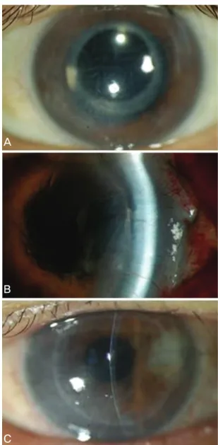
Figure 1. Preoperative and postoperative photographs (Case 3): Graft dislocation. (A) Preoperative picture.
(B) Picture which shows graft dislocation (Postoperative 10 days). (C) Picture after air injection and graft repo- sition (Postoperative 1 month).
놓고 Taco 방식으로 반으로 접어 윤부 절개창을 통해 전방 내에 삽입한 후 무균 공기를 전방에 주입하여 각막편의 생 착을 도왔다. 술 후 레보플록사신과 프레드니솔론 1% 안약 을 하루에 4번 점안토록 하였으며, 술 후 6시간 이상 앙와 위로 침상 안정을 하도록 하였다.
모든 환자의 평균 경과 관찰 기간은 4.14±1.35개월(3~
6개월)이었고, 모든 환자에서 수술 중 합병증은 발생하지 않았으나, 수술 후 2안에서 각각 술 후 10일째와 1일째에 수여 각막이식편의 부분적인 탈락이 관찰되었고, 이에 점안 마취하에 30-gauge 바늘을 통해 무균 공기를 전방내 재주 입하고 이식편의 위치를 조정하여 생착을 도왔다(Fig. 1).
그리고 2안에서 일시적인 각막 간질층의 부종 및 혼탁을 보 였으며, 1안에서는 치료 후 호전되었으나 나머지 1안은 결 국 이식실패를 보였다(Fig. 2). 이식 실패를 보인 1안을 제 외한 7안의 환자에서 이식된 각막편은 잘 유지되었고, 이식 실패 환자는 현재 각막이식 대기 중에 있다(Table 3). 수술 후 3개월 째, 이식 실패를 보인 1안을 제외하고 시력 호전 을 보였으며 평균 최대 교정시력(logMAR)은 수술 전 1.625±0.60에서 수술 후 0.825±0.35로 측정되었고, 각막 두께는 일시적인 거부반응을 보인 1안과 이식 실패한 1안 을 포함하여 총 3안에서 술 전에 비교하여 증가하였으나 나 머지 5안에서는 감소하여, 평균 각막두께는 술 전 785.63±
135.58 μm에서 692.00±150.85 μm로 호전되었다. 그러나 각막 내피세포는 2안을 제외하고 수술 후 3개월의 경과 관 찰기간 동안에는 측정되지 않았다(Table 4).
고 찰
데스메막박리 각막내피층판이식술은 미세각막절개도10를 이용하여 수여자의 각막 내피층과 데스메막을 제거하고, 공 여 각막의 내피층, 데스메막 그리고 각막 실질 일부를 이식 하는 방법이다. 비교적 작은 절개창을 만듦으로써 안구 형
A B
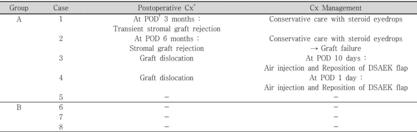
Figure 2. Preoperative and postoperative photographs (Case 2): Graft failure. (A) Preoperative picture. (B) Graft failure state (Postoperative 3 months).
Table 2. Characteristics of donor corneas
Group Case Recipient’s pathology Donor age (yrs) Death to preservation time Donor corneal ECC*(/mm2)
A 1 BK† 64 11 hrs 36 min 2974
2 BK 72 17 hrs 21 min 2521
3 BK 46 1 hr Unchecked
4 BK 61 2 hrs Unchecked
5 BK 54 11 hrs 44 min 2718
B 6 Graft failure after PKP‡ 65 8 hrs 5 min 3024
7 Graft failure after PKP 64 11 hrs 36 min 2974
8 Graft failure after DSAEK§ 45 2 hrs 30 min 2475
Mean 58.88±9.63 442.50±383.12 min 2781.00±244.44
*ECC=endothelial cell count; †BK=bullous keratopathy; ‡PKP=penetrating keratoplasty; §DSAEK=Descemet-stripping automated endothelial keratoplasty.
Table 3. Postoperative complication and management
Group Case Postoperative Cx* Cx Management
A 1 At POD†3 months :
Transient stromal graft rejection
Conservative care with steroid eyedrops
2 At POD 6 months :
Stromal graft rejection
Conservative care with steroid eyedrops
→ Graft failure
3 Graft dislocation At POD 10 days :
Air injection and Reposition of DSAEK flap
4 Graft dislocation At POD 1 day :
Air injection and Reposition of DSAEK flap
5 - -
B 6 - -
7 - -
8 - -
*Cx=complication; †POD=postoperative day.
태의 유지에 유리하고, 봉합을 시행하지 않아 봉합사로 인 한 합병증이 없으며, 시력 회복이 빠르며 굴절력 이상이 적 고 예측 가능하다고 알려져 있다. 그리고 비교적 비침습적 으로 이식 실패의 경우에 재수술을 시행한다고 하더라도 성공률이 더 높다.11-15그러나 미세각막절개도를 이용할 경우 공여 각막이식편의 제작 중 인공 전방위에 가해지는 높은 압력, 이식편을 접어 삽입하는 Taco 방식, 그리고 이식편
삽입 후 전방내 공기를 주입함으로써 야기되는 높은 안구 내 압력에 의해 공여 각막이식편의 손상이 예상될 수 있 다.16그리고 수술 후 이식편의 위치이상이 30% 이상 발생 한다는 보고가 있으며,17 기존의 전층각막이식술에 익숙한 술자에게 새로운 수술 과정에 대한 숙련과정이 필요하고, 이는 수술 결과에 영향을 미칠 수 있다.16
데스메막박리 각막내피층판이식술에서 수여 각막 및 공
Table 4. Results of grafted corneas
Group Case Preop BCVA Postop BCVA* Preop corneal thickness (μm)
Postop corneal thickness (μm)
Donor corneal ECC† (mm2)
Postop grafted corneal ECC (/mm2)
A 1 1.0 0.7 622 715 2974 Uncheckable
2 1.6 1.6 856 977 2521 Uncheckable
3 1.3 0.5 797 466 Unchecked Uncheckable
4 1.6 0.5 749 608 Unchecked 2123
5 3 1.0 734 614 2718 Uncheckable
B 6 1.6 0.8 967 771 3024 Uncheckable
7 1.6 0.7 607 633 2974 1865
8 1.3 0.8 953 752 2475 Uncheckable
Mean 1.625±0.60 0.825±0.35 785.63±135.58 692.00±150.85 2781.00±244.44
*BCVA=best corrected visual acuity; †ECC=endothelial cell count.
여 각막편 준비, 이식편의 삽입 방법 등에 다양한 시도가 이루어지고 있다. 첫째, 미세각막절개도를 대신하여 펨토초 레이저를 이용하여 쉽고 빠르게 그리고 균일한 이식편을 만들 수 있었다는 보고가 나오고 있다.18,19둘째, Taco 방식의 각막이식편의 전방내 삽입 방법은 이식편의 내피세포 손상 을 일으킬 수 있는데20이에 Massimo et al21은 Busin glide 를 이용했을 때 내피세포 감소는 26.4±2.7%로, 전층 각막 이식술(<30%) 및 기존 방식의 데스메막박리 각막내피층 판이식술(<50%)에 비해 비교적 내피세포 손상이 적었다 고 보고하였다. 마지막으로 최근 데스메막 및 각막내피세포 이식술(Descemet membrane and endothelial kera- toplasty)이 후부 층판 각막이식술의 역사에 한 단계 발전 된 새로운 수술법으로 시도되고 있다.22
본 연구에서는 대부분의 환자에서 큰 합병증 없이 이식 편이 비교적 잘 생착되었으나 수입각막이 8안 중 5안으로 사망에서 보존, 이식까지 경과된 시간이 길었고, 미세각막 절개도를 이용하면서 두께가 균일하지 못한 이식편이 만들 어져 예후에 영향을 주었다. 또한 대상 환자의 수가 적었고, 경과 관찰 기간이 다소 짧았기 때문에 시력 및 각막 두께, 내피세포의 변화를 충분히 관찰하지 못한 제한점이 있어 술 후 장기적 예후에 대해서는 더 연구가 필요할 것이다.
본 연구에서는 각막 내피세포 부전 환자에서 데스메막박리 각막내피층판이식술을 시행할 때, 보다 더 나은 수술 결과를 위해 수술 숙련도 향상 및 각막이식편의 제작과 수술 기법 개발에 더 많은 연구와 발전이 필요할 것으로 사료된다.
참고문헌
1) Thompson RW Jr, Price MO, Bowers PJ, Price FW Jr. Long-term graft survival after penetrating keratoplasty. Ophthalmology 2003;
110:1396-402.
2) Ing JJ, Ing HH, Nelson LR, et al. Ten-year postoperative results of penetrating keratoplasty. Ophthalmology 1998;105:1855-65.
3) Boisjoly HM, Tourigny R, Bazin R, et al. Risk factors of corneal
graft failure. Ophthalmology 1993;100:1728-35.
4) Al-Towerki AE, Gonnah el-S, Al-Rajhi A, Wagoner MD. Changing indications for corneal transplantation at the King Khaled Eye Specialist Hospital (1983–2002). Cornea 2004;23:584-8.
5) Al-Yousuf N, Mavrikakis I, Mavrikakis E, Daya SM. Penetrating keratoplasty: indications over a 10 year period. Br J Ophthalmol 2004;88:998-1001.
6) Kang PC, Klintworth GK, Kim T, et al. Trends in the indications for penetrating keratoplasty (1980-2001). Cornea 2005;24:801-3.
7) Melles GR, Eggink FA, Lander F, et al. A surgical technique for posterior lamellar keratoplasty. Cornea 1998;17:618-26.
8) Terry MA, Ousley PJ. Deep lamellar endothelial keratoplasty in the first United States patients: early clinical results. Cornea 2001;
20:239-43.
9) Price FW Jr, Price MO. Descemet’s stripping with endothelial keratoplasty in 50 eyes; a refractive neutral cornea transplant. J Refract Surg 2005;21:339-45.
10) Gorovoy MS. Descemet-stripping automated endothelial kerato- plasty. Cornea 2006;25:886-9.
11) Vincenzo S, Patricia T. Descemet-stripping automated endothelial keratoplasty by using suture for donor insertion. Cornea 2008;
27:825-9.
12) Koenig SB, Covert DJ. Early results of small incision Descemet’s stripping and automated endothelial keratoplasty. Ophthalmology 2007;114:221-6.
13) Koenig SB, Covert DJ, Dupps WJ Jr, Meisler DM. Visual acuity, refractive error, and endothelial cell density six months after DSAEK. Cornea 2007;26:670-4.
14) Price MO, Price FW Jr. DSEK: comparative outcomes with micro- keratome-dissected and manually dissected donor tissue. Ophthal- mology 2006;113:1936-42.
15) Tan DT, Mehta JS. Future directions in lamellar corneal trans- plantation. Cornea 2007;26:S21-8.
16) Olson RJ. Air and the corneal endothelium: an in vivo specular microscopy study in cats. Arch Ophthalmol 1980;98:1283-4.
17) Koenig GB, Covert DJ. Early results of small-incision Descemet’s stripping and automated endothelial keratoplasty. Ophthalmology 2006;114:221-6.
18) Seo WM, Kim HK. Early result of Femtosecond laser assisted Descemet’s membrane stripping endothelial keratoplasty. J Korean Ophthalmol Soc 2008;49:40-7.
19) Cheng YY, Pels E, Nuijts RM. Femtosecond laser assisted Descemet’s membrane stripping endothelial keratoplasty. J
=ABSTRACT=
Eight Cases of Descemet’s Stripping Automated Endothelial Keratoplasty in Eyes With Bullous Keratopathy
Hee Ok Jeong, MD, Hyun Soo Lee, MD, Kyung Sun Na, MD, Choun-Ki Joo, MD, PhD
Department of Ophthalmology and Visual Science, Seoul St. Mary’s Hospital, College of Medicine, The Catholic University of Korea, Seoul, Korea
Purpose: To investigate the postoperative results of Descemet’s stripping automated endothelial keratoplasty in bullous keratoplasty and graft failure cases.
Case summary: Eight eyes of eight patients who underwent DSAEK between September 2006 and August 2008 were followed-up for at least 3 months, and the charts of eight patients were reviewed retrospectively. Five eyes of the total eight had bullous keratopathy, and three eyes needed DSAEK due to graft failure. The best corrective visual acuity (logMAR) improved from 1.625±0.60 to 0.825±0.35 and corneal thickness decreased from 785.63±135.58 μm to 692.00±150.85 μm 3 months postopera- tively. Grafted corneal dislocation occurred in two eyes, and we repositioned the cornea in those participants by air injection.
We found graft rejection signs in two cases, and one case showed graft failure despite steroid therapy.
Conclusions: We think that DSAEK will be a good surgical procedure in bullous keratopathy or graft failure patients because of its favorable postoperative prognosis.
J Korean Ophthalmol Soc 2009;50(7):1115-1119
Key Words: Bullous keratoplasty, Descemet-stripping automated endothelial keratoplasty, Graft failure
Address reprint requests to Choun-Ki Joo, MD, PhD
Department of Ophthalmology, Seoul St. Mary’s Hospital, College of Medicine, The Catholic University of Korea
#505 Banpo-dong, Seocho-gu, Seoul 137-040, Korea
Tel: 82-2-2258-7620, Fax: 82-2-533-3801, E-mail: [email protected] Cataract Refract Surg 2007;33:152-5.
20) Mehta JS, Por YM, Poh R, et al. Comparison of donor insertion techniques for DSAEK. Arch Ophthalmol 2008;126:1383-8.
21) Busin M, Bhatt PR, Scorcia V. A modified technique for DSAEK
to minimize endothelial cell loss. Arch Ophthalmol 2008;126:
1133-7.
22) Melles GR. Posterior lamellar keratoplasty: DLEK to DSEK to DMEK. Cornea 2006;25:879-81.