pISSN: 0378-6471 eISSN: 2092-9374 DOI : 10.3341/jkos.2010.51.11.1431
= 증례보고 =
미세각막절개도를 이용한 데스메막박리 각막내피층판이식술의 국내 장기 임상 결과
이준성 박영걸 윤경철 전남대학교 의과대학 안과학교실
목적: 국내에서 데스메막박리 각막내피층판이식술을 시행받은 환자들의 장기 임상 결과를 알아보고자 하였다.
대상과 방법: 본원에서 수포각막병증으로 데스메막박리 각막내피층판이식술을 시행받은 환자 중 18개월 이상 경과 관찰하였던 7명(7안) 을 대상으로 수술 전, 후의 최대교정시력, 굴절이상 및 난시값, 각막내피세포수, 각막두께 등을 후향적으로 분석하였다.
결과: 평균 관찰 기간은 19.9 ± 2.9 (18~24)개월이었으며 평균 연령은 61.42 ± 10.13 (46~76)세였다. 전체 7안 중 6안(85.7%)에서 성 공적인 상태를 보였고 최대교정시력(logMAR)은 수술 전 1.62 (중앙값)에서 수술 후 1개월째 1.15 (중앙값) (p = 0.027)로 유의한 호전을 보였으며 최종 경과 관찰 시까지 유지되었다. 수술 후 18개월의 굴절이상 및 난시값은 수술 전과 비교하여 유의한 차이를 보이지 않았 다. 1안에서 이식실패를 보여 수술 후 12개월째 전층각막이식술을 시행하였다.
결론: 장기 경과 관찰 결과 데스메막박리 각막내피층판이식술은 빠른 시력 회복을 보였고 굴절 및 난시값의 변화가 적었으며, 각막내피 부전증 환자에 있어 선택할 수 있는 좋은 수술법이라 생각된다.
<대한안과학회지 2010;51(11):1431-1437>
접 수 일: 2010년 3월 10일 심사통과일: 2010년 8월 18일
책 임 저 자: 윤 경 철
광주광역시 동구 학동 8 전남대학교병원 안과
Tel: 062-220-6742, Fax: 062-227-1642 E-mail: [email protected]
1998년 Melles et al1에 의하여 후부층판 각막이식술 (posterior lamellar keratoplasty)이 소개된 후 각막내피세 포만을 선택적으로 이식하는 수술 기법은 전층각막이식술 을 대신하며 지속적으로 발전해 왔다. 그 동안 정상 각막지 형의 보존을 가능하게 한 심층판 각막내피이식술(deep la- mellar endothelial keratoplasty, DLEK)이 발표되었고, 이 후 매끄러운 수여각막편의 접촉면을 통해 더욱 안전한 생착 을 유도한 데스메막박리 각막내피층판이식술(Descemet- stripping endothelial keratoplasty, DSEK)이 소개되었다.2-4 또한 Gorovoy5는 미세각막절개도를 이용한 데스메막박리 각막내피층판이식술(Descemet-stripping automated en- dothelial keratoplasty, DSAEK)을 통해 보다 안전하고 일 정한 두께의 각막편을 만들 수 있도록 하였으며, 이는 현재 각막내피부전증 환자에서 우선적인 치료로 시행 되고 있다.
이후 DSAEK이 과거의 전층각막이식술(penetrating keratoplasty, PKP)이나 그 밖에 DLEK이나 DSEK와 비교 하여 더 빠른 시력 회복과 더 낮은 수술 후 난시값을 보인 다는 여러 보고들이 있었다.6-9또한 Bahar et al10은 DLEK
과 DSAEK을 동시에 시행받은 환자를 대상으로 연구한 결 과 최종 시력은 차이가 없었으나 낮은 고위수차 등의 이유 로 환자들은 DSAEK을 더 선호하였다고 하였다.
최근 국내에서 DSAEK 시행 후 3개월 경과 관찰 결과에 대한 보고가 있었으나 아직 1년 이상의 장기 임상 결과는 없는 실정으로, 이에 저자들은 본원에서 수포각막병증으로 진단 받고 DSAEK을 시행받은 환자 중 18개월 이상 경과 관찰이 가능하였던 7안에 대한 임상 결과를 분석하여 이를 보고하고자 한다.
대상과 방법
2008년 1월부터 본원 안과에 내원하여 수포각막병증으 로 진단받고 DSAEK을 시행받은 환자 중 18개월 이상 경 과 관찰이 가능하였던 환자를 대상으로 의무기록을 후향적 으로 분석하였다(Table 1). 대상 환자 모두에서 수술 전 검 사로 나안 시력, 최대 교정 시력(logMAR), 현성굴절검사, 안압, 중심각막두께(ultrasonic pachymeter, UP1000, Nidek, Japan), 세극등현미경검사, 경면현미경검사(specular mi- croscope, Noncon ROBO CA, Konan, Japan) 등을 시행하 였다.
공여 각막의 평균 나이는 44.86 ± 15.9 (18~63)세로 모 두 국내 기증 각막이었고, 사망 후 각막보존까지의 평균 시 간은 5.48 ± 1.71시간이었다. 2안에서 무수정체안에 대한
Case Age
(yrs) Sex Preoperative diagnosis Preoperative CCT* (μm)
Preoperative BCVA† (logMAR)
Endothelial cell count (cells/mm2)
Combined procedure
1 46 M‡ Bullous keratopathy 924 1.4 Error
2 55 M Bullous keratopathy 1,014 1.4 Error
3 56 M Bullous keratopathy, aphakia 941 1.4 371 Scleral fixation of IOLΠ
4 61 M Bullous keratopathy, aphakia 749 2.3 Error Scleral fixation of IOL
5 70 F§ Bullous keratopathy, cataract 723 1.7 Error Phaco# with IOL implantation
6 76 M Bullous keratopathy, pseudophakia 1,006 1.7 281
7 66 M Bullous keratopathy, pseudophakia 864 1.7 498
*CCT = central corneal thickness; †BCVA = best corrected visual acuity; ‡M = male; §F = female; ΠIOL = intraocular lens; #Phaco = phacoemulsification.
Table 1. Demographics of patients who underwent Descemet’s stripping automated endothelial keratoplasty
Figure 1. Boxplot showing changes of best
corrected visual acuity in the patients who underwent Descemet’s stripping automated endothelial keratoplasty (* W ilcoxon signed rank test, p<0.05 between preoperative and postoperative results).인공수정체 공막고정술을, 1안에서 백내장으로 인해 수정체 초음파유화술 및 인공수정체삽입술을 동시에 시행하였다.
모든 수술은 단일 수술자에 의하여 시행되었다. 구후마 취하에 공여 각막을 인공 전방(Moria, Antony, France)에 올려놓고, 평형염류용액(Balanced salt solution, BSS, Alcon, USA)을 통해 인공 전방의 압력을 유지하고 각막상 피를 벗긴 후, 350 μm head의 미세각막절개도(Moria, Antony, France)를 이용하여 전부각막절삭(anterior ker- atectomy)을 시행한 다음 남아 있는 각막내피판을 8.0 mm 직경의 원형절제기로 절제하여 공여각막편을 만들었다. 수 여자의 각막은 환자의 귀쪽 방향에서 4.5 mm의 윤부절개 창을, 머리쪽, 아래쪽, 코쪽 방향에 보조절개창을 만들고 BSS로 전방을 유지한 상태에서 reversed bent-Synskey hook을 이용하여 데스메막을 8.0 mm 직경으로 박리하고 scraper (DORC international)를 사용하여 벗겨냈다. 점탄 물질을 공여 각막편의 내피세포면에 올려놓고 6:4의 비율 로 내피세포면이 안으로 접히도록 Taco 방식으로 접은 후 Goosey forcep (Moria, Antony, France)으로 잡고 윤부절 개창을 통하여 삽입하였으며 BSS를 전방 내로 주입하여 펴 지도록 한 후 무균공기를 주입하여 생착을 유도하였다. 백 내장제거술 및 인공수정체삽입술이나 무수정체안에서 공막
고정술을 동시에 시행하는 경우에는 데스메막을 박리하기 전 귀쪽 방향의 윤부절개창을 통해 일반적인 백내장제거술 및 인공수정체삽입술 혹은 공막고정술과 같은 방법으로 진 행하였다.
술 후 2시간 이상 앙와위 상태를 유지하였고 레보플록사 신(Cravit, Santen, Japan), 1% 프레드니솔론(Pred forte, Allergan, USA), 1% 싸이클로스포린, 0.5% 아트로핀을 점 안하였다. 수술 후 1 주, 2주, 그리고 매 개월마다 외래 경 과 관찰을 시행하였고 12개월 이후에는 2개월의 간격으로 경과 관찰하였다. 외래 내원 시 모든 환자에서 수술 전과 같은 검사를 시행하였다.
DSAEK과 PKP의 수술 후 결과를 간접적으로 비교해 보 기 위해, 같은 기간 동안 동일한 술자에 의하여 PKP를 시 행받고 18개월 이상 경과관찰이 가능하였던 환자들의 수술 후 시력 회복 속도, 굴절 및 난시의 변화 정도, 각막두께, 각 막내피세포의 수, 합병증 및 성공율 등을 조사하였다.
수술 전후 임상 결과 비교를 위해 Wilcoxon signed rank 검정, paired t-test를 이용하였고, 모든 통계분석은 SPSS 14.0 통계 프로그램을 사용하였으며 유의수준은 p<0.05으 로 하였다.
Before operation median (range)
Postoperative 12 months median (range)
Postoperative 18 months median (range)
Sphere (D) 0.13 ( 1.5~2.75) +0.5 (0.25~0.75) +0.75 (0.25~1.25)
Cylinder (D) 0.0 ( 2.5~1.5) +1.25 (-1.25~2.0) +0.75 ( 1.25~2.0)
Spherical equivalent (D) 0.5 ( 1.5~3.5) +1.0 (0.0~1.5) +1.0 ( 0.25~2.25)
Central corneal thickness (μm) 932.5 (723~1014) 601* (600~716) 651* (611~701) Endothelial cell density (cells/mm2) Not applicable 1359 (1258~1587) 1125 (998~1262)
* Statistically significant difference (Wilcoxon signed rank test, p < 0.05) between preoperative and postoperative results.
Table 2. Refractive ametropia, astigmatism, central corneal thickness, and endothelial cell density before and after Descemet’s strip-
ping automated endothelial keratoplastyCase Age (yrs) Sex
Follow-up duration
(mon)
Postoperative BCVA* (logMAR)
at 18 mon after DSAEK#
Postoperative BCVA* (logMAR)
at last follow-up
Postoperative CCT† (μm) at 18 mon after
DSAEK
Endothelial cell count (cells/mm2) at 18 mon after
DSAEK
Complication and management
1 46 M‡ 18 0.7 0.7 611 1,154
2 55 M 18 0.7 0.7 646 1,262
3 56 M 24 0.9 0.8 628 998 Graft detachment →
Air injection
4 61 M 24 1.1 0.9 700 1,256
5 70 F§ 18 Graft failure → PKPΠ
6 76 M 18 0.4 0.4 698 1,096
7 66 M 20 0.7 0.7 656 1,011
*BCVA = best corrected visual acuity; †CCT = central corneal thickness; ‡M = male; §F = female; ΠPKP = penetrating keratoplasty; #DSAEK = Descemet’s stripping automated endothelial keratoplasty.
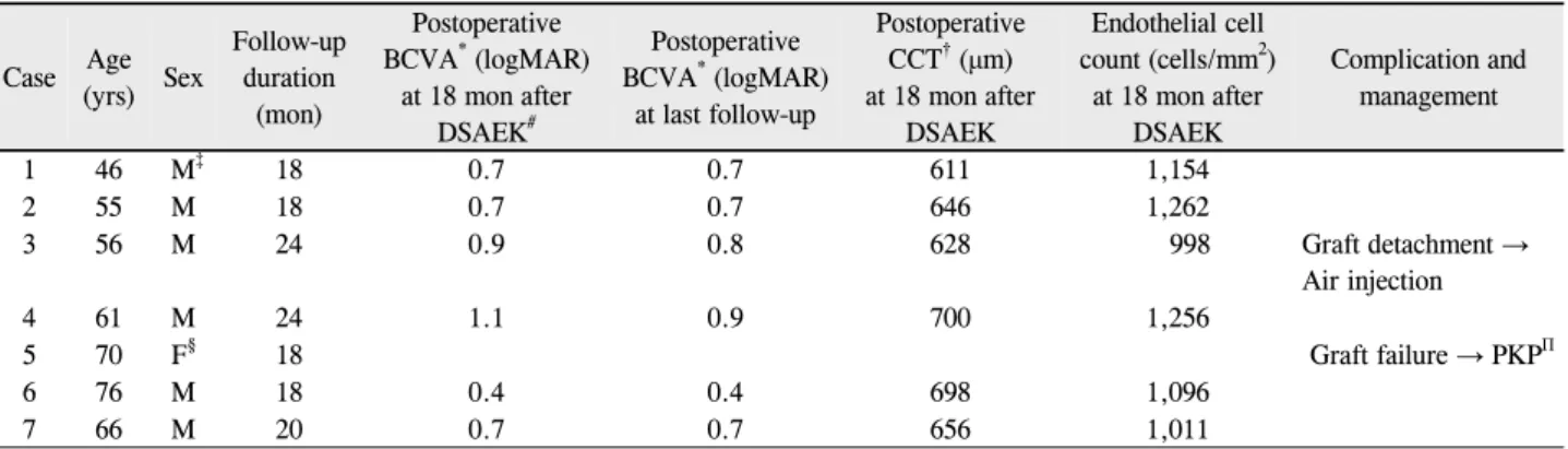
Table 3. Overall visual and surgical results at 18 months and at last follow-up after Descemet’s stripping automated endothelial kera-
toplasty결 과
DSAEK을 시행받은 환자는 7명 7안이었고 남자는 6명, 여자는 1명이었으며 평균 나이는 61.42 ± 10.1 (46~76) 세였다. 수술 중 합병증은 발생하지 않았으며 수술 후 평균 경과 관찰 기간은 19.9 ± 2.9 (18~24)개월이었다. 7안 중 1안에서 수술 후 1개월째부터 지속적인 각막부종 및 혼탁 을 보였다.
각막부종 및 혼탁을 보인 1안을 제외한 나머지 6안의 최 대교정시력(logMAR, 중앙값)은 수술 전 1.62에서 수술 후 1개월째 1.15으로 통계적으로 유의하게 호전되었으며 (Wilcoxon signed rank test, p=0.03), 이후 3개월째 0.90, 6개월째 0.90, 9개월째 0.75, 12개월째 및 18개월째 0.70 으로 유지되는 양상을 보였다(Fig. 1). 또한 각막두께(중앙 값)는 수술 전 932.5 μm에서 수술 후 12개월 601 μm (Wilcoxon signed rank test, p=0.03), 수술 후 18개월 651 μm (p=0.03)로 통계적으로 유의하게 감소하였다. 무 수정체안 2안을 제외한 나머지 4안의 수술 전과 수술 후 12개월 및 18개월의 굴절이상치와 난시값은 유의한 차이 는 없었고, 구면렌즈대응치는 약간의 원시성 변화를 보였으 나 이 또한 통계적인 유의성은 없었다(Table 2).
수술 후 합병증으로 1안에서 수술 후 6일째 각막이식편 의 비측 부위에 부분적인 탈락이 관찰되어 점안 마취하에 공기를 전방내 재주입함으로써 생착을 유도하였고 이후 안 정된 상태를 보였다. 또한 다른 1안에서 수술 후 1개월째 각막기질의 부종 및 혼탁을 보여 약물 치료 후 관찰하였으 나 수술 후 6개월까지 호전을 보이지 않아 이식 실패로 간 주하고 수술 후 12개월째 전층각막이식술을 시행하였다.
이식실패를 보인 1안의 수술 후 1주 및 1개월째의 각막두 께는 각각 987 μm 및 941 μm으로 나머지 6안보다 더 두꺼 운 것으로 나타났다. 18개월 이상 추적 관찰 시 6안에서 성 공적인 각막이식편의 소견을 보여, 전체적인 이식 성공율은 85.7% (6/7)였다(Table 3).
동일한 기간 PKP를 시행받은 환자의 수는 38명(남자 22 명, 여자 16명)이었고, 평균 나이는 52.61 ± 9.19세였으며 평균 경과 관찰 기간은 21.4개월(18~27개월)이었다. 수술 후 합병증으로 1안에서 수술 후 3일째 창상유출이 있어 봉 합교정술을 시행받은 후 안정된 상태를 유지하였다. 추적관 찰 기간 동안 4안에서 이식실패의 소견이 관찰되어 전체적 인 성공율은 89.5% (34/38명)이었다. 수술 전과 비교하여 수술 후 3개월째에 통계적으로 유의한 시력 호전을 나타내 었으며, 이는 최종 경과 관찰 시까지 유지되었다(Fig. 2).
Postoperative 12 mon Postoperative 18 mon DSAEK*
median (range)
PKP† mean ± SD
DSAEK median (range)
PKP mean ± SD‡
Sphere (D) +0.5 (0.25~0.75) -3.78 ± 1.64 +0.75 (0.25~1.25) -3.98 ± 1.86
Cylinder (D) +1.25 (-1.25~2.0) -4.26 ± 2.33 +0.75 (-1.25~2.0) -4.52 ± 1.82
Central corneal thickness (μm) 601 (600~716) 598.33 ± 46.81 651 (611~701) 609.18 ± 27.86 Endothelial cell density (cells/mm2) 1,359 (1258~1587) 1,386.28 ± 192.92 1,125 (998~1262) 1,298.00 ± 251.76
*DSAEK = Descemet-stripping automated endothelial keratoplasty; †PKP = penetrating keratoplasty; ‡SD=Standard deviation.
Table 4. Refractive ametropia, astigmatism, central corneal thickness, and endothelial cell density after Descemet’s stripping auto-
mated endothelial keratoplasty and penetrating keratoplastyFigure 2. Graphs showing changes in mean
best corrected visual acuity over time in the patients who underwent penetrating kerato- plasty (*Paired t-test, p< 0.05 between pre- operative and postoperative results).DSAEK을 시행받은 환자군에 비해 PKP를 시행받은 환자 들의 수술 후 12개월 및 18개월의 굴절이상치와 난시값은 좀 더 높은 수치를 나타냈고 다소 근시성 변화를 보였으며, 각막두께 및 내피세포 수에 있어 큰 차이가 없었으나 직접 적인 비교는 불가능하여 통계적인 유의성을 확인할 수는 없었다(Table 4).
증례
76세 남자 환자가 약 2년 전부터 발생한 우안 안통 및 시 력저하를 주소로내원하였다. 과거력상 3년 전 타 병원에서 초음파수정체유화술 및 인공수정체삽입술을 시행받은 기왕 력 있었고 이후 점진적으로 시력저하 및 안통이 발생하였 다. 내원시 시력(logMAR)은 1.7이었으며 안압은 18 mmHg 이었다. 세극등검사상 각막부종 및 혼탁이 관찰되었고, 경 면현미경검사상 내피세포수는 281 /mm2이었으며 각막두 께는 1006 μm로 증가되어 있었다. 내원 후 3개월째 DSAEK 을 시행하였다. DSAEK 시행 후 6개월째 시력(logMAR)은 0.7,각막내피세포수는 1115 /mm2, 각막두께는 721 μm였 고, 최종 경과 관찰 18개월째 이식편은 안정된 상태로 유지 되었으며 시력(logMAR)은 0.4, 내피세포수는 1096 /mm2, 각막두께는 698 μm로 호전되었다(Fig. 3).
고 찰
DSAEK은 이환된 각막내피와 데스메막만을 제거한 후 정상적인 각막내피와 데스메막을 선택적으로 이식하는 수 술로서, 현재 각막내피부전증 환자에서 각광받는 수술 기법 이 되었다.11 전층각막이식술에 비해 DSAEK은 봉합을 거 의 필요로 하지 않고, 각막의 고유의 구조를 상당 부분 유 지하여 수술에 의한 난시를 줄임으로써 더 빠른 시력 회복 을 가져다 준다고 알려져 있다.12,13
Hjortdal and Ehlers14는 푹스내피이상증(Fuchs’ endo- thelial dystrophy)으로 진단받고 DSAEK (20안) 혹은 PKP (20안)를 시행받은 후 12개월 이상 경과 관찰을 받은 환자를 대상으로 분석한 결과 PKP군에서는 이식실패 환자 가 없는 것에 비해 DSAEK군에서는 20안 중 2안에서 이식 실패를 보였으나 통계적인 유의성은 없었으며 DSAEK군에 서 더 낮은 굴절이상치와 난시값, 더 빠른 시력 회복을 나 타냈다고 하였다. 또한 Koenig et al15은 34명의 환자에서 DSAEK 시행 후 6개월 이상 경과 관찰 시 30명(88.2%)에 서 시력이 호전되었으며, 3명(9%)에서 이식실패를 보였다 고 하였다. 국내에서 DSAEK 수술 후 3개월간의 단기 결과 를 분석한 Jeong et al16의 보고에 의하면, 8안 중 7안에서 안정된 상태 및 시력호전을 보였으며 1안에서만 이식실패 가 발생하였다.
A B
C D
Figure 3. (A) Preoperative slit lamp photograph showing edema and diffuse haziness in the cornea. (B) At 18 months after
Descemet’s stripping automated endothelial keratoplasty, the corneal graft remains clear. (C) Pentacam demonstrates interface smoothness and regular thickness of the cornea. (D) Specular microscope reveals viable endothelial cells.본 연구에서 DSAEK을 시행 받고 18개월 이상 경과 관 찰이 가능하였던 7명의 환자를 조사한 결과, 6안(85.7%) 에서는 이식 각막편의 투명성이 유지되어 성공적인 결과를 보였으며, 1안(14.3%)에서는 이식실패를 보였다. 최대교 정시력의 경우 이식실패 1안을 제외한 나머지 모든 환자에 서 호전을 보였는데, 이는 수술 후 1개월째부터 통계적으로 유의한 차이를 보였고 이후 최종 경과 관찰 시까지 유지되 었다. 무수정체안 2안을 제외한 나머지 4안의 수술 전, 후 의 굴절이상 및 난시값은 통계적으로 유의한 차이를 보이 지 않았는데 이는 DSAEK에서 굴절이상이 적고 시력회복 이 빠르다는 기존의 보고들과 일치하였다.3,4,17-20 수술 후 12개월 및 18개월의 각막 두께는 수술 전과 비교하여 통계 적으로 유의하게 감소하였다. 또한 수술 전과 비교하여 DSAEK시행 후 6개월에 약 32 %에서 34 % 정도의 내피 세포 소실이 있다는 발표가 있었으나, 본 연구에서는 공여 각막의 내피세포수가 측정되어 있지 않아 비교할 수 없었 다.21,22
본 연구에서는DASEK과 PKP군 간의 수술 전 적응증, 각
막 및 환자의 상태 등이 달라 직접적인 통계적 비교를 시행 하기가 어려운 관계로, 간접적인 비교를 위해 같은 기간 동 안 PKP를 시행 받은 환자들의 수술 후 자료를 분석하였다.
그 결과 PKP를 시행 받은 군에서는 수술 전과 비교하여 수 술 후 3개월째 통계적으로 유의한 시력 호전을 보여 수술 후 1개월째 유의한 호전을 보인 DSAEK에 비해 시력 회복 속도가 좀 더 늦은 것으로 나타났다. 술 후 굴절이상치 및 난시값의 경우 또한 PKP군에서 더 높은 수치를 보였는데, 이는 DSAEK의 경우 각막의 외층에 대한 영향이 거의 없고 PKP에 비해 절개창이 작기 때문인 것으로 여겨진다.17,18 내 피세포의 경우 DSAEK 시행 시 PKP에 비해 상대적으로 잦 은 이식편의 조작으로 인해 보다 내피세포의 많은 소실이 있을 수 있다는 보고가 있다.14 본 연구에서는 수술 후 내 피세포의 수와 각막두께치는 두 군 간에 수치상으로 큰 차 이를 보이지는 않았으나, 통계적인 유의성 유무를 확인할 수는 없었으며 향후 많은 대상 수를 포함한 추가적인 연구 및 PKP와의 직접 비교 연구가 요구된다.
Jun et al23은 DSAEK 수술 후 44안에서 평균 0.88 디옵
터의 원시성 변화(hyperopic shift)를 보이고 이식편의 직 경이 클수록 원시성 변화의 정도 또한 커진다고 하였으며, DSAEK과 인공수정체삽입술을 병행할 시 이러한 원시성변 화를 고려하여 목표 굴절치(target refraction)를 정해야 한 다고 하였다. 본 연구의 경우 수술 전 무수정체안 2명을 제 외한 나머지 4안의 수술 전후의 구면렌즈대응치 비교에서 전반적인 원시성 변화를 보였으나 통계적인 유의성은 없었 으며 더 많은 대상 수를 포함하여 원시성 변화량에 대한 예 측 가능한 노모그램의 연구가 필요할 것으로 사료된다.
Koenig and Covert24는 26안에서 DSAEK 시행 후 9안 (34.6 %)에서 재고정이 필요할 정도의 각막이식편의 탈락 이 발생하여 무균공기를 재주입 하였으며, Jeong et al16또 한 8안 중 2안(25.0 %)에서 각막이식편의 탈락을 보여 무 균공기를 재주입함으로써 생착을 도왔다. 본 연구에서는 7 안 중 1안(14.3 %)에서 수술 후 비측의 부분적인 탈락을 보여 수술 후 6일째 전방내 공기재주입술을 시행하였으며 이후 안정적인 상태를 유지하였다.
아직까지 공여각막이나 수술 기법 이외에 DSAEK의 예 후에 영향을 미치는 다른 인자에 대한 보고는 없으나, Shih et al25은 세극등빛간섭단층촬영검사(slit-lamp optical coherence tomography)를 이용한 수술 후 1주째의 각막두 께의 측정치가 수술 성공 여부를 가늠할 수 있는 예측인자 가 될 수 있으며, 수술 후 1주째 이식편의 두께가 350 μm 이하인 경우 수술 성공율은 98%에 이른다고 보고하였다.
한편 Terry et al22은 DSAEK과 백내장수술을 병행한 149 안 중 이식실패는 발생하지 않아 초음파유화술이 DSAEK 의 예후에 유의하게 미치는 영향은 없는 것으로 발표하였 는데 본 연구에서는 수정체유화술과 병행한 DSAEK 수술 환자에서 술 후 이식 실패를 보였다. 이 환자의 경우 다른 환자들과 비교하여 초음파유화술 이외에 수술 전 기타 지 표나 공여각막에 있어 유의할 만한 차이점은 없었으나, 수 술 후 1주 및 1개월째의 각막두께는 나머지 6안보다 더 두 꺼운 것으로 나타났다. 초음파유화술이 DSAEK 수술 전에 시행되었으므로, 두꺼운 각막두께치 및 이식실패의 원인이 초음파유화술에 의한 것은 아닐 것으로 생각되나, 향후 초 음파유화술의 여부에 따른 군들에 대한 추가적인 비교 연 구가 필요할 것으로 여겨진다.
본 연구의 가장 큰 제한점으로는 위에서 언급한 것처럼, 적은 대상 환자 수로 인해 통계값의 검정력이 약하다는 것 과, 단일 수술만을 시행한 것이 아니라 공막고정술이나 수 정체유화술 등이 병행되어 동일 기법에 대한 비교를 하지 못한 것, DSAEK 시행 후 내피세포 소실율을 구하지 못한 것, 같은 기간 동안PKP를 시행받은 환자들의 자료를 참고 치로 제시하였으나 수술 전 적응증, 각막 및 환자의 상태가
달라 직접 비교를 시행하지는 못한 점 등이 있다. 향후 더 많은 수의 대상 환자를 통한 장기간의 연구 및 PKP를 포함 한 다른 수술 기법과의 직접 비교 연구가 필요할 것으로 생 각한다.
결론적으로, DSAEK 시행 후 18개월 이상의 장기 경과 관찰 결과 빠른 시력 호전 및 낮은 굴절 이상을 보였으며, 이는 각막내피부전증 환자에서 DSAEK이 효과적인 수술 방법임을 뒷받침한다.
참고문헌
1) Melles GR, Eggink FA, Lander F, et al. A surgical technique for posterior lamellar keratoplasty. Cornea 1998;17:618-26.
2) Terry MA, Ousley PJ. Deep lamellar endothelial keratoplasty in the first United States patients: early clinical results. Cornea 2001;
20:239-43.
3) Price FW Jr, Price MO. Descemet’s stripping with endothelial ker- atoplasty in 50 eyes: a refractive neutral cornea transplant. J Refract Surg 2005;21:339-45.
4) Price FW Jr, Price MO. Descemet's stripping with endothelial kera- toplasty in 200 eyes: early challenges and techniques to enhance donor adherence. J Cataract Refract Surg 2006;32:411-8.
5) Gorovoy MS. Descemet-stripping automated endothelial keratoplasty.
Cornea 2006;25:886-9.
6) Bahar I, Kaiserman I, McAllum P, et al. Comparison of posterior lamellar keratoplasty techniques to penetrating keratoplasty.
Ophthalmology 2008;115:1525-33.
7) Hyams M, Segev F, Yepes N, et al. Early postoperative complica- tions of deep lamellar endothelial keratoplasty. Cornea 2007;26:
650-3.
8) Yepes N, Segev F, Hyams M, et al. Five-millimeter-incision deep lamellar endothelial keratoplasty: one-year results. Cornea 2007;
26:530-3.
9) Melles GR, Lander F, Beekhuis WH, et al. Posterior lamellarker- atoplasty for a case of pseudophakic bullous keratopathy. Am J Ophthalmol 1999;127:340-1.
10) Bahar I, Kaiserman I, Srinivasan S, et al. Posterior lamellar kerato- plasty--comparison of deep lamellar endothelial keratoplasty and Descemet stripping automated endothelial keratoplasty in the same patients: a patient's perspective. Br J Ophthalmol 2009;93:186-90.
11) Covert DJ, Koenig SB. New triple procedure: Descemet's stripping and automated endothelial keratoplasty combined with phacoe- mulsification and intraocular lens implantation. Ophthalmology 2007;114:1272-7.
12) Covert DJ, Koenig SB. Descemet stripping and automated endo- thelial keratoplasty (DSAEK) in eyes with failed penetrating keratoplasty. Cornea 2007;26:692-6.
13) Price PO, Price FW Jr. Descemet’s stripping with endothelial kera- toplasty: comparative outcomes with microkeratome-dissected and manually dissected donor tissue. Ophthalmology 2006;113:
1936-42.
14) Hjortdal J, Ehlers N. Descemet's stripping automated endothelial keratoplasty and penetrating keratoplasty for Fuchs' endothelial dystrophy. Acta Ophthalmol 2009;87:310-4.
=ABSTRACT=
Long-Term Results of Descemet’s Stripping Automated Endothelial Keratoplasty in Korea
Jun Sung Lee, MD, Yeoung Geol Park, MD, Kyung Chul Yoon, MD
Department of Ophthalmology, Chonnam National University Medical School and Hospital, Gwangju, Korea
Purpose: To evaluate the long-term results of Descemet’s stripping automated endothelial keratoplasty in Korea (DSAEK).
Methods: Seven patients with bullous keratopathy who underwent DSAEK and who were followed-up for more than 18 months were reviewed retrospectively. Best corrected visual acuity, refraction, corneal thickness, and endothelial cell count were examined before and after surgery.
Results: The mean follow-up period was 19.9 ± 2.9 months (18 to 24 months), and the mean age was 61.42 ± 10.13 years (46 to 76 years). Six patients (85.7%) showed successful results after surgery. Best corrected visual acuity (logMAR) was significantly improved from 1.62 (median) to 1.15 (median) (p = 0.027) at one month after surgery and was maintained until the final follow-up period. There were no statistical differences in spherical ametropia or astigmatism before or 18 months after the operation. Graft failure was observed in one case, in which penetrating keratoplasty was performed 12 months af- ter DSAEK.
Conclusions: Long term results of DSAEK showed fast visual recovery, low ametropia and astigmatism. DSAEK may be a good option for the surgical management of corneal endothelial disease.
J Korean Ophthalmol Soc 2010;51(11):1431-1437 Key Words: DSAEK, Graft failure, Refractive change
Address reprint requests to Kyung Chul Yoon, MD
Department of Ophthalmology, Chonnam National University Hospital
#8 Hak-dong, Dong-gu, Gwang-ju 501-757, Korea
Tel: 82-62-220-6742, Fax: 82-62-227-1642, E-mail: [email protected] 15) Koenig SB, Covert DJ, Dupps WJ Jr, Meisler DM. Visual acuity,
refractive error, and endothelial cell density six months after Descemet stripping and automated endothelial keratoplasty (DSAEK). Cornea 2007;26:670-4.
16) Jeong HO, Lee HS, Na KS, Joo CK. Eight case of Descemet’s strip- ping automated endothelial keratoplasty in eyes with bullous keratopathy. J Korean Ophthalmol Soc 2009;50:1115-9.
17) Fogla R, Padmanabhan P. Initial results of small incision deep la- mellar endothelial keratoplasty (DLEK). Am J Ophthalmol 2006;141:346-51.
18) Koenig SB, Covert DJ, Dupps WJ Jr, et al. Visual acuity, refractive error, and endothelial cell density six months after Descemet strip- ping and automated endothelial keratoplasty (DSAEK). Cornea 2007;26:670-4.
19) Yoo SH, Kymionis GD, Deobhakta AA, et al. One-year results and anterior segment optical coherence tomography findings of desce- met stripping automated endothelial keratoplasty combined with Phacoemulsification. Arch Ophthalmol 2008;126:1052-5.
20) Rose L, Briceno CA, Stark WJ, et al. Assessment of eye bank-pre-
pared posterior lamellar corneal tissue for endothelial keratoplasty.
Ophthalmology 2008;115:279-86.
21) Terry MA, Chen ES, Shamie N, et al. Endothelial cell loss after Descemet's stripping endothelial keratoplasty in a large pro- spective series. Ophthalmology 2008;115:488-96.
22) Terry MA, Shamie N, Chen ES, et al. Endothelial keratoplasty for Fuchs' dystrophy with cataract: complications and clinical results with the new triple procedure. Ophthalmology 2009;116:631-9.
23) Jun B, Kuo AN, Afshari NA, et al. Refractive change after desce- met stripping automated endothelial keratoplasty surgery and its correlation with graft thickness and diameter. Cornea 2009;28:
19-23.
24) Koenig SB, Covert DJ. Early results of small-incision Descemet's stripping and automated endothelial keratoplasty. Ophthalmology 2007;114:221-6.
25) Shih CY, Ritterband DC, Palmiero PM, et al. The use of post- operative slit-lamp optical coherence tomography to predict pri- mary failure in descemet stripping automated endothelial keratoplasty. Am J Ophthalmol 2009;147:796-800.