pISSN: 0378-6471 eISSN: 2092-9374
http://dx.doi.org/10.3341/jkos.2013.54.2.210
= 증례보고 =
데스메막박리 각막내피층판이식술에서 공여자 각막내피절편 두께에 따른 시력 예후
한종철⋅배지현⋅정태영⋅정의상 성균관대학교 의과대학 삼성서울병원 안과학교실
목적: 데스메막박리 각막내피층판이식술(Descemet stripping automated endothelial keratoplasty, DSAEK)에서 공여자 각막내피절편 두께가 얇을수록 술 후 시력 예후가 좋을 것인지 알아보고자 한다.
대상과 방법: 2008년 1월부터 2010년 12월까지 본원에서 각막내피부전증으로 DSAEK 수술을 받은 22명 22안을 대상으로 공여자 각막 내피절편 두께 130 μm를 기준으로 이보다 얇은 경우 thin lenticule 군으로, 두꺼운 경우 thick lenticule 군으로 나누어 각각 1, 3, 6개월 에서의 시력(logMAR)을 비교하였다.
결과: Thin lenticule과 thick lenticule 군에서의 술 후 1개월 최대교정시력(logMAR)은 각각 0.46 ± 0.22, 0.71 ± 0.26 (p=0.025), 3개월 최대교정시력(logMAR)은 각각 0.36 ± 0.17, 0.54 ± 0.32 (p=0.129), 6개월 최대교정시력(logMAR)은 0.27 ± 0.15, 0.38 ± 0.18 (p=0.141)이었다.
결론: DSAEK 수술에서 공여자 각막내피절편 두께가 130 μm 보다 얇은 경우 술 후 1개월째만 내피의 두께가 130 μm 이상인 경우보다 좋아 공여자 각막내피절편 두께는 술 후 1개월 후의 단기 시력 예후에만 영향을 미치는 것을 알 수 있었다.
<대한안과학회지 2013;54(2):210-214>
■ 접 수 일: 2012년 4월 27일 ■ 심사통과일: 2012년 7월 31일
■ 게재허가일: 2012년 11월 24일
■ 책 임 저 자: 정 의 상
서울특별시 강남구 일원로 81 성균관대학교 삼성서울병원 안과 Tel: 02-3410-3565, Fax: 02-3410-0074 E-mail: [email protected]
* 이 논문의 요지는 2011년 대한안과학회 제106회 학술대회에서 구연으로 발표되었음.
지난 수십 년간 데스메막박리 각막내피층판 이식술(Descemet stripping automated endothelial keratoplasty, DSAEK)은 안과 수술 후 발생한 수포성 각막병증 및 푹스이영양증 또 는 포도막염, 안내염 등의 안과적 질환에 의한 각막내피부 전을 치료하는데 있어서 전층각막이식(Penetrating kera- toplasty, PKP)을 대체하고 있다. DSAEK은 PKP와 비교하 여 상대적으로 빠른 시력 회복, 적은 난시, 적은 봉합사 연 관 합병증, 빠른 수술 시간 및 적은 술 중 혹은 술 후 맥락 막상강 출혈 등이 장점으로 알려졌다.1
이후 DSAEK 수술 후 장기 임상 결과들이 발표되면서 시 력 예후에 영향을 미치는 요소들 중 공여자 각막 내피(donor corneal lenticule)과 연관된 보고들이 있었다. 내피 각막의 크기와 각막 내피의 생존과의 관계,2 각막내피의 중심부와 주변부 두께 변화에 의한 굴절률 변화,3각막 내피가 얇아 짐과 시력 호전의 비례 정도4 등의 보고가 있었다.
한편 최근 각막 실질을 포함하지 않고 각막내피만을 이 식하는 데스메막 각막내피이식술(Descemet membrane endothelia keratoplasty, DMEK) 수술의 시력 예후가 각막 실질의 일부를 포함하는 각막내피를 사용하는 DSAEK 수술 보다 좋다고 보고된 바가 있다.5-7이에 착안하여 DSAEK에 서도 공여자 각막내피절편두께가 얇을수록 시력 예후가 좋 을지 알아보고자 본 연구를 시행하였다.
대상과 방법
2008년 1월부터 2010년 12월까지 본원 안과에서 각막 내피부전증으로 DSAEK을 시행 받은 22명 22안을 대상으 로 의무기록을 후향적으로 분석하였다. 대상 환자 모두에서 수술 전후 검사로 최대 교정시력(logMAR), 중심각막두께 (ultrasonic pachymeter, UP1000, Nidek, Japan), 세극등 현미경검사, 경면현미경검사(specular microscope, Noncon ROBO CA, Konan, Japan) 등을 시행하였다. 술 전 검사상 각막 실질부의 혼탁이 심한 경우, 망막 질환이 동반된 경우 등 술 후 시력에 영향을 미칠 수 있는 요소가 동반된 경우 는 제외하였다.
공여각막은 Lions Eye Bank of Oregon (Portland OR, USA)에서 artificial anterior chamber내에서 Moria mi- crokeratome을 이용하여 가공되어 Optisol-GS solution
Table 1. Demographics for patients undergoing DSAEK
Thin lenticule (n = 11) Thick lenticule (n = 11)
Mean age ± standard deviation (years) 61 ± 10 64 ± 14
Sex (female(%)/male(%)) 2 (18)/9 (82) 2 (18)/9 (82)
Reason for transplantation (n (%))
Fuchs 3 (27) 2 (18)
PBK 7 (64) 8 (73)
Other endothelial dysfunction 1 (9) 1 (9)
PBK = pseudophakic bullous keratopathy.
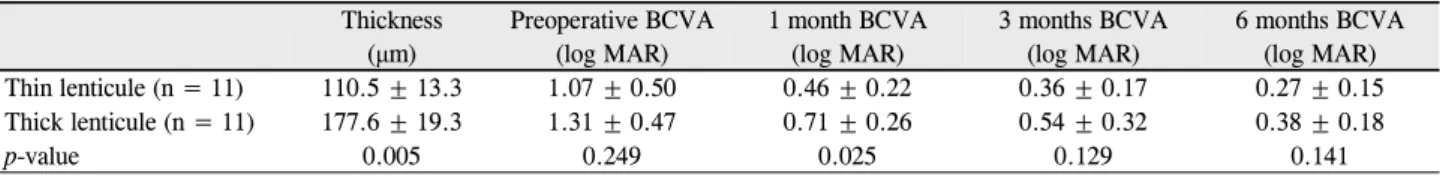
Table 2. Comparison of thickness and visual outcomes between two groups (thin lenticule vs thick lenticule)
Thickness(μm)
Preoperative BCVA (log MAR)
1 month BCVA (log MAR)
3 months BCVA (log MAR)
6 months BCVA (log MAR) Thin lenticule (n = 11) 110.5 ± 13.3 1.07 ± 0.50 0.46 ± 0.22 0.36 ± 0.17 0.27 ± 0.15 Thick lenticule (n = 11) 177.6 ± 19.3 1.31 ± 0.47 0.71 ± 0.26 0.54 ± 0.32 0.38 ± 0.18
p-value 0.005 0.249 0.025 0.129 0.141
Values are presented as mean ± SD.
BCVA = best corrected visual acuity.
(Chiron Ophthalmics, Irvine, CA)에 담겨 국내로 운송된 각막(Precut cornea)을 사용하였다. 공여각막 정보지에 적 혀있는 공여자 정보를 참고하여 HBV, HCV, HIV 등의 바 이러스 양성이거나 신경계 악성종양이 있던 경우는 제외하 였고, 술 전 공여자 각막내피의 수가 2600 cell/mm2이상인 경우에 수술에 사용하였다. Lions Eye Bank of Oregon에 서 제공한 공여자 각막정보지(Precut cornea information sheet)에 적힌 lenticule graft thickness를 기준으로 thin lenticule와 thick lenticule로 나누었으며, thin graft와 thick graft의 경계값으로는 본 연구에 이용된 공여각막내 피두께의 중간값에 가까운 130 μm로 정하였다.
수술은 전신마취를 한 후 수술을 시행하였고 수여각막에 서는 Reverse Sinskey hook (Moria, Inc., Doylestown, PA, USA)을 이용하여 데스메막을 8 mm 지름으로 표지하 고 Descemets stripper (Moria, Inc.)를 이용하여 데스메막 을 벗겨내었다. 환자의 귀쪽 방향에서 5 mm 윤부절개를 만 들었다. ICL injector를 사용하여 공여자 각막내피절편을 전방내로 주입하였으며, 전방내로 공기를 주입하여 공여각 막편이 수여각막후면에 고정되도록 하였다. 윤부절개창에 hydrosealing 후 수술을 마쳤다. 술 후 2시간 이상 앙와위 상태를 유지하였고 환자들은 감염 예방을 위해 항생제 안 약(Levofloxacin, Cravit, Santen, Japan) 및 스테로이드 안 약(Pred forte, Allergan, USA)을 점안하였다. 술 후 1주, 2주, 1개월 경과 관찰을 시행하였으며 이후에는 환자의 상 태에 따라 1개월 혹은 2개월마다 경과관찰을 하였다.
통계적 분석방법으로 두 군간의 교정시력, 각막두께, 각 막내피수의 차이에 대해 independent t-test를 시행하였으 며, 통계분석은 SPSS 14.0 통계 프로그램을 사용하였으며
유의수준은 p<0.05으로 하였다.
결 과
Thin lenticule 군 11명 11안의 평균 연령은 61 ±10세, thick lenticule 군 11명 11안의 평균 연령은 64 ±14세였 으며, 두 군 모두 남녀 비율은 82%, 18%로 남자의 비율이 높았다. 원인 질환으로는 양군에서 모두 인공수정체성수포 각막병증이 각각 64%, 73%로 가장 많았으며, 푹스각막이 영양증이 27%, 18%로 다음으로 흔한 원인이었고, 양군 모 두 각각 9%에서 헤르페스각막염 등의 다른 원인으로 DSAEK 수술을 받았으며, 인공수정체성수포각막병증의 경 우를 제외한 다른 경우는 백내장 변화가 없어서 DSAEK 수 술만 시행하였다(Table 1). Thin lenticule 군과 thick len- ticule 군에서 사용된 lenticule thickness의 평균은 각각 110.5 ±13.3 μm, 177.6 ±19.3 μm로 통계적으로 유의한 차이가 있었으며(p=0.005), 각 군의 술 전 최대교정시력 (logMAR)은 각각 1.07 ±0.50, 1.31 ±0.47이었으며 두 군간 통계적으로 유의한 차이는 없었다(p=0.249). 술 후 1 개월 최대교정시력은 thin lenticule과 thick lenticule 군이 각각 0.46 ±0.22, 0.71 ±0.26이었으며 두 군간 차이는 통계적으로 유의했다(p=0.025). 술 후 3개월 최대교정시 력은 두 군에서 각각 0.36 ±0.17, 0.54 ±0.32, 술 후 6개 월 최대교정시력은 두 군에서 각각 0.27 ±0.15, 0.38 ± 0.18로 thin lenticule을 사용한 군의 시력예후가 좋았으나 통계적으로 유의하지는 않았다(p=0.129; p=0.141) (Table 2).
술 전 각막내피수는 thin lenticule 군과 thick lenticule 군 에서 각각 2746 ±236 cells/mm2, 2708 ±395 cells/mm2로
Table 3. Comparison of endothelial cell Count between two groups (thin lenticule vs thick lenticule)
Preoperative (number/mm2) 6 months (number/mm2)
Thin lenticule (n = 9) 2746 ± 236 2033 ± 614
Thick lenticule (n = 9) 2708 ± 395 1729 ± 509
p-value 0.790 0.270
Values are presented as mean ± SD.
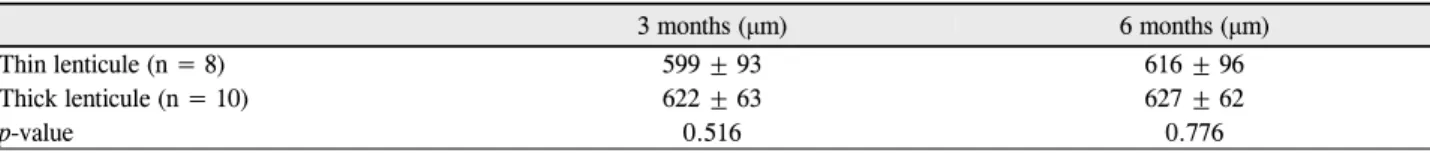
Table 4. Comparison of central corneal chickness measured by pachymetry between two groups (thin lenticule vs thick lenticule)
3 months (μm) 6 months (μm)
Thin lenticule (n = 8) 599 ± 93 616 ± 96
Thick lenticule (n = 10) 622 ± 63 627 ± 62
p-value 0.516 0.776
Values are presented as mean ± SD.
통계적으로 유의한 차이가 없었다(p=0.790). 술 후 6개월 각막내피수는 thin lenticule 군 2033 ± 614 cells/mm2, thick lenticule 군 1729 ±509 cells/mm2이었으며 두 군간 통계적으로 유의한 차이는 없었다(p=0.270) (Table 3).
각막두께는 술 후 3개월 및 6개월 측정하였으며 thin len- ticule 군 8안, thick lenticule 군 10안이 포함되었다. 술 후 3개월 측정된 각막두께는 각각 599.0 ±92.7 μm, 622.0 ± 63.0 μm이었으며, 술 후 6개월 측정된 각막두께는 각각 616.0 ±96.2 μm, 627.0 ±61.9 μm로 양군 모두 통계적 으로 유의한 차이는 없었다(p=0.516; p=0.776) (Table 4).
고 찰
데스메막박리 각막내피층판 이식술(Descemet stripping automated endothelial keratoplasty; DSAEK)은 2005년 Price and Price8에 의해 처음으로 소개되었다. 이는 전층 각막이식술(Penetrating keratoplasty; PKP)보다 수여자 각막의 실질이 보존되므로 외상에 안정적이고 시력 회복 속도가 상대적으로 빠르며 봉합이 필요 없기 때문에 이로 인해 난시가 발생하는 가능성이 적고 봉합사로 인한 합병 증이 없는 장점이 있다.9,10Chen et al11은 327안의 DSAEK 수술의 6개월 경과 관찰 결과를 발표한 바 있으며 교정시력 은 술 전 20/80에서 술 후 6개월 20/36으로 호전되었으며, 술 전 각막내피세포 단위면적 (mm2) 당 2873개에서 1925 개로 줄어들었다고 보고하였다. Price et al12은 DSAEK 173안과 PKP 410안을 비교한 논문에서 DSAEK의 경우 술 전 단위면적당 2778개에서 6개월 후 1838개로 줄었다 고 보고하였다. 본 연구의 경우에는 6개월 최종 시력이 thin lenticule 군과 thick lenticule 군에서 각각 20/40, 20/50으 로 이전 보고와 큰 차이가 없었으며, 각막내피세포수는 thin lenticule에서 술 전 단위면적당 2746개에서 술 후 6개 월 2033개, thick lenticule에서는 술 전 단위면적당 2708
개에서 술 후 6개월 1729개로 측정되었고 이는 과거 문헌 과 비교하여 비슷한 결과를 보였다.
그러나 데스메막박리 각막내피층판 이식술이 많은 장점 을 가지고 있음에도 불구하고 이식된 각막편의 후층판으로 인해 발생할 수 있는 굴절이상과 고위 수차의 변화 등이 최 종적인 시력 예후에 영향을 미치는 것으로 알려지면서 이 식 각막편의 두께와 시력예후의 관계가 임상적으로 중요한 의미를 갖게 되었다.3,13,14
Neff et al15은 DSAEK 수술 후 최소 6개월 이상 경과된 환자를 대상으로 anterior segment optical coherence to- mography (AS-OCT)를 이용하여 측정한 각막내피이식편 과 교정시력의 연관성을 연구한 보고에서 이식편의 두께가 131 μm 보다 얇은 경우 이보다 두꺼운 경우보다 최종 교정 시력이 더 좋다고 보고하였다. Pogorelov et al4은 6개월간 DSAEK 수술 환자 15안에 대해 AS-OCT를 이용하여 술 후 이식편의 두께변화와 시력변화와의 관계를 연구하였으 며, 술 후 이식된 각막 내피 두께가 감소할수록 시력도 호 전된다고 보고하였다.
그러나 수술 후 AS-OCT를 이용하여 이식편의 두께를 측정하는 데는 측정자에 따라 차이가 날 수 있고, 수술 후 각막상태에 따라서 부정확한 두께 측정이 가능하기 때문에 AS-OCT를 이용하여 시행한 이전 연구4,15와는 달리 본 연 구에서는 후층판이 절삭되어 제공되는 precut tissue를 이 용하여 시행한 경우만 포함시켰다. Chen et al16과 Terry et al17은 DSAEK에서 precut tissue를 사용하는 경우와 수술 장에서 술자가 직접 각막내피를 얻어 수술을 하는 경우 시력 예후에서 이전과 큰 차이가 없었음을 보고하였으며, precut tissue를 얻는 경우 오히려 함께 제공되는 각막정보지를 통 해 술 전 각막내피두께를 정확히 측정할 수 있기에 본 연구 에는 precut tissue를 이용하게 되었다.
Terry et al17은 본 연구와 같이 precut dissected tissue 를 이용하여 데스메막박리 각막내피층판 이식술을 받은
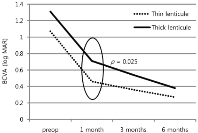
0 0.2 0.4 0.6 0.8 1 1.2 1.4
preop 1 month 3 months 6 months Thin lenticule Thick lenticule
BCVA (log MAR)
p = 0.025
Figure 1. The comparison of visual prognosis after DSAEK
surgery between thin lenticule group and thick lenticule group.100명을 분석하였다. 이들 보고에 의하면 술 전 측정된 공 여자 각막내피 두께와 DSAEK 수술 후 6개월, 12개월에 측 정한 교정시력간에 통계적으로 유의한 연관성은 관찰되지 않았다. 그러나 본 연구에서는 술 전 공여자 각막내피의 두 께가 얇을수록 술 후 1개월 교정시력이 통계적으로 유의하 게 좋은 것으로 보였으며, 술 후 3개월 및 6개월 교정시력 도 얇은 공여자 각막내피의 두께가 좋은 시력 예후를 보이 는 경향성을 가졌으나, 통계적으로 유의한 연관성은 없었다 (Fig. 1). 본 연구에서 술 후 6개월 시력과 공여자 각막내피 의 두께와 연관성이 없다는 사실은 Terry et al17이 보고한 결과와 일치하였으나, 수술 직후 1개월 이내의 단기 시력 예후는 통계적으로 유의한 차이가 있다는 사실을 보임으로 써, 얇은 공여자 각막내피를 사용하는 것이 최종적인 시력 예후에 영향을 미치기 보다는 최종적인 시력까지 도달하는 시간을 좀더 단축시켜주는 장점이 있음을 알 수 있었다.
본 연구의 제한점은 표본수가 22명22안으로 크지 않아 통계값의 검정력이 약하다는 점과 경과 관찰 기간이 6개월 로 충분히 길지 않았다는 점, 후향적 연구라는 점이 있다.
또한 최종 시력에 영향을 주는 요소로는 이식된 각막 내피 두께 외에 정난시 혹은 부정난시, 고위수차, 술 전 각막 혼 탁 정도의 차이 등이 있겠으나 이 요인들에 대해서는 두 군 간에 미치는 영향 차이가 없을 것이라는 가정 하에 연구를 진행했다는 제한점이 있다.
결론적으로 DSAEK 수술에서 공여자 각막내피절편 두께 가 130 μm 보다 얇은 경우 술 후 1개월째 내피절편의 두께 가 130 μm 이상인 경우보다 좋지만 3개월 이후에는 통계 적으로 유의한 차이가 없음을 알 수 있었으며 이에 대해서 는 향후 많은 환자군을 대상으로 장기간 경과 관찰을 통한 전향적 연구가 추가적으로 필요할 것으로 보인다.
참고문헌
1) Lee WB, Jacobs DS, Musch DC, et al. Descemet's stripping endo- thelial keratoplasty: safety and outcomes: a report by the American Academy of Ophthalmology. Ophthalmology 2009;116:1818-30.
2) Terry MA, Li J, Goshe J, Davis-Boozer D. Endothelial kerato- plasty: the relationship between donor tissue size and donor endo- thelial survival. Ophthalmology 2011;118:1944-9.
3) Scorcia V, Matteoni S, Scorcia GB, et al. Pentacam assessment of posterior lamellar grafts to explain hyperopization after Descemet's stripping automated endothelial keratoplasty. Ophthalmology 2009;
116:1651-5.
4) Pogorelov P, Cursiefen C, Bachmann BO, Kruse FE. Changes in donor corneal lenticule thickness after Descemet's stripping auto- mated endothelial keratoplasty (DSAEK) with organ-cultured corneas. Br J Ophthalmol 2009;93:825-9.
5) Dapena I, Ham L, Melles GR. Endothelial keratoplasty: DSEK/
DSAEK or DMEK--the thinner the better? Curr Opin Ophthalmol 2009;20:299-307.
6) Price MO, Giebel AW, Fairchild KM, Price FW Jr. Descemet's membrane endothelial keratoplasty: prospective multicenter study of visual and refractive outcomes and endothelial survival.
Ophthalmology 2009;116:2361-8.
7) McCauley MB, Price MO, Fairchild KM, et al. Prospective study of visual outcomes and endothelial survival with Descemet mem- brane automated endothelial keratoplasty. Cornea 2011;30:315-9.
8) Price FW Jr, Price MO. Descemet's stripping with endothelial kera- toplasty in 50 eyes: a refractive neutral corneal transplant. J Refract Surg 2005;21:339-45.
9) Covert DJ, Koenig SB. Descemet stripping and automated endo- thelial keratoplasty (DSAEK) in eyes with failed penetrating keratoplasty. Cornea 2007;26:692-6.
10) Price MO, Price FW Jr. Descemet's stripping with endothelial kera- toplasty: comparative outcomes with microkeratome-dissected and manually dissected donor tissue. Ophthalmology 2006;113:
1936-42.
11) Chen ES, Terry MA, Shamie N, et al. Endothelial keratoplasty: vi- sion, endothelial survival, and complications in a comparative case series of fellows vs attending surgeons. Am J Ophthalmol 2009;
148:26-31.e2.
12) Price MO, Gorovoy M, Benetz BA, et al. Descemet's stripping au- tomated endothelial keratoplasty outcomes compared with pene- trating keratoplasty from the Cornea Donor Study. Ophthalmology 2010;117:438-44.
13) Bahar I, Kaiserman I, Livny E, Slomovic A. Changes in corneal curvatures and anterior segment parameters after descemet strip- ping automated endothelial keratoplasty. Curr Eye Res 2010;35:
961-6.
14) Lombardo M, Terry MA, Lombardo G, et al. Analysis of posterior donor corneal parameters 1 year after Descemet stripping auto- mated endothelial keratoplasty (DSAEK) triple procedure. Graefes Arch Clin Exp Ophthalmol 2010;248:421-7.
15) Neff KD, Biber JM, Holland EJ. Comparison of central corneal graft thickness to visual acuity outcomes in endothelial keratoplasty.
Cornea 2011;30:388-91.
16) Chen ES, Terry MA, Shamie N, et al. Precut tissue in Descemet's stripping automated endothelial keratoplasty donor characteristics
=ABSTRACT=
The Correlations between Donor Endothelial Lenticule Thickness and Visual Prognosis in DSAEK
Jong Chul Han, MD, Ji Hyun Bae, MD, Tae Young Chung, MD, PhD, Eui Sang Chung, MD, PhD
Department of Ophthalmology, Samsung Medical Center, Sungkyunkwan University School of Medicine, Seoul, Korea
Purpose: To determine the correlations between donor endothelial lenticule thickness and visual prognosis in Descemet’s stripping automated endothelial keratoplasty (DSAEK).
Methods: The present study included 22 patients (22 eyes), who underwent DSAEK surgery in our clinic due to endothelial decompensation. BCVA (log MAR) was compared at 1 month, 3 months and 6 months postoperatively between the thin lenticule group and thick lenticule group (≥130 μm).
Results: The BCVA (log MAR) at 1 month postoperatively was 0.46 ± 0.22 in the thin lenticule group, and 0.71 ± 0.26 in the thick lenticule group, and significant statistical correlations between donor lenticule thickness and visual acuity were ob- served (p = 0.025). However, no significant correlations were observed at 3 months (p = 0.129) and 6 months (p = 0.141) postoperatively.
Conclusions: The thin donor lenticule (<130 μm) can result in better visual acuity at 1 month postoperatively than the thick donor lenticule (≥130 μm), however, there is no difference in visual acuity between the 2 groups at 3 and 6 months postoperatively.
J Korean Ophthalmol Soc 2013;54(2):210-214
Key Words: DSAEK, DSAEK visual prognosis, Endothelial thickness, Lenticule
Address reprint requests to Eui Sang Chung, MD, PhD Department of Ophthalmology, Samsung Medical Center
#81 Irwon-ro, Gangnam-gu, Seoul 135-710, Korea
Tel: 82-2-3410-3565, Fax: 82-2-3410-0074, E-mail: [email protected] and early postoperative complications. Ophthalmology 2008;115:
497-502.
17) Terry MA, Shamie N, Chen ES, et al. Precut tissue for Descemet's
stripping automated endothelial keratoplasty: vision, astigmatism, and endothelial survival. Ophthalmology 2009;116:248-56.