1388
대한안과학회지 2014년 제 55 권 제 9 호 J Korean Ophthalmol Soc 2014;55(9):1388-1391 pISSN: 0378-6471⋅eISSN: 2092-9374
http://dx.doi.org/10.3341/jkos.2014.55.9.1388
Case Report
홍채앞유착이 있는 환자에서 전층각막이식 시 각막 부분층 박리를 이용한 유착박리술 1예
A Case of Anterior Synechiolysis with Lamellar Corneal Dissection in Penetrating Keratoplasty
성윤미⋅김은철
Yoon Mi Sung, MD, Eun Chul Kim, MD, PhD
가톨릭대학교 의과대학 부천성모병원 안과학교실
Department of Ophthalmology, Bucheon St. Mary’s Hospital, The Catholic University of Korea College of Medicine, Bucheon, Korea
Purpose: To report a case of anterior synechiolysis with lamellar corneal dissection in penetrating keratoplasty.
Case summary: In an eye with graft failure and anterior synechiae, we performed anterior synechiolysis with a healon needle af- ter lamellar dissection using vacuum trephine to visualize the anterior chamber during penetrating keratoplasty. Additionally, the remnant corneal layer was removed using corneal scissors. A donor cornea was harvested using a vacuum trephine and the cor- neal button was sewn in place with 10-0 nylon. We observed a well grafted cornea and well formed anterior chamber with no an- terior synechiae observed on follow-up.
Conclusions: In patients with anterior synechiae, the cornea can be dissected from the anterior synechiae completely using la- mellar corneal dissection in penetrating keratoplasty to visualize the anterior chamber.
J Korean Ophthalmol Soc 2014;55(9):1388-1391
Key Words: Anterior synechiae, Lamellar corneal dissection, Penetrating keratoplasty, Synechiolysis
■Received: 2014. 4. 26. ■ Revised: 2014. 7. 25.
■Accepted: 2014. 8. 1.
■Address reprint requests to Eun Chul Kim, MD, PhD Department of Ophthalmology, The Catholic University of Korea, Bucheon St. Mary’s Hospital, #327 Sosa-ro, Wonmi-gu, Bucheon 420-717, Korea
Tel: 82-32-340-2125, Fax: 82-32-340-2544 E-mail: [email protected]
ⓒ2014 The Korean Ophthalmological Society
This is an Open Access article distributed under the terms of the Creative Commons Attribution Non-Commercial License (http://creativecommons.org/licenses/by-nc/3.0/) which permits unrestricted non-commercial use, distribution, and reproduction in any medium, provided the original work is properly cited.
각막이식은 혼탁된 각막으로 인한 시력저하를 개선하기 위해 투명한 각막편을 이식하는 방법이다. 원추각막, 수포 성 각막염이 각막이식의 주된 원인 질환이지만, 각막염, 트 라코마, 외상 등으로 인한 각막 혼탁도 드물지 않다.1 후자 의 경우 홍채앞유착이 동반되는 경우가 많고 수술 전후 녹 내장으로 인해 수술 경과가 좋지 않다. 또한, 이러한 경우
각막 혼탁으로 인해 전방을 관찰하기가 힘들어 전층각막이 식 시 수여 각막을 trephination할 때 홍채에 손상을 일으킬 수 있다.
이렇게 손상된 홍채는 각막에 유착되기 쉽고 수술 후 다 시 홍채앞유착을 야기하게 된다. 이러한 홍채앞유착은 지 속적인 이식편 부종 및 속발 폐쇄각 녹내장, 각막 혈관화, 이식 거부반응, 이식편 기능상실의 원인이 된다.2 이식편 기 능상실로 재이식을 하는 경우에 앞에서 언급한 원인 이외 에도 초기 수술 시 이식편이 크거나 수술 후 창상 누출이 있거나 포도막염 등으로 인해 홍채앞유착이 있는 경우가 많다.
각막 부분층 박리를 이용한 전낭 원형절개술 후 백내장 수정체 초음파 유화술 및 인공 수정체 삽입술을 시행한 사 례는 보고되었다. 이에 저자는 홍채앞유착이 동반된 각막
1389 - 성윤미⋅김은철 : 각막 부분층 박리를 이용한 유착박리술 -
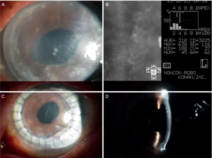
Figure 1. (A) Host cornea before surgery. (B) Corneal endothelium microscopy before surgery (the figure 3225 in the result was miscounted). (C) Anterior segment photography on 1 month postoperatively. (D) Well formed anterior chamber after surgery on 1 month postoperatively.
이식편 기능상실 환자에서 전층각막이식 시 각막 부분층 박리를 통해 유착박리술을 함께 시행한 1예를 보고하고자 한다.
증례보고
84세 여자환자가 일주일 전부터 발생한 좌안 시력 저하 및 통증으로 내원하였다. 3년 전 양안 수포성 각막병증 진 단받고 타병원에서 3년 전 좌안 전층각막이식, 2년 전 우 안 양막 이식 받은 자로 내원 시 시력 우안 광각무, 좌안 0.02, 안압 우안 46, 좌안 13이었다. 우안은 각막 전층 혼 탁 및 혈관화를 보였고 전방은 각막홍채 유착으로 인해 collapse된 소견을 보였다. 좌안은 각막 부종과 2시부터 11시 방향으로 아래쪽에 270도 이상의 홍채앞유착을 보였 고 각막두께검사에서 775 μm로 각막이식편 기능상실 소 견을 보였다(Fig. 1A, B). 안초음파에서는 양안 특이소견 보이지 않았다. 타병원에서 좌안 각막 부종 및 양안 안압
상승 소견으로 우안에 Dorzolamide-timolol (Cosopt®, Merck
& Co., Inc., USA) 2회, bromonidine (Alphagan-P®, Allergan, Inc., Irvine, California) 2회, latanoprost (Xalatan®, Pfizer Inc., New York, USA) 취침 전 1회 점안, 좌안에 0.3% gati- floxacin (Gatiflo®, Handok, Seoul, Korea) 2회, 0.1% Fluor- ometholone (Ocumetholone, Samil, Seoul, Korea) 2회, Cosopt® 2회, Alphagan-P® 2회, Xalatan® 취침 전 1회 점안, acetazolamide (Diamox®) 2T 복용 중이었다. 좌안 각막이식 편 기능상실로 전신마취하에 전층각막이식술을 계획하였다.
먼저 전층각막이식에 사용하는 진공 trephine (7.75 mm) 을 이용하여 수용각막의 75%를 trephination하고(Fig. 2A, B), beaver blade를 이용하여 각막실질부위에서 박리를 시 행하였다(Fig. 2C, D). 남아 있는 실질의 일부, 데스메막, 내 피 층은 어느 정도의 투명도를 유지하고 있어 전방을 관찰 할 수 있었다. 1 mm knife로 전방 천자를 한 후, 전방에 vis- coat를 주입하였다. 힐론 바늘로 힐론을 넣어가며 각막에 붙어 있는 홍채를 부드럽게 박리하여 주변부 우각 360도까
A B
C D
1390
- 대한안과학회지 2014년 제 55 권 제 9 호 -
A B
C
E
D
F
Figure 2. (A) Initial cut on the host cornea using vacuum trephine. (B) Cross-sectional view of (A). (C) Lamellar dis- section using beaver blade. (D) Cross-sectional view after lamellar dissection. (E) Anterior synechiolysis using healon needle. (F) Cross-sectional view of (E).
지 완전히 제거하였다(Fig. 2E, F). 이후 각막 가위를 이용하 여 수용각막의 나머지 부분을 절제하였다. 공여 각막을 8.0 mm 크기로 trephine하여 수용각막 위에 올려놓고 10-0 나일 론 봉합사를 이용하여 4개의 단속 봉합 후 12 개의 단속봉합 을 추가하였다. 평형염액으로 전방을 채워 전방각을 유지한 후 수술을 마무리하였다. 수술 후 0.3% gatifloxacin 4회, 1% prednisolone (Predforte, Allergan, Irvine, California) 4 회, Cosopt® 2회 점안, Prednisolone (Solondo, Yuhan® Pharmacy, Seoul, Korea) 30 mg을 복용하도록 하였다. 수술 후 5개월, 좌안 시력 0.8, 안압 14, 각막은 투명하게 유지되었고, 전방
깊이 중심부 4 CT 이상, 주변부 1 CT로 잘 형성되었고, 홍 채앞유착 없이 동공은 round하였다(Fig. 1C, D).
고 찰
각막이식을 필요로 하는 환자 중에는 각막염이나 트라코 마, 외상, 이전의 각막이식 등의 원인으로 홍채앞유착이 있 는 경우가 많다. 홍채앞유착은 수술 후, 지속적인 이식편 부종 및 속발 폐쇄각 녹내장, 각막 혈관화, 이식 거부반응, 이식편 기능상실의 원인이 된다.2 따라서 완전한 유착박리
1391
= 국문초록 =
홍채앞유착이 있는 환자에서 전층각막이식 시 각막 부분층 박리를 이용한 유착박리술 1예
목적: 홍채앞유착이 동반된 각막 질환 환자에서 전층각막이식 시 각막 부분층 박리를 통하여 유착박리술을 함께 시행한 1예를 보고하 고자 한다.
증례요약: 홍채앞유착이 동반된 각막이식편 기능상실안에서 전층각막이식 시, 진공 trephine으로 수여각막 부분층 박리를 시행하여 전방 시야 확보 후, 힐론 바늘로 홍채 유착 박리를 시행하였다. 이후, 남은 층을 각막가위로 제거하였고 공여 각막을 진공 trephine으 로 천공하여 10-0 나일론으로 봉합하였다. 수술 후 수개월 동안 전방 홍채유착 소견 보이지 않으며 전방 잘 형성되었으며 이식편은 잘 생착되었다.
결론: 전층각막이식 시 각막 부분층 박리를 통하여 전방의 시야를 좋게 하여 홍채앞유착을 주변부까지 완벽히 제거할 수 있었다.
<대한안과학회지 2014;55(9):1388-1391>
- 성윤미⋅김은철 : 각막 부분층 박리를 이용한 유착박리술 -
가 이루어져야 한다. 하지만 각막의 투명도가 떨어진 상태 에서 홍채앞유착 박리를 시행하는 것이 어렵고, 홍채의 상 태를 완전히 평가하기 어렵기 때문에 전층각막이식에서 수 용각막을 trephination할 때 홍채를 손상시킬 수가 있다. 이 러한 경우 수술 중 출혈, 수술 후 홍채결손이나 홍채분리가 발생할 수 있다. 또한 손상된 홍채가 이식된 각막 절편에 재유착될 수 있어 각막이식 후 성공률은 낮을 수밖에 없다.
본 증례는 3년 전 시행한 좌안 전층각막이식수술 후 180 도 이상의 홍채앞유착을 보였고 이식편 실패 및 안압 상승 소견을 보여 재이식을 계획하였다. 완전한 홍채앞유착 박 리가 필요하였지만 심한 각막 부종으로 전방 관찰이 어려 운 상태였다. 따라서 각막의 부분층 박리를 시행하여 전방 시야를 좋게 하였고, 힐론 바늘로 쉽게 완전한 홍채앞유착 박리를 시행할 수 있었다.
지금까지 각막으로부터 홍채를 분리하려는 시도는 계속되 어 왔고 Castroviejo3는 각막이식 후 윤부 절개를 하고 iris spatula, Barraquer’s sweep 혹은 synechiotomy scissor를 이 용하였다. 하지만 전방이 좁아짐으로 인해 이식편의 손상이 있을 수 있고 술 후 절개부위를 봉합하여야 한다. Stallard and Roper-Hall4는 Castroviejo’s synechiae 가위를 이용하여 유착된 부위를 자르는 방법을 제안하였다. 이를 이용하여 수용 각막을 제거하면서 가위를 이용하여 홍채를 각막으로 부터 떼어내는 방법이 일반적으로 시행되고 있다. 하지만 이는 적은 범위의 홍채앞유착에서는 유용하나 넓은 범위의 경우에는 출혈이나 홍채분리가 발생할 수 있어 어려움이 있다.5 또한 충분히 보면서 시행할 수 없기 때문에 상당한 홍채의 손상은 불가피하다.
Ardjomand et al6은 Triple procedure를 위해 각막부분층 을 박리하고 연속전낭절개 및 수정체 초음파 유화를 시행 하였다. 저자들은 이에 착안하여 각막부분층 박리를 통해 혼탁이 있는 상피, 실질의 일부를 제거하였다. 각막두께의 60-80%를 목표로 하기 때문에 천공의 위험이 낮고 쉽게 박 리해낼 수 있다. 이로써 각막의 투명도가 증가하기 때문에 전방 시야가 좋게 되고 주변부까지 완전히 박리를 시행할 수 있다. 뿐만 아니라, 이는 semi-closed chamber 수술로 안 압 하강에 따른 합병증의 위험이 낮다.
저자들이 전층각막이식 시 각막 부분층 박리를 통하여 전방의 시야를 좋게 하여 홍채앞유착을 주변부까지 완벽히 제거할 수 있었던 첫 번째 보고이고, 수술 후 5개월까지 재 유착 소견 없이 이식편은 잘 유지되었다.
REFERENCES
1) Chen WL, Hu FR, Wang IJ. Changing indications for penetrating ker- atoplasty in taiwan from 1987 to 1999. Cornea 2001;20:141-4.
2) Wilson SE, Kaufman HE. Graft failure after penetrating keratoplasty.
Surv Ophthalmol 1990;34:325-56.
3) Castroviejo R. Surgical treatment of anterior synechiae before and after keratoplasty. Arch Ophthalmol 1964;72:240-5.
4) Stallard HB, Roper-Hall MJ. Stallard's eye surgery, 6th ed. Bristol [Eng.]: John Wiley & Sons,1980;392-445.
5) Castroviejo R. Atlas of keratectomy and keratoplasty, 1st ed.
Philadelphia: WB Saunders, 1966;350.
6) Ardjomand N, Fellner P, Moray M, et al. Lamellar corneal dis- section for visualization of the anterior chamber before triple procedure. Eye (Lond) 2007;21:1151-4.