© 2016 The Korean Ophthalmological Society
This is an Open Access article distributed under the terms of the Creative Commons Attribution Non-Commercial License (http://creativecommons.org/licenses /by-nc/3.0/) which permits unrestricted non-commercial use, distribution, and reproduction in any medium, provided the original work is properly cited.
Original Article
Comparison of Long-term Clinical Outcomes between Descemet’s Stripping Automated Endothelial Keratoplasty and Penetrating
Keratoplasty in Patients with Bullous Keratopathy
Sung Eun Kim1, Sung A Lim1, Yong-Soo Byun1, Choun-Ki Joo1,2
1Department of Ophthalmology and Visual Science, Seoul St. Mary’s Hospital, The Catholic University of Korea College of Medicine, Seoul, Korea
2Catholic Institute for Visual Science, The Catholic University of Korea College of Medicine, Seoul, Korea
Purpose: To compare 2-year clinical outcomes of Descemet’s stripping automated endothelial keratoplasty (DSAEK) and penetrating keratoplasty (PK) in patients with bullous keratopathy.
Methods: A retrospective chart review was performed to obtain 2 years of follow-up data of DSAEK or PK at a single center from March 2009 to September 2012. The study comprised 15 eyes of DSAEK and 11 eyes of PK. Outcome measures included best-corrected visual acuity (BCVA), spherical and keratometric changes, central corneal thickness, endothelial cell density, intraocular pressure, and postoperative complications. Graft survival rate was assessed by Kaplan-Meier survival analysis.
Results: There were no differences in patient baseline characteristics between the two groups. At postoper- ative 2 years, better BCVA of 0.69 ± 0.51 logarithm of the minimum angle of resolution (logMAR) was found after DSAEK compared to 0.88 ± 0.48 logMAR after PK. Refractive cylinder in DSAEK and PK was -2.60 ± 1.53 and -6.00 ± 1.05 diopters (D), respectively, and keratometric cylinder was 3.27 ± 3.70 and 6.34 ± 3.51 D, respectively, at postoperative 2 years. The difference of mean spherical equivalents between postoperative 1 month and 2 years was 0.84 D after DSAEK and 2.05 D after PK. A hyperopic shift of 1.17 D was present after 2 years of DSAEK. The mean endothelial cell density at postoperative 2 years was 1,548 ± 456 cells/mm2 for DSAEK and 1,052 ± 567 cells/mm² for PK, with a cell loss of 19.96% vs. 52.38%, respectively when compared to postoperative 1 month. No significant difference in central corneal thickness was found between DSAEK and PK (592 ± 75 vs. 563 ± 90 µm, respectively). Finally, the 2-year survival rate did not differ significantly be- tween DSAEK and PK (93.3% vs. 81.8%, respectively, p = 0.344).
Conclusions: Compared to PK, DSAEK provided more stable refractive errors with better visual outcome, lower endothelial cell loss, and a lower rate of graft rejection at postoperative 2 years in patients with bullous kera- topathy.
Key Words: Bullous keratopathy, Clinical outcomes, Descemet stripping endothelial keratoplasty, Penetrating keratoplasty
Received: August 24, 2015 Accepted: January 11, 2016
Corresponding Author: Choun-Ki Joo, MD, PhD. Department of Ophthalmology and Visual Science, Seoul St. Mary’s Hospital, The Catholic Uni- versity of Korea College of Medicine, #222 Banpo-daero, Seocho-gu, Seoul 06591, Korea. Tel: 82-2-2258-1188, Fax: 82-2-599-7405, E-mail: ckjoo@
catholic.ac.kr
Bullous keratopathy accounts for the majority of all cor- neal transplants. Although penetrating keratoplasty (PK) has been the gold standard procedure for bullous keratopa- thy in the past, Descemet’s stripping automated endothelial keratoplasty (DSAEK) has entered the limelight of corneal transplantation because it only replaces the endothelium where the lesion is presented. In addition, studies have shown that DSAEK results in less astigmatism and mini- mal refractive errors as well as providing for faster visual recovery [1-4], elimination of suture related problems, and decreased frequency of wound healing related complica- tions due to the fact that the procedure can be performed through a self-sealing limbal or sclera tunnel incision [5].
Theoretically, DSAEK is also associated with decreased risk of immune rejection of transplanted corneal tissue compared with endothelial keratoplasty because a smaller amount of tissue is transplanted and also because the endo- thelium is located in what is normally an immune privi- leged location [6]. While a few long-term clinical studies have been conducted in South Korea, this study is the first attempt to compare long-term clinical outcomes between DSAEK and PK in patients with single preoperative diag- nosis of bullous keratopathy.
Materials and Methods
Patients and study design
The present study adhered to the tenets of the Declara- tion of Helsinki and was approved by the institutional re- view board of the Catholic University of Korea (KC 15RISI0845). A retrospective medical chart review of pa- tients who underwent DSAEK and PK due to bullous kera- topathy at Seoul Saint Mary’s Hospital was performed to obtain follow-up data for 2 years between March 2009 and September 2012. Outcome measures included best-correct- ed visual acuity (BCVA), mean spherical equivalent, spherical and keratometric changes, central corneal thick- ness as measured with a Tomey pachymeter SP-3000 (Tomey, Nagoya, Japan), endothelial cell density (ECD) measured using a noncontact specular microscope (Konan ROBO-CA, Konan Medical, Hyogo, Japan), and postoper- ative complications. Intraocular pressure (IOP) was mea- sured using a handheld applanation tonometer Tono-Pen AVIA (Reichert, Depew, NY, USA). Preoperative anterior
chamber (AC) depth was measured in each eye with a Tomey ultrasonic biometry UD-6000. PK was performed between the years 2009 and 2011 and DSAEK was per- formed between 2011 and 2012. Patients with a history of ocular trauma, presence of corneal stromal opacities, un- controlled glaucoma, or uncontrolled uveitis or other ocu- lar diseases that may have influenced visual outcomes were excluded from our study.
Surgical technique
All DSAEK and PK procedures were performed by CKJ using either general or local retrobulbar anesthesia. All do- nor tissues were stored in corneal storage solution (Optisol;
Bausch & Lomb surgical, Irvine, CA, USA). For prepara- tion of DSAEK lenticules, precut tissue or surgeon-cut tis- sue was used. In the surgeon-cut method, donor corneas were obtained from the Eye bank of Korea at Seoul Saint Mary’s hospital. Precut tissue was imported from Midwest Eye bank, having been prepared by a professional engineer with a microkeratome. In both cases, an 8-mm marker was used for epithelial marking in a circular pattern. Superior corneal incisions of 1 mm were made and after air injec- tion, Descemet’s membrane was dissected using a Modi- fied Price-Sinskey hook. A 4-mm temporal scleral tunnel was then made and Descemet’s membrane and endotheli- um were removed from the planned graft area using an I
& A system. Next, the donor cornea was dissected with a microkeratome equipped with a 300-μm head and associ- ated artificial AC and was cut to a diameter of 8.25 to 8.75 mm, depending on the recipient corneal diameter. After dissection and punch with a corneal trephine, an anchoring 10-0 Prolene stitch on a long curved needle was placed on the donor disc at the 6 o’clock position. The donor cornea was then placed using the Tan-endoglide method and in- serted into the AC. The AC was filled with air for 10 min- utes and then part of the air was removed and replaced with balanced salt solution.
The PK procedure was using either general or local ret- robulbar anesthesia. An 8-line marker was applied at cor- nea surface and the donor cornea was trephined using a sharp disposable blade in a guillotine punch block appara- tus. The host cornea was trephined to partial thickness us- ing a vacuum trephine and the AC was filled with visco- elastics. The donor corneal tissue was then placed in the host bed properly and four cardinal sutures were placed at
the 12, 3, 6, and 9 o’clock positions. Next, 16-bite interrupt- ed sutures were placed with 10-0 nylon. Selective suture removal along the steepest meridian was performed if the astigmatism was greater than 4 diopters (D) in that meridi- an, beginning 2 months after the surgery.
Postoperative care regimens were the same in both sur- gical groups, consisting of 0.1% prednisolone (Pred Forte;
Allergan, Irvine, CA, USA) and 0.3% gatifloxacin antibiot- ic (Gatiflo; Handok, Seoul, Korea) eye drops administered four times daily for 2 months. Antibiotic eye drops were discontinued and 0.1% prednisolone eye drops were ta- pered from twice daily to once daily over a 3-month peri- od.
Data collection and analysis
Study outcome measures consisted of BCVA, spherical and keratometric changes, central corneal thickness, ECD, IOP up to 24 months, and postoperative complications.
The data are presented as the mean ± standard deviation and were compared using two-tailed Student’s t-tests. The Wilcoxon signed rank test was used to assess the differ- ences between follow-up periods in each group.
Mann-Whitney U-tests were used to compare parameters between two procedures. Graft success was assessed by Kaplan-Meier survival analysis. A p-value of less than 0.05 was selected for the threshold of statistical significance.
Statistical analyses were performed with IBM SPSS ver.
19.0 (IBM Co., Armonk, NY, USA).
Results
Among a total of 26 patients (19 males and 7 females) with bullous keratopathy, 15 eyes of 15 patients underwent DSAEK and 11 eyes of 11 patients underwent PK. The mean age was 60.48 ± 10 and 60.17 ± 13 years in DSAEK and PK, respectively. Twelve eyes that underwent DSAEK and 11 eyes of PK were pseudophakic with posterior cham- ber intraocular lenses. Three eyes that underwent DSAEK were phakic without cataracts at the time of keratoplasty and cataract surgery was not performed during the 2-year follow-up period. Due to the nature of bullous keratopathy, refractive values such as sphere, cylinder, spherical equiv- alent and ECD were unmeasurable preoperatively in most subjects. There were no comparable differences in preop- erative demographics between groups (Table 1). In addi- tion, because of the characteristics of bullous keratopathy, most patients had a greater or lesser degree of corneal opacity before surgery and minor corneal opacity that per- sisted for the entire follow-up period. Baseline donor char- acteristics for the two groups are summarized in Table 2.
There were no significant differences in mean donor age, death to preservation time, death to operation time, and
Table 1. Demographics of patients undergoing DSAEK and PK procedures
DSAEK (n = 15) PK (n = 11) p-value*
Age (yr) 60.48 ± 10 60.17 ± 13 0.347
Sex (male / female) 11 (73) / 4 (27) 8 (73) / 3 (27) 0.455
Preoperative BCVA (logMAR) 1.89 ± 0.48 1.95 ± 0.63 0.241
Sphere (D) Error (>82%) Error (>86%)
Refractive cylinder (D) Error (>82%) Error (>86%)
Spherical equivalent (D) Error (>82%) Error (>86%)
Keratometric astigmatism (D) 3.51 ± 2.89 3.88 ± 3.76 0.877
Central corneal thickness (mm) 750.00 ± 115.67 811.72 ± 161.47 0.113
ECD (cells/mm2) Error (>83%) Error (>84%)
Intraocular pressure (mmHg) 13.14 ± 4.42 14.07 ± 6.09 0.511
Anterior chamber depth (mm) 3.04 ± 0.94 2.98 ± 0.69 0.415
Values are presented as the mean ± standard deviation.
DSAEK = Descemet’s stripping automated endothelial keratoplasty; PK = penetrating keratoplasty; BCVA = best-corrected visual acuity;
logMAR = logarithm of the minimum angle of resolution; D = diopters; ECD = endothelial cell density.
*Statistics by Mann-Whitney U-test.
donor endothelial cell counts between the DSAEK and PK groups.
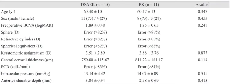
The BCVA before keratoplasty and postoperative 1, 3, 6, 12, and 24 months is presented in Fig. 1. The mean preop- erative BCVA was similar in DSAEK and PK (1.89 ± 0.48 vs. 1.95 ± 0.63 logarithm of the minimum angle of resolu- tion [logMAR], respectively, p = 0.241). Both groups showed improvement in visual outcomes after surgery, with better BCVA in the DSAEK group compared with the PK group at all follow-up periods, although the differ- ence was not statistically significant (0.69 ± 0.51 vs. 0.88 ± 0.48 logMAR, respectively at 24 months, p = 0.231).
Postoperative refractive and keratometric cylinder mea- sures (D) are shown in Table 3. At postoperative 2 years, the refractive cylinder in DSAEK and PK was -2.60 ± 1.53 and -6.00 ± 1.05 D (p = 0.002), respectively, and the ker- atometric cylinder was 3.27 ± 3.70 and 6.34 ± 3.51 D (p = 0.01), respectively. Fig. 2 shows the mean spherical equiva- lent (D) measured at postoperative 1, 3, 6, 12, and 24 months. The differences in mean spherical equivalent be- tween postoperative 1 month and 24 months was 0.84 and 2.05 D in the DSAEK and PK groups, respectively, indica- tive of comparably stable refractive changes for DSAEK.
In addition, a hyperopic shift of 1.17 D was followed after
Table 2. Baseline donor characteristics between DSAEK and PK procedures
DSAEK (n = 15) PK (n = 11) p-value*
Age (yr, range) 55.8 ± 12.7 (42-75) 57.1 ± 14.2 (45-72) 0.347
Death to preservation time (hr) 8.7 ± 7.5 8.5 ± 7.3 0.241
Death to operation time (hr) 50.9 ± 24.0 53.2 ± 18.6 0.553
Donor endothelial cell counts (cell/mm2) 2,570 ± 462 2,720 ± 448 0.372
Values are presented as mean ± standard deviation unless otherwise indicated.
DSAEK = Descemet’s stripping automated endothelial keratoplasty; PK = penetrating keratoplasty.
*Statistics by independent t-test.
Table 3. Comparison of refractive and keratometric cylinders between DSAEK and PK procedures
Refractive cylinder Keratometric cylinder
1 mon 6 mon 12 mon 24 mon 1 mon 6 mon 12 mon 24 mon
DSAEK (D) -3.60 ± 3.36 -3.43 ± 2.88 -2.98 ± 2.02 -2.60 ± 1.53 3.29 ± 2.24 3.11 ± 2.37 3.18 ± 2.44 3.27 ± 3.70 PK (D) -6.87 ± 2.81 -6.90 ± 1.47 -6.52 ± 1.51 -6.00 ± 1.05 6.85 ± 4.80 6.15 ± 4.65 6.55 ± 3.42 6.34 ± 3.51
p-value 0.067 0.031* 0.025* 0.002* 0.03* 0.02* 0.02* 0.01*
Values are presented as mean ± standard deviation.
DSAEK = Descemet’s stripping automated endothelial keratoplasty; PK = penetrating keratoplasty; D = diopters.
*Statistics by Mann-Whitney U-test.
Fig. 1. Comparison of best-corrected visual acuity (BCVA) be- tween Descemet’s stripping automated endothelial keratoplasty (DSAEK) and penetrating keratoplasty (PK).
Preoperative
BCVA (logMAR)
1 mon 3
2.5 2 1.5 1 0.5
0 3 mon 6 mon 12 mon 24 mon
DSAEK PK
Fig. 2. Comparison of mean spherical equivalent between De- scemet’s stripping automated endothelial keratoplasty (DSAEK) and penetrating keratoplasty (PK).
Spherical equivalent (D)
1 mon 0 -2 -4 -6 2 4 6
3 mon 6 mon 12 mon 24 mon
DSAEK PK
DSAEK at 24 months.
Table 4 shows the postoperative ECD changes over the 24-month follow-up period in eyes of clear grafts in which no complications had occurred. The mean preoperative donor ECD was 2,570 ± 462 cells/mm2 in the DSAEK group and 2,720 ± 448 cells/mm2 in the PK group (Table 2).
In the DSAEK group, endothelial cell loss was 25% during the first month, 31% at 6 months, and 40% at postoperative 2 years. In the PK group, endothelial cell loss was 19%
during the first month, 27% at 6 months, and 61% at post- operative 2 years. Postoperative ECD was higher in the PK group up to 6 months, but this trend reversed and was higher in the DSAEK group after postoperative 6 months.
Endothelial cell loss was relatively stationary in the DSAEK group and seemed to gradually plateau, whereas the PK group exhibited an abrupt decrease in ECD after 6 months. When percent cell loss was calculated by subtract- ing postoperative ECD at 24 months from 1 month ECD and dividing by 1 month ECD, the DSAEK group was found to have undergone a 19.96 % cell loss compared to the PK group, which had a 52.38% cell loss.
No significant difference in central corneal thickness at postoperative 24 months was found between the DSAEK and PK groups (592 ± 75 vs. 563 ± 90 mm, respectively), as shown in Table 5.
Postoperative complications
Two eyes that underwent DSAEK had graft dislocation on postoperative day 1, which was corrected with reposi- tioning by injecting an air bubble into the AC in order to press the donor tissue against the recipient cornea. In addi- tion, one subject in the DSAEK group had postoperative pupillary block. In the DSAEK and PK groups, two and three eyes had transient graft rejection, respectively, which was treated with intensive topical steroids and followed with close monitoring until all signs of rejection had re- solved. However, there was graft failure in one eye that underwent DSAEK and two eyes that underwent PK, which all required additional keratoplasty. The main cause of graft failure found in this study was immunologic rejec- tion. Two eyes from DSAEK and one eye from PK had high IOP during the early postoperative period, which were treated with IOP-lowering eye drops that returned the pressured to a normal range. A corneal ulcer occurred in one eye of each treatment group; however, there were no cases of endophthalmitis.
Graft survival rate
The 2-year graft survival rates for DSAEK and PK pro- cedures used to treat bullous keratopathy were compara- ble. Specifically, the 2-year graft survival rates for DSAEK and PK were 93.3% and 81.8%, respectively. According to Table 4. Comparison of endothelial cell density and cell loss between DSAEK and PK procedures
1 mon 3 mon 6 mon 12 mon 24 mon
DSAEK (cells/mm2) 1,934 ± 459 1,900 ± 444 1,773 ± 637 1,405 ± 475 1,548 ± 456
PK (cells/mm2) 2,209 ± 955 2,018 ± 331 1,992 ± 655 1,132 ± 661 1,052 ± 567
p-value* 0.080 0.806 0.355 0.186 0.090
Values are presented as mean ± standard deviation.
DSAEK = Descemet’s stripping automated endothelial keratoplasty; PK = penetrating keratoplasty.
*Statistics by Mann-Whitney U-test.
Table 5. Comparison of central corneal thickness between DSAEK and PK
Preoperative 1 mon 6 mon 12 mon 24 mon
DSAEK (mm) 750 ± 116 629 ± 114 601 ± 79 605 ± 84 592 ± 75
PK (mm) 811 ± 161 570 ± 105 553 ± 70 556 ± 76 563 ± 90
p-value 0.113 0.032* 0.063 0.053 0.080
Values are presented as mean ± standard deviation.
DSAEK = Descemet’s stripping automated endothelial keratoplasty; PK = penetrating keratoplasty.
*Statistics by Mann-Whitney U-test.
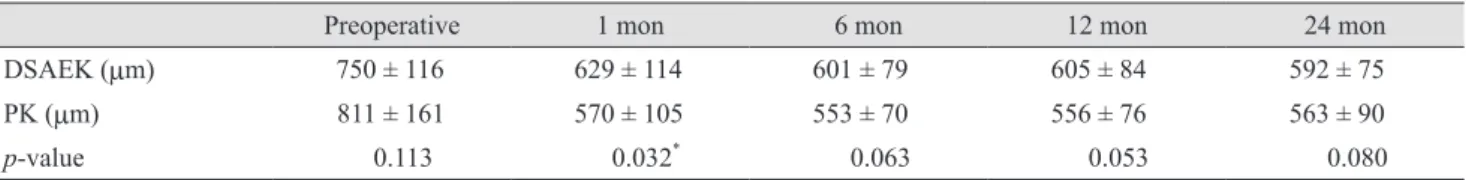
Kaplan-Meier survival analysis, the mean graft survival time after DSAEK and PK was 56 and 44 months, respec- tively (p = 0.344) (Fig. 3).
Discussion
This is the first study to report long-term outcomes of DSAEK and PK performed in South Korea for the same preoperative diagnosis of bullous keratopathy. The results of the present study identified a comparably stable refrac- tive change and better visual outcome in DSAEK. Accord- ing to Jun et al. [7], the mean change in refraction at an av- erage of 5 months after in pseudophakic DSAEK is +0.71
± 1.11 D (range, -1.75 to 3.0 D), and the overall refractive change achieved by DSAEK is +0.88 ± 1.02 D (range, -1.75 to 3.0 D), which includes a DSAEK triple surgery group.
Another study by Koenig et al. [1] reported a hyperopic shift in refraction of 1.19 ± 1.32 D for patients of DSAEK not undergoing simultaneous cataract surgery. In addition, the same study indicated that refractive astigmatism, topo- graphic astigmatism, and keratometry were not signifi- cantly different between preoperative and postoperative periods after DSAEK. In the present study, in which only bullous keratopathy was evaluated, improvement of visual acuity was achieved with only a mild tendency toward a
hyperopic shift of +1.17 D and without significant induc- tion of astigmatism after DSAEK.
All of the DSAEK and PK procedures performed in this study were carried out by an experienced surgeon, and the ECD results of the present study were consistent with data published by experienced surgeons in Western countries.
Specifically, we noted an ECD of 1,548 ± 456 cells/mm2 in DSAEK group and 1,052 ± 567 cells/mm2 in PK group at postoperative 2 years. Wacker et al. [8] conducted a 5-year study of Descemet Stripping Endothelial Keratoplasty for Fuchs’ endothelial corneal dystrophy, and reported an ECD at postoperative 2 and 5 years of 1,837 ± 551 cells/
mm2 and 1,322 ± 487 cells/mm2, respectively. Likewise, a study by Price et al. [9] reported that at postoperative 3 years, the median ECD was 1,763 cells/mm2 following DSAEK and 1,636 cells/mm2 following PK, with a median 3-year cell loss for DSAEK and PK of 59% and 61% (p = 0.70), respectively. Another study reported that the percent endothelial cell loss was lower in eyes that underwent DSAEK compared with PK at 1 (30 ± 22% vs. 37 ± 25%, p
= 0.045), 2 (36 ± 23% vs. 45 ± 33%, p = 0.018) and 3 years (39 ± 24% vs. 47 ± 28%, p = 0.022) postoperatively [10]. In our study, compared with PK, DSAEK was associated with a greater cell loss in the first 6 months, which we at- tributed to surgical trauma and manipulation during donor preparation and insertion, as the procedure requires skill- ful technique [5,11]. In subsequent months, however, the DSAEK group exhibited a slowing of endothelial cell loss compared to PK. These findings were consistent with a number of studies that collectively suggest that 6-month cell loss is significantly higher after EK than PK [1,11-13], and also that there is a high level of cell loss initially fol- lowing DSAEK in the first year, but less cell loss in subse- quent years [13]. In addition, according to a large prospec- tive series by Terry et al. [11], 30% to 40% of endothelial cell loss occurs within the first year of transplantation (without significant loss between 6 months and 1 year), but appears to plateau thereafter. Likewise, the specular Mi- croscopy ancillary study of the Cornea Donor Study [14]
identified a relatively higher rate of endothelial cell loss in the early postoperative (1 year) period, but a relatively sta- ble degree of cell loss thereafter compared to a modest in- crease in cell loss associated with PK.
Many corneal surgeons favor PK because of the relative ease of the procedure. However, PK carries with it a num- ber of postoperative complications such as high and irreg- Fig. 3. Kaplan-Meier survival curves comparing Descemet’s
stripping automated endothelial keratoplasty (DSAEK) and pen- etrating keratoplasty (PK).
Cum survival
Survival functions
DSAEK PK
1.0 0.8 0.6 0.4 0.2
0.0
0.0 10.00 20.00 30.00 40.00 50.00 60.00 Survival time (months)
ular astigmatic changes and prolonged visual rehabilita- tion, ocular surface problems, and long-term endothelial cell loss. As endothelial keratoplasty techniques have evolved, DSAEK has become a widely used method whose major advantages include stability of refraction and faster visual rehabilitation. In the present study, we found that preoperative BCVA results were similar for DSAEK and PK, while DSAEK had a 0.21 average logMAR better BCVA than PK throughout the follow-up period. In addi- tion, visual acuity was stabilized at postoperative 1 month after DSAEK and no fluctuations in visual acuity were ob- served.
Despite its advantages, certain complications are unique to the DSAEK procedure. Specifically, there can be graft detachment in DSAEK as a result of the graft being held in place initially with an air bubble rather than sutures. In our study, two eyes (13.3%) had postoperative graft dislo- cation that was corrected by an additional injection of air into the AC. The principal cause of graft failure within 2 years after DSAEK and PK was immunologic graft rejec- tion (incidence rate of 6.6% vs. 18.1%, respectively). The 2-year survival rate did not differ significantly between DSAEK and PK procedures performed for bullous kera- topathy (93.3% vs. 81.8%, respectively, p = 0.344) and the Kaplan-Meier probabilities of survival time were not sig- nificantly different. This result was comparable to that of a previous study reporting a 3-year graft survival rate of 86% for DSAEK vs. 84% for PK (p = 0.41) in eyes mainly with pseudophakic/aphakic cornea edema [9]. Another study reported a primary graft failure rate of 3.4% for PK compared with 0.8% for DSAEK, although there was no difference in Kaplan-Meier survival probabilities for PK and DSAEK at 1 (89.7% vs. 94.1%), 2 (85.0% vs. 88.2%) and 3 years (85.0% vs. 86.5%, log-rank p = 0.671) [10].
There were some limitations to the present study. First, the subjects were not randomized, although the character- istics of the subjects were similar in that only eyes of bul- lous keratopathy were studied. The ideal approach to de- termine any statistically and, more important, clinically significant differences in clinical outcomes and graft suc- cess would be with a prospective, randomized study. A second limitation of this study was that it was difficult to directly compare astigmatism before and after the stitches were removed due to the use of selective stitch removal in some patients in the PK group, as all 11 patients had at least some remnant sutures at postoperative 2 years. Anal-
ysis of the final astigmatic outcomes of the patients in our study will require a longer follow-up period and complete removal of sutures.
In conclusion, despite a relatively large initial postopera- tive endothelial cell loss as well as a wide range of cell counts, most of the grafts in the study were maintained clear. To the best of our knowledge, there have been no comparative studies involving subjects of only bullous ker- atopathy. Therefore, this study is important in that it com- pared the clinical outcomes of two procedures in patients with the same diagnosis.
Conflict of Interest
No potential conflict of interest relevant to this article was reported.
Acknowledgements
This study was supported by Basic Science Research Program through the National Research Foundation of Ko- rea (NRF) f unded by the Ministry of Education (2016R1A6A1A03010528).
References
1. Koenig SB, Covert DJ, Dupps WJ Jr, Meisler DM. Visual acuity, refractive error, and endothelial cell density six months after Descemet stripping and automated endotheli- al keratoplasty (DSAEK). Cornea 2007;26:670-4.
2. Ratanasit A, Gorovoy MS. Long-term results of Descemet stripping automated endothelial keratoplasty. Cornea 2011;30:1414-8.
3. Chen ES, Terry MA, Shamie N, et al. Descemet-stripping automated endothelial keratoplasty: six-month results in a prospective study of 100 eyes. Cornea 2008;27:514-20.
4. Terry MA, Shamie N, Chen ES, et al. Precut tissue for De- scemet’s stripping automated endothelial keratoplasty: vi- sion, astigmatism, and endothelial survival. Ophthalmolo- gy 2009;116:248-56.
5. Anshu A, Price MO, Tan DT, Price FW Jr. Endothelial ker- atoplasty: a revolution in evolution. Surv Ophthalmol 2012;57:236-52.
6. Nanavaty MA, Wang X, Shortt AJ. Endothelial keratoplasty versus penetrating keratoplasty for Fuchs endothelial dys- trophy. Cochrane Database Syst Rev 2014;(2):CD008420.
7. Jun B, Kuo AN, Afshari NA, et al. Refractive change after Descemet stripping automated endothelial keratoplasty surgery and its correlation with graft thickness and diame- ter. Cornea 2009;28:19-23.
8. Wacker K, Baratz KH, Maguire LJ, et al. Descemet strip- ping endothelial keratoplasty for Fuchs’ endothelial corneal dystrophy: five-year results of a prospective study. Oph- thalmology 2016;123:154-60.
9. Price MO, Gorovoy M, Price FW Jr, et al. Descemet’s strip- ping automated endothelial keratoplasty: three-year graft and endothelial cell survival compared with penetrating keratoplasty. Ophthalmology 2013;120:246-51.
10. Ang M, Mehta JS, Lim F, et al. Endothelial cell loss and graft survival after Descemet’s stripping automated endo-
thelial keratoplasty and penetrating keratoplasty. Ophthal- mology 2012;119:2239-44.
11. Terry MA, Chen ES, Shamie N, et al. Endothelial cell loss after Descemet’s stripping endothelial keratoplasty in a large prospective series. Ophthalmology 2008;115:488-96.e3.
12. Price MO, Price FW Jr. Endothelial cell loss after Descem- et stripping with endothelial keratoplasty influencing fac- tors and 2-year trend. Ophthalmology 2008;115:857-65.
13. Busin M, Bhatt PR, Scorcia V. A modified technique for Descemet membrane stripping automated endothelial kera- toplasty to minimize endothelial cell loss. Arch Ophthal- mol 2008;126:1133-7.
14. Price MO, Gorovoy M, Benetz BA, et al. Descemet’s strip- ping automated endothelial keratoplasty outcomes com- pared with penetrating keratoplasty from the Cornea Do- nor Study. Ophthalmology 2010;117:438-44.