대한안과학회지 2017년 제 58 권 제 11 호 J Korean Ophthalmol Soc 2017;58(11):1307-1312 ISSN 0378-6471 (Print)⋅ISSN 2092-9374 (Online)
https://doi.org/10.3341/jkos.2017.58.11.1307
Case Report
큰 황반원공 환자에서 내경계막 자가이식술 후 발생한 교세포 과증식
Hyper-proliferation of Glial Tissues Following Autologous Internal Limiting Membrane Transplantation in Idiopathic Large Macular Holes
박종호1,2⋅정재우1⋅이승민1,2⋅박성후3,4,5⋅이지은3,4,5⋅변익수1,2,5
Jong Ho Park, MD1,2, Jae Woo Jung, MD1, Seung Min Lee, MD1,2, Sung Who Park, MD3,4,5, Ji Eun Lee, MD, PhD3,4,5, Ik Soo Byon, MD1,2,5
양산부산대학교병원 안과1, 양산부산대학교병원 의생명융합연구소2, 부산대학교병원 안과3, 부산대학교병원 의생명연구소4, 부산대학교 의과대학 안과학교실5
Department of Ophthalmology, Pusan National University Yangsan Hospital1, Yangsan, Korea
Research Institute for Convergence of Biomedical Science and Technology, Pusan National University Yangsan Hospital2, Yangsan, Korea Department of Ophthalmology, Pusan National University Hospital3, Busan, Korea
Medical Research Institute, Pusan National University Hospital4, Busan, Korea Department of Ophthalmology, Pusan National University School of Medicine5, Yangsan, Korea
Purpose: To report three cases of glial hyper-proliferation after autologous internal limiting membrane (ILM) transplantation in idiopathic large macular holes.
Case summary: Three eyes with full thickness macular holes >500 μm underwent autologous ILM transplantation. After surgery, the macular hole was closed and foveal contour was U-shaped. Optical coherence tomography revealed long-lasting pro- liferation of glial cells in the fovea after the hole closure. This glial proliferation continued for 6 months, with improved visual acui- ty, and bump-like features of the fovea.
Conclusions: Autologous transplantation of ILM effectively induced long-lasting proliferation of glial cells, thereby achieving the closure of large macular holes. However, an abnormality of the foveal contour may develop after the hole closure in some cases.
J Korean Ophthalmol Soc 2017;58(11):1307-1312
Keywords: Autologous internal limiting membrane transplantation, Glial cell, Hyper-proliferation, Macular hole
■Received: 2017. 7. 20. ■ Revised: 2017. 9. 20.
■Accepted: 2017. 10. 26.
■Address reprint requests to Ik Soo Byon, MD
Department of Ophthalmology, Pusan National University Yangsan Hospital, #20 Geumo-ro, Mulgeum-eup, Yangsan 50612, Korea
Tel: 82-55-360-2592, Fax: 82-55-360-2161 E-mail: [email protected]
*Conflicts of Interest: The authors have no conflicts to disclose.
ⓒ2017 The Korean Ophthalmological Society
This is an Open Access article distributed under the terms of the Creative Commons Attribution Non-Commercial License (http://creativecommons.org/licenses/by-nc/3.0/) which permits unrestricted non-commercial use, distribution, and reproduction in any medium, provided the original work is properly cited.
전층 황반원공은 중심와의 망막결손과 중심와주변의 낭 포황반변성을 일으켜 심한 시력 저하와 변형시를 일으키는 질환이다.1,2 원공을 폐쇄하기 위해서 유리체절제술과 함께
내경계막을 추가로 제거하며 원공 주변의 견인력을 제거하 고 교세포 증식을 유도하며, 가스를 눈 속에 충전하여 원공 속으로 액체가 유입되지 않게 한다.1,2 내경계막 제거술이 소개된 이후로 약 80-90%의 높은 원공폐쇄율을 달성할 수 있게 되었으나,3 크기가 큰 황반원공에서는 성공률이 약 60% 정도로 낮다고 보고되고 있다.4
최근에는 새로운 술기를 이용하여 크기가 큰 황반원공 에서도 높은 수술 성공률을 달성한 연구들이 보고되었다. 내경계막을 제거하지 않고 절편(flap)을 만든 뒤 뒤집어 원 공 위를 덮거나, 벗겨낸 내경계막 절편조각을 원공 속에 이식하여, 교세포의 증식을 유도하여 큰 원공을 폐쇄할 수 있게 하는 것이다.5,6 본 저자들도 500 μm 이상의 큰 원공
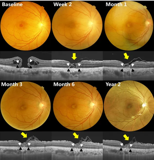
Figure 1. A Case of 67-year-old woman with full thickness macular hole. Two weeks after autologous internal limiting membrane
transplantation, optical coherence tomography images showed a glial tissues in the fovea without a complete hole closure. The long-lasting proliferation of glial cells (yellow arrows) resulted in the closure of hole, but the abnormal contour of fovea was post- operatively observed from 3 months to 24 months. The defects of ellipsoid zone (black arrowheads) and external limiting membrane (white arrowheads) remained. The foveal depigmentation was seen in the postoperative fundus photographs.을 가진 환자에서 내경계막 자가이식술을 시행하여 성공 적인 원공폐쇄를 보고한 적이 있다.6 이러한 새로운 수술 술기에 대한 경험이 늘어나고 수술 후 경과 관찰 기간이 길어지면서 새로운 소견들을 경험하게 되었다. 저자들은 특발성 황반원공 환자에서 내경계막 자가이식술 후 성공 적으로 큰 원공이 폐쇄된 뒤에도 지속적으로 교세포가 증 식하여 중심와 구조이상이 발생한 증례들을 경험하여 보 고하고자 한다.
증례보고
수술술기 및 중심오목 구조의 평가
백내장수술과 함께 25게이지 유리체절제술을 시행하였다.
0.025% brilliant blue G (BBG; Sigma-Aldrich, St. Louis, MO, USA)로 내경계막을 염색한 뒤, 중심와를 중심으로 시 신경유두 지름의 2배 정도의 크기로 벗겨내었다. 벗겨낸 내 경계막 절편 일부를 원공의 크기에 맞게 잘라낸 뒤 원공 속 에 이식하였다. 액체-공기 교환을 아주 천천히 시행하여 절 편이 원공 속에 유지될 수 있게 하였다. 이후 18% 육불화
- 박종호 외 : 내경계막 자가이식술 후 교세포 과증식 -
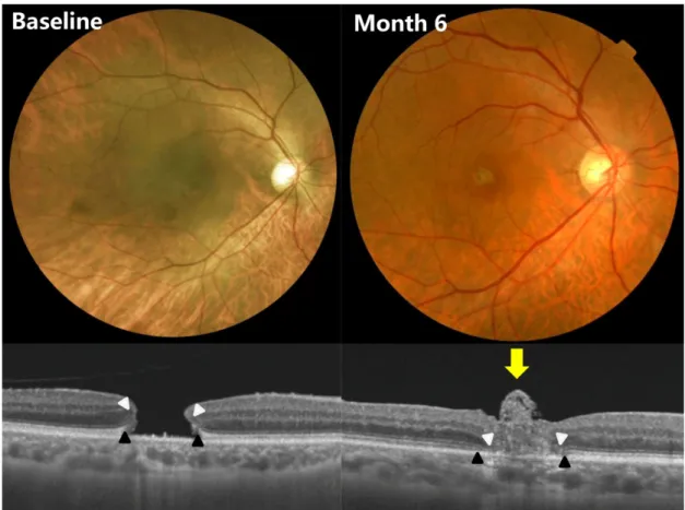
Figure 2. A case of 70-year-old woman with full thickness macular hole. One month after autologous internal limiting membrane
transplantation, the closure of the hole and glial tissues in fovea (yellow arrow) were observed in optical coherence tomography.Foveal depigmentation was seen in fundus photography. At 6 months, the protrusion of fovea developed. The defects of ellipsoid zone (black arrowheads) and external limiting membrane (white arrowheads) remained.
유황가스(SF6)를 충전하고 수술을 종료하였다. 수술 후에는 7일간 엎드린 자세를 유지하도록 하였다.
수술 전후 파장가변 빛간섭단층촬영(DRI OCT-1, Topcon, Tokyo, Japan)을 이용하여 원공의 크기, 시세포 내/외절 경 계의 결손, 외경계막의 결손을 측정하였다. 시세포 내/외절 경계의 결손, 외경계막의 결손은 수직, 수평 경선을 측정하 여 평균을 계산하였다.
증례 1
67세 여자 환자가 우안의 심한 시력저하를 주소로 내원 하였다. 최대교정시력 0.1로 측정되었다. 안저검사에서 전 층 황반원공을 확인할 수 있었다. 빛간섭단층촬영으로 측 정한 원공의 최소직경은 667 µm, 시세포 내/외절 경계의 결손은 1,200.5 µm, 외경계막의 결손은 1,082 µm였다.
내경계막 이식술 후 2주째 측정한 빛간섭단층촬영에서 원공 안에 이식된 내경계막 절편을 확인할 수 있었다. 1개 월째는 교세포 증식으로 중심오목의 모양이 더욱 개선된 상태였다. 3개월째에는 교세포 증식이 중심오목 밖으로 진 행된 것이 관찰되었으며 6개월째까지 지속되었다. 수술 후
6개월에는 시세포 내/외절 경계의 결손은 681.5 µm, 외경 계막의 결손은 557 µm로 감소하였으나 완전히 회복되지는 못하였다(Fig. 1). 시력은 술 후 3개월째 0.2로 다소 회복되 었고, 2년 경과관찰기간 동안 유지되었다.
증례 2
70세 여자 환자가 2년 전부터 시작된 우안 시력저하로 내원하여 전층 황반원공으로 진단되었다. 최대교정시력은 0.15였다. 원공의 최소직경은 694 µm, 시세포 내/외절 경계 의 결손은 951 µm, 외경계막의 결손은 899.5 µm로 측정되 었다. 내경계막 자가이식술 시행 후 7일째 성공적인 원공의 폐쇄가 확인되었다. 수술 후 1개월 파장가변 빛간섭단층촬 영 영상에서는 중심오목의 형태가 평평해지고 있었다. 수 술 후 3개월째는 중심오목의 돌출된 구조가 관찰되었으며, 이러한 증식은 6개월째까지 지속되었다. 수술 후 6개월째 시세포 내/외절 경계의 결손은 711.5 µm, 외경계막의 결손 은 622 µm로 감소하였으며, 시력은 3개월째 0.3으로 호전 되어 6개월 경과관찰기간 동안 유지되었다(Fig. 2).
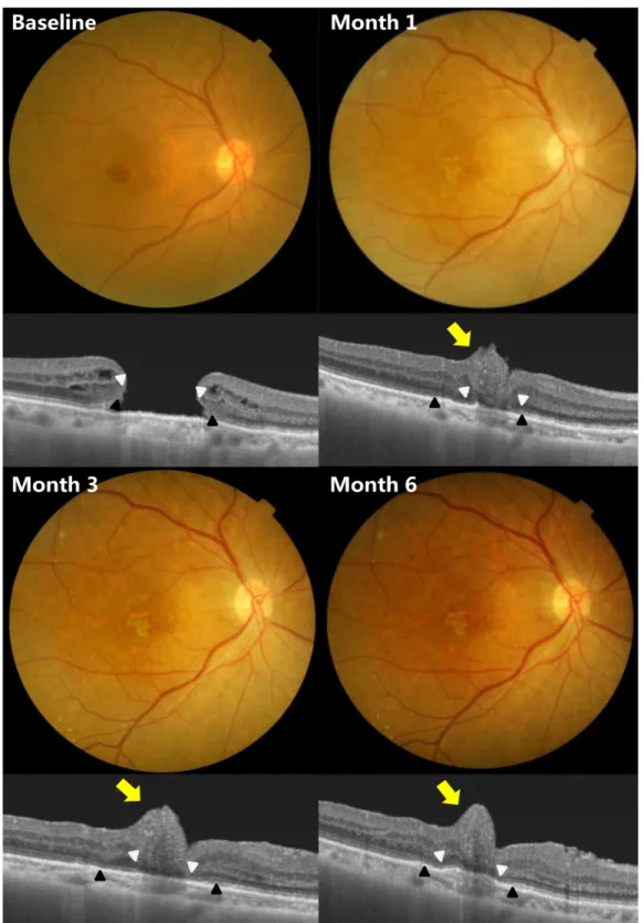
Figure 3. A case of 75-year-old woman with full thickness macular hole. One month after autologous internal limiting membrane
transplantation, irregular contour of fovea (yellow arrows) was detected in optical coherence tomography. At 3 months, the bump-like fovea developed. The depigmentation of fovea was seen in postoperative fundus photograph. The defects of ellipsoid zone (black arrowheads) and external limiting membrane (white arrowheads) remained. And it lasted until 6 months.증례 3
75세 여자 환자가 1년 전부터 시작된 우안 시력저하로 내원하여 전층 황반원공으로 진단받았다. 수술 전 최대교
정시력은 0.15, 원공의 최소직경은 818.5 μm, 시세포 내/외절 경계의 결손은 1,241.5 μm, 외경계막의 결손은 1,126.5 μm였 다. 내경계막 자가이식술을 시행 받고 성공적으로 원공이
- 박종호 외 : 내경계막 자가이식술 후 교세포 과증식 -
폐쇄되었으나, 수술 후 1개월의 파장가변 빛간섭단층촬영 영상에서는 중심오목의 돌출이 관찰되었다. 수술 후 6개월 째 영상에서는 더욱 증가된 중심오목의 돌출이 관찰되었으 나, 시세포 내/외절 경계의 결손은 938 μm, 외경계막의 결 손은 824 μm로 수술 전에 비해 호전된 상태였다(Fig. 3).
최대교정시력은 술 후 3개월에 0.2로 회복되어 6개월 경과 관찰기간 동안 유지되었다.
고 찰
유리체절제술과 내경계막 제거술을 통해 황반원공이 폐 쇄되는 기전은 전후 방향 및 접선 방향의 견인력이 제거되 어 원공의 경계면이 서로 접근하게 되고, 교세포의 증식이 자극되어 신경망막의 결손된 부분을 메우게 되는 것이다.7 눈속 가스 충전은 액체가 황반원공에 접촉하는 것을 차단 하고 교세포의 이주를 촉진하는 역할을 하여 치유과정을 돕게 된다. 하지만 크기가 큰 원공에서는 이런 술기만으로 는 원공폐쇄에 부족한 경우가 많다.8 최근에는 원공 위로 내경계막 절편을 덮는 방법5 및 내경계막 절편을 원공 속에 이식하는 방법9 등의 새로운 술기를 이용하여 큰 황반원공 에서 성공적인 원공의 폐쇄를 달성할 수 있게 되었다.
본 증례에서 시행된 내경계막 자가이식술은 내경계막 절 편과 함께 내경계막 표면에 존재하는 뮬러세포가 원공 속 으로 이주되게 되며, 원공 속의 내경계막이 뮬러세포 등의 교세포가 증식하는 데 골격체 역할을 하여 큰 원공을 성공 적으로 폐쇄되게 하는 것으로 여겨진다.10,11 하지만 내경계 막 자가이식술에서 원공폐쇄에 필요한 적절한 내경계막 절 편의 크기에 대해서는 아직 알려진 바가 없다. 저자들은 원 공의 최소 직경을 고려하여 내경계막 절편의 크기를 결정 하였는데, 본 증례들에서는 원공 폐쇄에 필요한 것보다 내 경계막 절편의 크기가 커서 교세포의 과증식이 유도되었을 거라 추측된다. Oh et al12은 내경계막 제거술을 시행 받은 황반원공 환자들 중에서 크기가 큰 경우에 교세포 증식이 더 흔하게 발생하였다고 보고한 바 있다. 크기가 큰 원공에 서는 상대적으로 벗겨낸 내경계막이 크고 이로 인해 교세 포의 증식이 더욱 유도되었을 것이라 생각된다. 이러한 교 세포의 과도한 증식은 술 후 정상적인 시세포 배열의 회복 을 막아 시력회복이 불량하다고 하였는데,12 내경계막 이식 후 교세포 과증식이 발생한 본 증례들에서도 비록 원공폐 쇄 후 시력회복이 있었지만, 시세포 내절/외절 경계와 외경 계막 결손이 완전히 회복되지 못하였다. 이러한 불완전한 시세포층의 회복은 추가적인 술 후 시력개선에 제한이 되 었을 가능성도 있다.
Imai et al13은 황반원공 수술 후 중심오목을 정상적인 구
조의 U-type, 가파른 구조의 V-type, 그리고 불규칙적인 형 태를 보이는 W-type으로 구분하였고, 시력예후는 U-type, V-type, W-type 순으로 우수하다고 보고하였다.본 증례들 은 U-type의 원공의 폐쇄를 달성하였으나 이후 중심오목의 돌출이 발생하였다. 돌출된 중심오목의 구조는 W-type에 해당된다고 보인다. 비록 세 증례 모두 중심오목의 돌출이 발생하였음에도 불구하고 시력은 호전되었으나, 이러한 중 심오목의 구조변화도 수술 후 더 많은 시력 개선에 제한이 되었을 가능성도 배제할 수 없다.
결론적으로 황반원공 수술에서 내경계막 자가이식술은 크기가 큰 원공에서 성공적인 원공 폐쇄를 달성할 수 있으 나 일부 환자에서는 교세포 과증식으로 인해 중심오목의 구조이상이 발생할 수 있음을 고려해야 할 것이다. 향후 내 경계막 자가이식술 후 중심와 구조의 변화가 수술 후 시력 에 미치는 영향에 대해서는 더 많은 환자를 대상으로 연구 가 필요할 것으로 생각된다.
REFERENCES
1) Johnson RN, Gass JD. Idiopathic macular holes. Observations, stages of formation, and implications for surgical intervention.
Ophthalmology 1988;95:917-24.
2) la Cour M, Friis J. Macular holes: classification, epidemiology, nat- ural history and treatment. Acta Ophthalmol Scand 2002;80:579-87.
3) Brooks HL Jr. Macular hole surgery with and without internal lim- iting membrane peeling. Ophthalmology 2000;107:1939-48; dis- cussion 1948-9.
4) Salter AB, Folgar FA, Weissbrot J, Wald KJ. Macular hole surgery prognostic success rates based on macular hole size. Ophthalmic Surg Lasers Imaging 2012;43:184-9.
5) Michalewska Z, Michalewski J, Adelman RA, Nawrocki J. Inverted internal limiting membrane flap technique for large macular holes.
Ophthalmology 2010;117:2018-25.
6) Kim KH, Jung JW, Park SW, et al. Autologous transplantation of internal limiting membrane for the treatment of large macular hole.
J Korean Ophthalmol Soc 2015;56:1899-905.
7) Wender J, Iida T, Del Priore LV. Morphologic analysis of stage 3 and stage 4 macular holes: implications for treatment. Am J Ophthalmol 2005;139:1-10.
8) Stec LA, Ross RD, Williams GA, et al. Vitrectomy for chronic macular holes. Retina 2004;24:341-7.
9) De Novelli FJ, Preti RC, Ribeiro Monteiro ML, et al. Autologous internal limiting membrane fragment transplantation for large, chronic, and refractory macular holes. Ophthalmic Res 2015;55:
45-52.
10) Smiddy WE, Feuer W, Cordahi G. Internal limiting membrane peeling in macular hole surgery. Ophthalmology 2001;108:1471-6;
discussion 1477-8.
11) Costa RA, Cardillo JA, Morales PH, et al. Optical coherence to- mography evaluation of idiopathic macular hole treatment by gas-assisted posterior vitreous detachment. Am J Ophthalmol 2001;132:264-6.
= 국문초록 =
큰 황반원공 환자에서 내경계막 자가이식술 후 발생한 교세포 과증식
목적: 내경계막 자가이식술을 시행 받은 큰 황반원공 환자에서 교세포 과증식이 발생한 증례 3안을 경험하여 보고하고자 한다.
증례요약: 크기가 500 μm 이상인 전층 황반원공으로 내경계막 자가이식술을 시행 받은 3안은 원공이 성공적으로 폐쇄되고 U 모양의 중심와를 회복하였다. 빛간섭단층촬영에서 원공 폐쇄 이후에도 지속적인 교세포 증식이 관찰되었으며, 수술 후 약 3개월에 중심와 구조가 소실되었다. 이러한 교세포의 증식은 수술 6개월째까지 지속되어 최종적으로 중심와 일부가 돌출된 구조를 나타내었다. 교세 포의 과증식으로 인한 중심와의 구조적 이상의 발생에도 불구하고, 수술 후 호전된 시력이 유지되었다.
결론: 내경계막 자가이식술은 지속적인 교세포의 증식을 유도하여 큰 황반원공을 폐쇄하는 데 효과적이나, 일부에서는 원공 폐쇄 후 중심와 구조의 이상이 발생할 수 있었다.
<대한안과학회지 2017;58(11):1307-1312>
12) Oh J, Yang SM, Choi YM, et al. Glial proliferation after vitrectomy for a macular hole: a spectral domain optical coherence tomog- raphy study. Graefes Arch Clin Exp Ophthalmol 2013;251:477-84.
13) Imai M, Iijima H, Gotoh T, Tsukahara S. Optical coherence tomog- raphy of successfully repaired idiopathic macular holes. Am J Ophthalmol 1999;128:621-7.