Capillary hemangiomas are benign vascular tumors that are believed to be hamartomatous proliferations of vascular endothelial cells. They are bright red to blue in color, sharply demarcated from the surrounding skin or mucosal tissue, and usually located on the head and neck in childhood (1). Spinal intradural extramedullary capillary hemangiomas are extremely rare and only a few cases have been reported (2-8). They appear isoin- tense or hypointense relative to the spinal cord on T1- weighted images, isointense or slightly hyperintense on T2-weighted images, and exhibit a strong homogenous enhancement on contrast-enhanced T1-weighted im- ages. To our knowledge, intradural extramedullary cap- illary hemangiomas with a long segment of transient T2 hyperintensity in the spinal cord have not previously
been reported. We present a rare case of thoracic in- tradural extramedullary capillary hemangioma that was accompanied by a long segment of transient T2 hyperin- tensity in the spinal cord.
Case Report
A 48-year-old man was admitted to the hospital be- cause of decreased body sensations below the level of 10 cm above the umbilicus. The patient had been well until a month earlier, when he experienced the sudden onset of back pain at the level of the thoracic spine. About two weeks before admission, he experienced a tingling sen- sation and radiating pain in both lower extremities (the left more than the right), and a week before admission, hypesthesia developed below the level of 10 cm above the umbilicus.
Physical examination revealed decreased motor strength in both lower extremities and hypesthesia be- low the T8 dermatome level.
MR imaging of the thoracic spine performed with a 3-
Intradural Extramedullary Capillary Hemangioma with Long Segment of Transient Cord Edema: A Case Report1
Hyeon Yu, M.D., Yong Chul Lee, M.D., Sung Nam Hwang, M.D.2, Seung Min Yoo, M.D., Hwa Yeon Lee, M.D., In Sup Song, M.D., Jong Beum Lee, M.D., Kun Sang Kim, M.D.
1Department of Radiology, Chung-Ang University Yongsan Hospital
2Department of Neurosurgery, Chung-Ang University Yongsan Hospital Received December 20, 2005 ; Accepted January 10, 2006
Address reprint requests to : Hyeon Yu, M.D., Department of Radiology, Chung-Ang University Yongsan Hospital, 65-207, Hangangro 3-ga, Yongsan-gu, Seoul 140-757, Korea.
Tel. 82-2-748-9682 Fax. 82-2-798-4745 E-mail: [email protected]
We present a case of intradural extramedullary capillary hemangioma of the thoracic spine with a long segment of transient cord edema. Spinal capillary hemangiomas are extremely rare vascular tumors and only a few cases have been reported. On the MR images, the mass showed hypointensity on the T1-weighted images, hyperintensity on the T2-weighted images relative to the spinal cord, and strong homogeneous enhance- ment on the contrast-enhanced T1-weighted images. The T2-weighted images showed a long segment of ill-defined hyperintense area in the spinal cord which was complete- ly resolved after surgery.
Index words :Angioma
Magnetic resonance (MR) Spinal cord, MR
T Achieva system (Philips Medical Systems, Best, The Netherlands) revealed a well-circumscribed intradural extramedullary mass at the T6-T7 level. The mass was located in the dural sac, posterolateral to the spinal cord.
The spinal cord was displaced and compressed by the mass. On the T2-weighted images, the mass showed slight hyperintensity with peripheral dark signal intensi- ty relative to the spinal cord. The T2-weighted sagittal images of the spinal cord showed a long segment of dif- fuse and ill-defined hyperintense area extending from the T2 to T9 level. The T1-weighted images showed a hypointense mass with a peripheral hyperintense por- tion attached to the spinal cord in the dural sac. The con- trast-enhanced T1-weighted images revealed strong ho- mogeneous enhancement of the mass associated with a short enhancing vessel on the dorsal inner surface of the dura.
The preoperative diagnosis was a neurogenic tumor and the differential diagnosis included meningioma.
The patient underwent a T6-7 total laminectomy in the prone position. When the dura was opened, a 10×
10×7 mm3dark reddish mass was seen to be compress- ing the spinal cord ventrally. The mass was able to be dissected from the cord quite well, except for one dorsal root which was firmly adhered to the mass. The mass was removed along with the dorsal root, at which time a tiny portion of the tumor was seen in the parenchyma of the dorsal spinal cord. The remaining tumor was re- moved and the wound was closed.
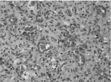
Histologic examination revealed numerous small, cap- illary-sized vascular channels, lined by monotonous en- dothelial cells. This finding was consistent with a capil- lary hemangioma. The capillaries were relatively uni- form in size, with no centrally placed larger vessels, and there were associated organizing hemorrhagic lesions.
The MR images of the thoracic spine obtained two months after surgery showed the disappearance of the hyperintense signal area in the spinal cord on the T2- weighted images. The patient’s back pain was im- proved and the strength of both extremities was recov- ered at the time of discharge.
Discussion
Capillary hemangiomas are benign vascular neo- plasms which are composed of capillary-sized blood ves- sels. They are bright red to blue in color, and situated on a level with the surface of the skin, or slightly elevated.
They are commonly found in the skin, subcutaneous tis-
sues, and oral mucosa. They usually appear on the head and neck in infancy and childhood (1).
Spinal capillary hemangiomas involving the intradural space are rare. Only a few cases of spinal intradural ex- tramedullary capillary hemangiomas have previously been reported (2-8). They are considered as hamar- tomatous proliferations of the vessels in the spine (2).
The sites where intradural extramedullary heman- giomas have been found to originate include the blood vessels of the nerve roots in the cauda equina, the inner surface of the dura, and the pial surface of the spinal cord (3, 4). Our case turned out to develop from the pial surface of the spinal cord and the blood vessels of the T6 nerve root.
The MR imaging findings in our case were similar to those of previous case reports of intradural ex- tramedullary capillary hemangiomas (2-8). Our case showed heterogeneous hypointensity on the T1-weight- ed images and hyperintensity on the T2-weighted im- ages. On the contrast-enhanced T1-weighted images, the mass showed strong homogeneous enhancement (Figs.
1A-1C). Since the MR features of our case were not specific, the mass could not be accurately differentiated from other intradural extramedullary tumors. The dif- ferential diagnosis for intradural extramedullary tumors and hamartomatous lesions includes schwannoma, meningioma, ependymoma, cavernous angioma, he- mangioblastoma, hemangiopericytoma, hemangioen- dothelioma, angiolipoma, solitary fibrous tumor, and metastasis (5). Among these intradural extramedullary lesions, schwannoma and meningioma are the most common neoplasms which are included in the differen- tial diagnosis for capillary hemangioma. Schwannomas show hypo- or isointensity on the T1-weighted images, and hyperintensity on the T2-weighted images. The sig- nal characteristics in meningiomas are usually iso- or hypointense on the T1-weighted images, and iso- or slightly hyperintense on the T2-weighted images.
Furthermore, both schwannomas and meningiomas are strongly enhanced on the contrast-enhanced T1-weight- ed images. Schwannomas frequently show cystic change or necrosis, and if they did not do so, it would be difficult to differentiate them from capillary heman- gioma (6). Choi et al (6) described three cases of in- tradural extramedullary capillary hemangiomas, two of which showed dural attachment and dural tail sign on the contrast-enhanced T1-weighted images. Although one of the most characteristic MR imaging findings of meningiomas is their dural attachment with dural tail
sign on the contrast enhanced study, in differentiating meningiomas from capillary hemangiomas.
Zander et al (3) reported an intradural extramedullary capillary hemangioma showing heterogeneous hyperin- tensity on the T1-weighted images and homogeneous hypointensity on the T2-weighted images, which corre- sponded to a hemorrhagic focus that was confirmed on histologic examination. In our case, focal hyperintensity was seen at the lower lateral aspect of the mass on the T1-weighted images and the mass was hypointense on the T2-weighted images (Figs. 1A, 1B). This finding was
consistent with a subacute or chronic hemorrhage.
These MR features are suggestive of hemangioma or other vascular neoplasms, although they are not specif- ic.
Abdullah et al (7) reported a thoracic intradural ex- tramedullary capillary hemangioma associated with en- larged draining perimedullary veins that were enhanced on the contrast-enhanced T1-weighted images. They suggested that when enlarged draining veins are found along with an intraspinal mass on the MR images, one should consider a highly vascular tumor or malforma-
A B C
Fig. 1. A 48-year old man with thoracic intradural extramedullary capillary hemangioma.
A. Sagittal T2-weighted image demon- strates a round well-circumscribed nodular lesion with heterogeneous hy- perintensity relative to the spinal cord in the thecal sac at the T6 level (arrow- head). At the posterior lower edge of the mass, a crescent-shaped hy- pointense rim can be seen (arrow). A long segment of ill-defined hyperinten- sity is seen in the spinal cord.
B. On the T1-weighted sagittal image, the mass is heterogeneously hy- pointense relative to the spinal cord (arrowhead) and shows a crescent- shaped hyperintense peripheral rim at the lower edge (arrow), which corre- sponds to the hypointense portion on the T2-weighted sagittal image.
C. The contrast-enhanced T1-weight- ed sagittal image demonstrates a mass with strong homogeneous enhance- ment (arrowhead) and a short linear enhancing structure on the posterior inner surface of the dura adjacent to the mass, which is presumably due to a draining vein rather than a dural tail sign (arrow).
D. On the T2-weighted axial image, the mass compresses the spinal cord in which hyperintense edema is seen (arrowhead). At the posterolateral edge of the mass, a crescent-shaped hypointense area can be seen, which corresponds to the hemorrhagic focus (arrow).
E. The T2-weighted sagittal image ob- tained 2 months after surgery demon- strates the complete disappearance of the long segment hyperintensity in the spinal cord.
D E
tion and perform preoperative spinal angiography to avoid the risk of hemorrhage. Choi et al (6) also reported a case of intradural extramedullary capillary heman- gioma showing a vertical linear enhancing structure within the thecal sac, presumably due to a radicu- lomedullary vein. In our case, an enhancing short drain- ing vein was seen on the dorsal inner surface of the dura on the contrast-enhanced T1-weighted images (Fig. 1C).
Rivierez et al (9) reported a case of intramedullary cap- illary hemangioma associated with a transient hypersig- nal in the spinal cord on the T2-weighted images. This finding disappeared on the follow-up MR images ob- tained three months later. Shin et al (4) reported a spinal intradural capillary hemangioma with both ex- tramedullary and intramedullary components, showing diffuse edema in the spinal cord which was hyperin- tense on the T2-weighted images. However, no postop- erative MR images were obtained, so the cord hyperin- tensity could not be followed up in this case. In our case, the T2-weighted images showed a long segment of hy- perintense area with an ill-defined outer margin in the spinal cord extending from the T2 to T9 level, which was completely resolved on the follow-up MR images obtained two months after surgery (Figs. 1D, 1E).
Although the exact pathophysiology of this MR imaging finding associated with capillary hemangioma is still un- known, it is thought that the reversible T2 hyperintensi- ty in the spinal cord may represent the cord edema and is most likely caused by disturbed local venous circula- tion induced by chronic spinal cord compression (10).
To our knowledge, pure intradural extramedullary cap- illary hemangioma without an intramedullary compo- nent showing a long segment of transient cord edema has not previously been reported.
The treatment of choice for intradural extramedullary capillary hemangioma is complete surgical resection (2, 8). Follow-up MR imaging is a reasonable choice in cas- es where there is a T2 hyperintense signal in the spinal cord, in order to differentiate transient cord edema from a permanent cord injury. Roncaroli et al (8) reported no recurrence at 10 years after the complete surgical resec- tion of spinal capillary hemangioma in two cases.
However, if complete surgical resection of the mass is not possible, postoperative follow-up MR imaging is warranted (8).
In conclusion, intradural extramedullary capillary he- mangiomas are rare, but they should be taken into ac- count when making a differential diagnosis of intradural extramedullary neoplasms. Although a long segment of transient edema in the spinal cord is not specific, we hope it provides an additional MR imaging finding of rare spinal capillary hemangiomas.
References
1. Benign tumors and tumor-like conditions: Blood Vessels. In. Kumar V, Abbas AK, Fausto N. Robbins and Cotran Pathologic Basis of Disease. 7th ed. Philadelphia: Elsevier Saunders, 2005:545-546 2. Nowak DA, Widenka DC. Spinal intradural capillary haeman-
gioma: a review. Eur Spine J 2001;10:464-472
3. Zander DR, Lander P, Just N, Albrecht S, Mohr G. Magnetic reso- nance imaging features of a nerve root capillary hemangioma of the spinal cord: case report. Can Assoc Radiol J 1998;49:398-400 4. Shin JH, Lee HK, Jeon SR, Park SH. Spinal intradural capillary he-
mangioma: MR findings. AJNR Am J Neuroradiol 2000;21:954-956 5. Andaluz N, Balko MG, Stanek J, Morgan C, Schwetschenau PR.
Lobular capillary hemangioma of the spinal cord: case report and review of the literature. J Neuroonco 2002;56:261-264
6. Choi BY, Chang KH, Choe GY, Han MH, Park SW, Yu IK, et al.
Spinal Intradural Extramedullary Capillary Hemangioma: MR findings. AJNR Am J Neuroradiol 2001;22:799-802
7. Abdullah DC, Raghuram K, Philips CD, Jane JA Jr, Miller B.
Thoracic intradural extramedullary capillary hemangioma. AJNR Am J Neuroradiol 2004;25:1294-1296
8. Roncaroli F, Scheithauer BW, Krauss WE. Capillary hemangioma of the spinal cord-report of four cases. J Neurosurg 2000;93 Suppl:
148-151
9. Rivierez M, Heyman D, Jouannelle A, Arfi S. Capillary heman- gioma of the spinal cord. A new case. Neurochirurgie 2002;48:440- 444
10. Lee J, Koyanagi I, Hida K, Seki T, Iwasaki Y, Mitsumori K. Spinal cord edema: unusual magnetic resonance imaging findings in cer- vical spondylosis. J Neurosurg 2003;99 Suppl:8-13
Fig. 2. Hematoxyllin and eosin-stained microscopic image (original magnification ×200) shows the capillary-sized spaces lined by monotonous endothelial cells in a collageneous fi- brous stroma.
대한영상의학회지 2006;54:343-347
척수의 일시적 부종을 동반한 경막내 척수외 모세 혈관종: 증례 보고1
1중앙대학교부속 용산병원 영상의학과
2중앙대학교부속 용산병원 신경외과
유 현・이용철・황성남2・유승민・이화연・송인섭・이종범・김건상
척추에서 발생한 모세 혈관종은 드문 혈관 종양으로서 현재까지 매우 적은 증례들만이 보고되고 있다. 저자들은 최근 흉추의 경막내 척수외 공간에서 발생한 모세 혈관종으로서 척수내에 긴 분절의 부종을 동반한 증례를 경험하 였기에 이를 보고하고자 한다. 자기공명영상촬영에서 이 종양은 T1 강조영상에서는 저신호강도를, T2 강조영상에 서는 고신호강도를 보였고, 조영제 주입 후에는 균질하며 강하게 조영증강이 되는 소견을 보였다. T2 강조영상에 서 척수내에 경계가 불분명한 긴 분절의 고신호강도가 보였는데 이는 일시적인 부종으로 추정되며 수술 후 완전히 소실되었다.