서 론
비결핵 마이코박테리아(nontuberculous mycobacteria, NTM;
atypical mycobacteria)는 토양이나 물 등 생활환경에 널리 존재 하는 비병원성 균으로 인식되어 왔다. 그러나 1980년대 후천성면 역결핍증이 유행하면서 이와 관련한 NTM 유발 질환이 증가하 였고[1, 2], 임상적 중요성에 대한 인식과 배양법의 발달에 따라 HIV sero-negative 환자에서도 폐질환, 궤양성질환, 염증성 장 질환 등을 유발함이 보고되었다[3-5]. 결핵의 유병률이 높은 우리 나라의 경우, NTM이 임상검체에서 분리될 때 결핵으로 오인되 거나 단순 오염균으로 인식되어, 결핵 과거력이라는 NTM 폐질 환의 위험요인이 많음에도 불구하고 정확한 균동정을 요구하지 않는 경우가 많다.
153 153
16S Ribosomal RNA, Heat-shock Protein 65 및 RNA Polymerase -Subunit 유전자 염기서열분석을 통한 비결핵 마이코박테리아의 동정
Identification of Nontuberculous Mycobacteria by Sequence Analysis of the 16S Ribosomal RNA, the Heat-shock Protein 65 and the RNA Polymerase -Subunit Genes
Sue Shin, M.D.
1,3, Eui Chong Kim, M.D.
2,3, and Jong-Hyun Yoon, M.D.
1,3Departments of Laboratory Medicine, Boramae Hospital1; Seoul National University Hospital2; Seoul National University College of Medicine3, Seoul, Korea
신 수1,3∙김의종2,3∙윤종현1,3
보라매병원 진단검사의학과1, 서울대학교병원 진단검사의학과2, 서울대학교 의과대학 검사의학교실3
153 153
접 수: 2005년 9월 23일 접수번호:KJLM1887 게재승인일: 2005년 12월 12일
교 신저 자: 윤 종 현
우 156-707 서울시 동작구 신대방동 425 서울대학교 보라매병원 진단검사의학과 전화: 02-840-2280, Fax: 02-840-2742 E-mail : [email protected]
*본 연구는 2004년도 서울대학교 보라매병원 임상공동연구비(03-2004-04) 의 지원에 의해 이루어진 것임.
Background :The diagnosis of diseases caused by nontuberculous mycobacteria (NTM) is diffi- cult, because NTM are prevalent in the environment such as soil and water, and because they have fastidious properties. In this study we investigated clinical isolates of NTM for their distribution pattern and accurate species identification.
Methods :We selected presumptive NTM isolates negative for probe hybridization for M. tubercu- losis complex, cultured in a third referral hospital from 21 January 2003 to 20 January 2004. Ninety seven-isolates were identified to the species level by direct sequencing of fragments of 16S rRNA, hsp65 and rpoB genes. A total of 120 isolates were studied for the distribution analysis.
Results :Frequently identified NTM species were M. avium (30.8%), M. intracellulare (23.3%) and M. abscessus (18.3%). Others were M. gordonae, M. senegalense, M. fortuitum, M. peregrinum, M.
kansasii, M. terrae complex, M. lentiflavum, M. chelonae, and M. szulgai. Three M. tuberculosis com- plex (2.5%) were also identified among the presumptive NTM isolates. The identification rate by se- quencing of 16S rRNA, rpoB, and hsp65 were 65%, 82% and 87%, respectively. The hsp65 or rpoB gene was more efficient than 16S rRNA for the identification of NTM by sequencing.
Conclusions :Some NTM are increasingly considered to be the causative organisms in clinical diseases. Thus, direct sequencing could be adapted to routine work of clinical laboratories for accu- rate identification of NTM to the species level. (Korean J Lab Med 2006;26:153-60)
Key Words : Sequence analysis, Atypical mycobacteria, rpoB, 16S rRNA, Heat-shock protein
우리나라에서는 1981년
Mycobacterium avium
complex (MAC)에 의한 폐항산균증이 처음으로 보고되었으며[6], 이후 NTM 폐질환에 대한 국내 보고의 빈도는 늘고 있지만, 대상 환자 군 및 시기, 동정방법 등이 달라 직접적인 비교가 힘들뿐 아니라, 정확한 균 동정을 기초로 한 분리율 및 분포 등에 대한 연구는 부 족한 실정이다[7-12].NTM은 전통적으로 성장속도, 균 집락의 모양, 색 등의 미생물 학적 특징에 따라 구별된다. 그러나 생화학적 동정으로는 균종 구 분이 힘들고, 방법상 시간과 숙련도가 필요하며 새롭게 발견되는 종에 있어서는 충분한 기초 자료가 정립되어 있지 않다는 단점이 있다[13, 14]. 그러므로 신속하고 정확한 동정을 위하여 mycolic acid의 분석이나, 핵산소식자, 핵산증폭, 염기서열분석 등의 핵산 을 이용한 방법을 이용하고 있다. 일반적으로 검사실에서는 임상 적으로 중요한
M. tuberculosis
(MTB)와 NTM의 구별에 신속 감별법 중 하나를 도입하고 있다.PCR-based sequencing방법은 마이코박테리아 동정의 최종적 인 방법이 되며, 이때 이용되는 유전자는 16S rRNA, 16S-23S rRNA ITS (internally transcribed spacer), 65 kDa heat-shock protein gene (
hsp65
), RNA polymerase -subunit gene (rpoB
) 등이 연구되고 있다[15-18]. 16S rRNA 유전자는 모든 박테리아 에 존재하며, 보존부위 및 변이부위를 가지고 있어 미생물 계통분 류에 이상적인 유전자로 마이코박테리아 동정에도 많이 연구되었 다. 특히 16S rRNA 유전자의 5’쪽 500 bp를 이용한 RIDOM (Ribosomal differentiation of medical microorganisms system:ridom-rdna.de) database는 사용자의 접근이 용이하고, 고찰을 거 친 비교적 정확한 자료로써 서열 비교에 많이 이용된다[19].
hsp65
유전자 역시 모든 마이코박테리아에 존재하고, 종간 변 이가 16S rDNA 보다 커서M. tuberculosis
염기서열 396부터 836 사이의 염기서열을 분석하는 경우 16S rRNA로 구별할 수 없는M. abscessus
와M. chelonae
를 정확히 구분할 수 있다. 종 내 변이는 2% 이내이다[18, 20, 21].rpoB
유전자를 NTM 동정에 이용하는 경우 rifampin 내성을 갖는 분절(306 bp)의 NTM 균종간 차이는 0.7-15%, 균종내 변 이는 1% (3 bp) 이내이다[22]. 전체rpoB
유전자를 분석시 신 속성장균에서는 균종간 상동성은 84.3-96.6%이며, 균종내 상동성 은 98.2-99.9%로 동정에 이용할 경우 reference strain과의 차이 는 3%를 넘지 않으면서 균종간의 구별을 확실히 할 수 있다[23].본 연구에서는 염기서열분석을 통한 NTM의 동정에서 16S rDNA,
hsp65
및rpoB
유전자 분절의 효용을 비교하고자 하였다.대상 및 방법
1.대상
2003년 1월 21일부터 2004년 1월 20일까지 서울대병원 결핵균
검사실에서 Ogawa 배지(신양화학, 한국)에 배양된 균주 중 Ac- cuProbe Mycobacterium tuberculosis complex culture identi- fication test kit (Gen-Probe, San Diego, California, USA) 검 사상 음성인 균주를 대상으로 하였다. 가능한 단순 오염을 제외하 기 위하여 집락수 10개 이상인 검체와, 집락수가 10개 미만이라도 같은 환자에서 두 번 이상 배양된 경우는 포함하였다.
2.
방법
1) 중합효소 연쇄반응
배양균주에서의 핵산 추출은 resin이 포함된 DNA extraction buffer (Bioseum, Seoul, Korea)를 이용하였다. 중합효소 연쇄반 응은 총 50 L의 반응액에 3 L의 추출된 균 핵산액과 0.2 M 의 각 시발체(Bioneer, Daejon, Korea), 200 M의 dNTP (Roche, Penzberg, Germany), 1.5 unit의 Taq polymerase (Roche), 1×
PCR buffer (1.5 mM MgCl2 포함)를 첨가하였다. PCR은 94
℃ 5분간의 denaturation 단계 후, 94℃, 각 시발체에 해당하는 붙임온도(annealing temperature, Ta), 72℃ 각 1분씩 35회 시 행 후 최종 extension을 72℃ 10분간 시행하여 4℃에 보관하고 (Table 1), 5 L의 PCR 산물을 ethidium bromide가 첨가된 1% 아가로오스에서 100 V, 25분간 전기영동하여 확인하였다.
2) 염기서열분석 및 동정
PCR 산물은 AMPure (Agencourt, Beverly, MA, USA) pu- rification kit를 이용하였다. 정제된 PCR산물을 50 ng/ L의 농 도로 희석한 후 총 10 L의 반응량에 150 ng의 PCR 산물과 3.2 pmol의 각 reverse (16S rDNA) 혹은 forward 시발체 (
rpoB
,hsp65
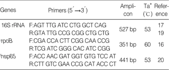
)를 넣고 Big Dye Terminator Sequencing kit (Applied Biosystems, CA, USA)를 이용하여 반응시키고, Applied Biosys- tems automatic sequencer를 이용하여 염기 서열을 분석하고 Ch- romas 2 program을 이용하여 정렬하였다. 정렬된 염기서열은 Basic Local Alignment Search Tool (BLAST) 혹은 RIDOM database와 비교하였다.*Ta, annealing temperature.
Genes Primers (5′3′) Ampli-
con Ta*
(℃) Refer-
ence
16S rRNA F:AGT TTG ATC CTG GCT CAG 17
R:GTA TTG CCG CGG CTG CTG 527 bp 53 19 rpoB F:CGA CCA CTT CGG CAA CCG
351 bp 60 16 R:TCG ATC GGG CAC ATC CGG
hsp65 F:ACC AAC GAT GGT GTG TCC AT
441 bp 53 20 R:CTT GTC GAA CCG CAT ACC CT
Table 1.Primers for amplification of 16S rRNA, rpoB and hsp65 genes
결 과
1.임상검체에서의
NTM빈도
1년간 서울대학교병원 결핵검사실에서 Ogawa 배지(신양화학) 에 배양된 검체 수는 총 1,851검체였으며 이 중 46검체에 대해서 는 검체오염을 이유로 Accuprobe MTBC culture identification (Gen-Probe) 선별 검사가 시행되지 않았다. 선별 검사된 1,805 배양균주 중 MTBC probe 음성인 검체는 388검체(21.5%)였으 며 총 228명의 환자에서 배출되었다. 그러나 228명 중 6명의 환자 에서는 같은 시기에 배출된 다른 균주로 반복시 MTBC probe 양 성을 보였으므로, 순수 NTM으로 생각되는 환자는 222명이었다.
약제 감수성 검사 등을 위하여 결핵연구원에 의뢰된 것을 제외하 고 97환자의 균주만을 얻을 수 있어, 이에 대하여 염기서열을 분 석하였다.
2.
염기서열분석을 통한 임상분리균주의 동정
1) 16S rDNA
총 97개의 검체 모두가 증폭되었고 구강내 오염균 2개를 제외한 95개의 균주 중 62개(65%)에서 성공적으로 동정되었다(Table 2).
16S rDNA 5’쪽 490번째 이하부위의 399-434개의 염기를 분석 하였을 때 기존 서열과 99% 이상 일치되었고, RIDOM 데이터와 비교시에도 99% 이상 일치하였다. 증폭된 5’쪽 서열이 동일하여
동정할 수 없었던 균종들은
M. kansasii
와M. gastri
;M. sene- galense
와M. farcinogenes
와M. fortuitum
3rd biovar (sorbitol positive);M. abscessus
와M. chelonae
;M. septicum
과M.
peregrinum
이었다. Bacillus 속 3개와 주형의 혼재 혹은 불분명 양상을 보인 8개 균주는 동정이 불가능 하였는데 Bacillus sp.와 유 사했던 균주는M. avium
혹은M. abscessus
이었고, 서열자체가 불명확하였던 균주는M. abscessus
,M. gordonae
,M. avium
,M.
peregrinum
및M. fortuitum
으로 판명되었다(Table 3).2)
rpoB
M. tuberculosis
rpoB 유전자 2,174-2,524번째 염기를 증폭하였 을 때 구강내 오염균 2균주를 제외한 95개의 균주 중 83개(87%) 만 증폭되었다. 증폭된rpoB
유전자 분절 241-300개 염기를 비교 하였을 때 98.2-100% 일치율을 보이고 같은 균종내 차이는 5염 기 이내로 78균주가 성공적으로 동정되었다(82%). 12개의 균주 는 증폭산물을 보이지 않았는데 여기에는M. abscessus
,M. gor- donae
,M. intracellulare
,M. kansasii
가 포함되었다(Table 3). 기 존 보고된 서열과 98% 이하의 상동성을 보인 5개의 균주는 rpoB 유전자로는 동정할 수 없었다. 여기에는M. avium
(92%),M.
intracellulare
(95.8%),M. gordonae
(96.7%) 및 두 개의M. non- chromogenicum
(96.5%) 유사균주가 포함되었다(Table 4).*M. abscessus and M. chelonae share an identical sequence in the 5′- 16S rDNA sequence; �M. kansasii and M. gastri share an identical 16S rDNA; �M. peregrinum and M. septicum showed an identical 5′-16S rDNA sequence; �M. senegalense and M. farcinogenes and M. fortuitum third biovar (sorbitol +) share an identical 5′-16S rDNA sequence.
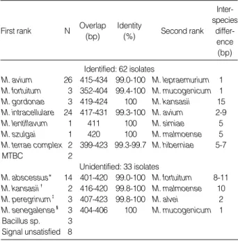
First rank N Overlap (bp)
Identity
(%) Second rank Inter- species
differ- ence (bp) Identified: 62 isolates
M. avium 26 415-434 99.0-100 M. lepraemurium 1 M. fortuitum 3 352-404 99.4-100 M. mucogenicum 1 M. gordonae 3 419-424 100 M. kansasii 15 M. intracellulare 24 417-431 99.3-100 M. avium 2-9
M. lentiflavum 1 411 100 M. simiae 5
M. szulgai 1 420 100 M. malmoense 5
M. terrae complex 2 399-423 99.3-99.7 M. hiberniae 5-7
MTBC 2
Unidentified: 33 isolates
M. abscessus* 14 401-420 99.0-100 M. fortuitum 8-11 M. kansasii� 2 416-420 99.8-100 M. malmoense 10 M. peregrinum� 3 407-423 99.8-100 M. alvei 2 M. senegalense� 3 404-406 100 M. mucogenicum 1 Bacillus sp. 3
Signal unsatisfied 8
Table 2.Similarity search results using 16S rRNA gene sequence
*MS, mixed sequence; �NA, no amplicon; �M. gordonae like organism with 96.7% similarity.
Final identification
Genes
16S rDNA rpoB hsp65
M. avium Bacillus sp M. avium NA�
M. abscessus Bacillus sp M. abscessus M. abscessus M. avium Bacillus sp M. avium M. avium M. abscessus M. abscessus NA� M. abscessus M. abscessus M. abscessus NA� M. abscessus M. abscessus M. abscessus NA� M. abscessus M. abscessus M. abscessus NA� M. abscessus M. abscessus M. abscessus NA� M. abscessus M. abscessus M. abscessus NA� M. abscessus M. abscessus M. abscessus NA� M. abscessus
M. abscessus MS* NA� M. abscessus
unidentifiable MS* M. gordonae� NA�
M. gordonae MS* M. gordonae M. gordonae
M. avium MS* M. avium M. avium
M. avium MS* M. avium M. avium
M. fortuitum MS* M. fortuitum M. fortuitum M. fortuitum MS* M. fortuitum M. fortuitum M. peregrinum MS* M. peregrinum M. peregrinum M. gordonae M. gordonae NA� M. gordonae M. gordonae M. gordonae NA� M. gordonae M. intracellulare M. intracellulare NA� MS*
M. kansasii M. kansasii NA� NA�
M. avium M. avium M. avium NA�
M. avium M. avium M. avium MS*
Table 3.Final identification of the isolates difficult to identify with a single gene fragment
3)
hsp65
증폭된 441염기중 352-406개의 염기를 비교 분석하였는데, 98.2-100% 일치하여 동정할 수 있었다(Table 5). 오염균을 제외 한 95개의 검체 중에서 91개(96%)가 증폭되었고 67개(71%)가 성공적으로 동정되었다.
M. senegalense
와M. farcinogenes
는 분 절 서열이 일치하였으며,M. abscessus
,M. chelonae
중 16균주또한 BLAST 검색으로는 동정이 어려웠다. Ringuet 등[18]의 분 석서열과 비교하였을 때
M. abscessus
와M. chelonae
두 균종 간 염기서열의 큰 차이로(30 bp) 정확히 동정할 수 있었고, 이에 따 라 동정 성공률을 87% (83/95)로 높일 수 있었다(Fig. 1). 증폭 되지 않았던 네 균주 및 서열이 불분명했던 두 균주는M. avium
,M. intracellulare
,M. kansasii
였다(Table 3).4) 균주 분포
두 번 이상 혹은 집락이 10개 이상 배양된 120환자에서 균종별 빈도는 대한결핵협회 결핵연구원에서의 동정 결과를 포함할 때,
M.
avium
30.8 %,M. intracellulare
23.3%,M. abscessus
18.3%,M.
fortuitum
6.7%,M. peregrinum
3.3%,M. gordonae
2.5%,M.
senegalense
2.5%,M. kansasii
2.5%,M. terrae
complex 1.7%,M. lentiflavum
0.8%,M. chelonae
0.8%,M. szulgai
0.8%였다.MTB complex로 동정된 경우도 2.5%나 되었고, 오염균은 1.7%, 동정할 수 없었던 경우는 1예(0.8%)가 있었으며 이는
M. gordon- ae
-like organism이었다(Table 6).고 찰
임상검체에서 배양된 항산균은 우선 AccuProbe (Gen-Probe) 등으로
Mycobacterium tuberculosis
complex (MTBC)와 비결 핵성마이코박테리아(NTM)를 구별하는데, 본 연구에서는 95개의 AccuProbe 음성인 마이코박테리아 균주 중 2균주(2.1%)에서, 결*M. gordonae like organism (similarity 96.7%).
Abbreviations: MTBC, Mycobacteriun tuberculosis complex; NTM, nontuberculous mycobacteria.
Organism No. case Percent (%)
M. avium 37 30.8
M. intracellulare 28 23.3
M. abscessus 22 18.3
M. fortuitum 8 6.7
M. peregrinum 4 3.3
M. gordonae 3 2.5
M. senegalense 3 2.5
M. kansasii 2 1.7
M. terrae complex 3 1.7
M. chelonae 1 0.8
M. celatum 1 0.8
M. lentiflavum 1 0.8
M. szulgai 1 0.8
MTB complex 3 2.5
Oral contaminant 2 1.7
unidentifiable* 1 0.8
Total 120 100
Table 6.Distribution of MTBC probe negative, presumptive NTM isolates
First rank N Overlap (bp)
Identity
(%) Second rank Inter- species
differ- ence (bp) Identified: 78 isolates
M. abscessus 7 277-300 98.3-100 M. immunogen 3-7 M. avium 30 232-294 99.0-100 M. scrofulaceum 3-15
M. chelonae 1 277 100 M. abscessus 7
M. fortuitum 5 279-296 98.9-100 M. farcinogenes 5 M. gordonae 2 285 98.6-99.5 M. asiaticum 12 M. intracellulare 22 277-289 98.2-100 M. asiaticum 9-18
M. kansasii 1 277 100 M. haemophilum 8
M. peregrinum 4 277-296 99.3-100 M. porcinum 7 M. senegalense 3 274-277 99.6 M. porcinum 3
M. szulgai 1 279 98.9 M. gordonae 13
MTB complex 2
Unidentified: 17 isolates
M. avium 1 313 92.0
M. intracellulare 1 283 95.8 M. avium 9-18
M. gordonae 1 210 96.7 M. asiaticum 8
M. nonchromo- 2 282 96.5 M. terrae complex 1 genicum
No amplicon 12
Table 4.Similarity search results using rpoB sequence
*M. senegalense and M. farcinogenes showed an identical hsp65 se- quence.
First rank N Overlap (bp)
Identity
(%) Second rank Inter- species
differ- ence (bp) Identified: 83 isolates
M. abscessus 15 374-396 99.2-100 M. chelonae 25-28 M. avium 27 352-406 99.7-100 M. intracellulare 3-10
M. chelonae 1 396 99.7 M. abscessus 28
M. fortuitum 5 381-392 100 M. senegalense 3 M. gordonae 4 389-403 97.5-99.5 M. asiaticum 8-16 M. intracellulare 22 380-406 99.2-100 M. avium 7-16
M. kansasii 1 386 100 M. gastri 8
M. lentiflavum 1 382 100 M. triplex 9
M. peregrinum 4 374-381 100 M. septicum 3
M. szulgai 1 383 100 M. lentiflavum 14
MTB complex 2
Unidentified: 12 isolates
M. senegalense* 3 374-396 98.2-98.5 M. fortuitum 1 M. avium 1 396 98.0 M. intracellulare 3 M. terrae complex 2 398 97.0 M. abscessus 8-16 No amplicon 4
Signal unsatisfied 2
Table 5.Similarity search results using hsp65 sequence
핵연구원에서 동정된 결과를 포함할 경우 2.5% (3/120)에서 MT- BC가 확인되었다. 일 년간의 검체를 분류하는 과정에서도 MTBC probe 음성인 환자 228명 중 6명(2.6%)은 그 환자가 배출한 다 른 검체에서 MTBC probe 양성으로써 probe 음성결과를 전적으 로 신뢰할 수는 없었다. 그러므로 결과를 접하는 임상의는 MTBC probe 음성이라도 결핵을 완전히 배제할 수 없음에 유의해야 하 며, 검사실에서도 보다 정확한 검사를 위한 노력 및 검체 처리과 정에 주의를 기울여야 할 것이다. 즉 마이코박테리아가 배양되는 경우, 결핵과의 감별을 위하여 가능하면 최종 동정과 약제감수성 을 확인하는 것이 권장된다.
본 검사실에서 마이코박테리아 배양 후에 probe 교잡반응을 이 용하여 선별검사를 시행하였을 때 일 년간 NTM이 분리된 환자 는 228명이고 검체 수로는 388개로 환자당 평균 1.7번의 배양 양 성인데 비하여, 결핵균은 409명의 환자에서 1,417개의 검체가 분 리되어 환자당 평균 3.5번의 배양 양성 횟수를 보였다. 이는 NTM이 실제로 오염균으로써 추적 검사상 배양되지 않았거나, NTM으로 보고된 경우 임상에서 오염균으로 간과하여 추적 검사 의뢰가 적었을 가능성이 있다. 마이코박테리아가 배양되는 경우 결 핵균과 비결핵균의 빈도는 각 78.5% 및 21.5%였는데, 1999년의 아산병원 결과 78.1:21.9%와는 비슷하였으나, 삼성서울병원의
2001까지 4년여간의 분리비율은 89.7:10.3%으로 환자 혹은 연구 시기 등에 따라 차이가 있음을 알 수 있었다[11, 24].
미국의 1980년 조사에 의하면 NTM은 검체에서 분리된 마이코 박테리아 중 35%를 차지하며, 이 중
M. avium
complex (MAC) 61%,M. fortuitum
complex를 포함한 신속성장균 19%,M. kan- sasii
10%의 빈도로 분리되었고, 1981-83년까지 2년간의 조사에 서는M. avium
complex는 62%,M. kansasii
가 24%,M. for- tuitum
5%로 보고되었으며, 1992년을 기점으로 결핵균보다 NTM 의 분리율이 더 높아졌다[25-27]. 우리나라에서는 1980년부터 1994년까지 대한결핵협회 결핵연구원에서 균이 확인된 환자를 대 상으로 하여 균종 분포를 조사하였을 때M. avium
complex 65.2%,
M. fortuitum
2.7%,M. chelonae
9.5%,M. gordonae
4.4%,M. terrae
3.2%,M. scrofulaceum
1.9%,M. kansasii
1.3%,M.
szulgai
1.3%, MAC &M. terrae
complex 0.6%를 차지하여 빈 도순으로 가장 많이 분리된 세 균종은 본 연구와 동일하였다[28].특히 1990년대 이후의 NTM이 전체 158예 중 96예로 84.2%를 차 지하였는데 이러한 증가추세는 유병률 자체의 변화뿐 아니라 진단 방법의 발전 및 임상의들의 관심도 변화에 따른 것으로 생각된다.
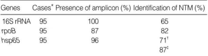
16S rDNA,
rpoB
및hsp65
유전자의 균종 구별력은 본 연구에 포함된 임상분리 균주의 증폭상태와 비교 서열 부재 등의 문제로 Fig. 1.Nucleotide sequences of the partial hsp65 gene from 15 M. abscessus (HSP2-HSP107) and one M. chelonae (HSP74) strains aligned with reference sequences from Genebank (AF071128 and AF071139, M. abscessus strains; AF071130, AF071141 and AF 071142, M. chelonae strains).인하여 동정 성공률은 각 65%, 82%, 87%였다(Table 7). 본 연 구에서 어느 한 가지 유전자만을 가지고 동정이 불가능하였던 원 인을 유전자 별로 분석하면, 16S rDNA 유전자는 이미 알려진 바 와 같이 몇 균종에서 5’쪽 비교 서열이 동일하였고 모든 미생물에 존재하는 16S rDNA가 같이 증폭이 되어 서열이 혼재되어있는 형 태로 나타나는 경우도 있었다. 이러한 균종의 혼재양상은 보관되 었던 고체배지위의 배양균주를 면봉으로 채취하는 과정에서 한 집 락만을 채취하지 못한 때문으로 생각되며, 순수배양이 아닌 임상검 체에서는 항상 다른 균종의 오염문제가 항상 존재하므로[21], 검 사실에서 실제 배양되는 NTM를 동정하고자 할 때는 16S rDNA 한 가지 만으로는 부족할 것으로 생각된다. 그러나 16S rDNA는 많은 비교분석자료가 존재하여
hsp65
나rpoB
로는 동정할 수 없 었던, 드물게 분리되는 균 동정에는 유용하였다. 본 연구에서 한 균주가rpoB
와hsp65
로는 각 95%M. intracellulare
, 98%M.
avium
유사균으로 생각되었는데, 16S rDNA의 RIDOM 데이터 와의 비교에서M. intracellulare
sqv. Ⅳ와 100% 일치되어 동정 된 예가 있었다.rpoB
유전자는 각 균종을 효과적으로 구분하였으나M. lenti- flavum
등은 비교서열이 없어 동정하지 못하였고,M. terrae
complex의 경우는 96.5%밖에 일치하지 않으면서M. nonchro- mogenicum
과의 구별이 어려웠다.rpoB
분석에서 증폭이 되지 않았던 12개 균주는M. tuberculosis
서열을 기준으로 한 시발체 [16]로는 증폭되지 않았지만rpoB
유전자의 5번째 변이부위[23]780 bp를 증폭하였을 때 11개 균주에서 증폭산물을 볼 수 있었다.
그러나 이 서열을 이용할 경우
M. abscessus
와M. chelonae
는 감 별가능 했으나M. abscessus
와M. immunogenum
구별은 어려웠 다.hsp65
유전자의 경우는 BLAST 검색을 통해서는 신속성장균 에 대한 신뢰할 수 없는 자료가 많아 쉽게 동정되지 않았고M.
farcinogenes
와M. senegalense
가 비교서열내에서 동일한 단점이 있지만, 검증된 표준균주와 비교했을 경우M. abscessus
와M.
chelonae
를 효과적으로 구분할 수 있었다[18].각 유전자별로 증폭되지 않은 검체가 있었고 이 때문에 검체 동 정률이 차이를 보였는데, 핵산 추출에 사용하였던 resin이나 이물 질이 증폭을 방해하였거나, 시발체 부위의 변이 등의 영향일 수 있
다. 다른 연구자들도
hsp65
를 이용한 동정에서 각 batch 별 증폭 양성률이 50-80% 정도 차이가 있음을 보고한 바 있는데[29], 본 연구에서도hsp65
에서 증폭되지 않은 네 균주는 모두 같은 작업 batch에서 시행되어 부적절한 검체채취에 따른 배지오염의 문제가 있었을 것으로 생각되었고, 같은 검체에서도hsp65
보다는rpoB
가 증폭이 안 되었던 것은 방해물질 뿐 아니라 시발체 부위의 변 이를 의심할 수 있으나 이는 전체 rpoB 유전자의 서열분석 등 더 연구가 필요할 것이다. 검사에 포함되었던 97개 균주 중 2균주는 서열분석상 구강내 오염균만을 발견할 수 있었는데, 이는 probe 검사와 항산성 염색은 배양 즉시 시행하고 염기서열분석을 위해 서는 보관 후 채취한 시간상의 차이 때문으로, 앞서 언급한 집락 채취상 오류 중 하나로 생각되며 실제 검사실에서 유전자를 이용 한 동정을 실시할 경우 주의해야 할 것으로 생각된다.최근에는 마이코박테리아 핵산을 이용한 신속진단법이 이용되 고 있는데, 이는 배양균주 뿐 아니라 검체에서 직접 이용할 수 있 는 장점이 있다. 특히 NTM을 동정하기 위해서는 probe법, PCR- RFLP법 등을 이용하는데 probe법은 간편하나 동정할 수 있는 균 종이 제한되며 비용이 비싸다. PCR-RFLP법은 특히
rpoB
유전 자를 이용하는 경우는 약제 저항성까지 알 수 있는 장점이 있지만 아직까지 pattern analysis에 필요한 비교서열이 완전하지 않아서 새롭게 발견되는 균이나 균종내 변이가 있는 경우는 동정에 어려 움이 있다. 염기서열분석법은 다양한 NTM의 동정에 가장 최종적 인 방법이 되며 실제로 PCR-RFLP 법으로M. szulgai
로 보고 된 것이 본 연구에서M. lentiflavum
으로 동정된 1예가 있었다.염기서열분석법을 이용하여 NTM의 임상분리 균주를 동정할 때 는 검체혼합, 채취상의 문제, 분석대상서열의 구별력 등을 고려하 여,
hsp65
나rpoB
중 하나를 먼저 분석하여 검증된 database와 비교하되, 균종 감별이 힘든 경우 16S rDNA를 부가적으로 이용 하는 것이 좋을 것으로 생각된다.요 약
배경 :
비결핵 마이코박테리아(NTM)는 환경분포균으로 환자 검체에서 분리되는 경우라도 질환과 관계없는 오염균으로 생각하 기 쉽고, 일반적인 미생물학적 동정방법으로는 정확한 동정이 어 렵다. 이에 저자들은 임상검체에서 배양된 NTM에 대하여 염기서 열분석법으로 정확하게 동정하여 균종 분포를 연구하고자 하였다.방법 :
2003년 1월 21부터 2004년 1월 20일까지 일개 3차 의료 기관의 결핵검사실에서 배양된 균주 중에서 결핵균 소식자에 음성 인 NTM를 대상으로 하였다. 이들 중 집락이 10개 이상 배양되거 나 10집락 미만이라도 동일 환자에서 두번 이상 배양된 97개에서 16S rRNA,hsp65
및rpoB
유전자 서열을 분석하였고, 결핵연구원 의 결과를 포함하여 120환자에서 균종 분포를 연구하였다.결과 :
임상검체에서 배양된 NTM 균종은M. avium
,M. in- tracellulare
,M. abscessus
가 각각 30.8%, 23.3%, 18.3%를 차지*Of the studied 97 isolates, two oral contaminants were excluded because they were not mycobacteria; �The percent was lowered to 70% when the two MTBC isolates were also excluded. But other values were not changed; �We could get the percentage when the sequences were com- pared with those in reference 18.
Abbreviation: NTM, nontuberculous mycobacteria.
Genes Cases* Presence of amplicon (%) Identification of NTM (%)
16S rRNA 95 100 65
rpoB 95 87 82
hsp65 95 96 71�
87�
Table 7.Comparison of the three gene segments used in identi- fication of presumptive NTM isolates
하였다. 이외에
M. gordonae
,M. senegalense
,M. fortuitum
,M.
peregrinum
,M. kansasii
,M. terrae complex
,M. lentiflavum
,M. chelonae
,M. szulgai
가 분리되었고 MTB complex도 2.5%에 서 확인되었다. 유전자별 동정 성공률은 16S rRNA,rpoB
,hsp65
각 65%, 82%, 87%로써, NTM을 염기서열분석법으로 동정할 때 에는 5’-16S rDNA보다는hsp65
나rpoB
를 이용하는 것이 성공 률을 높일 수 있었다.결론 :
임상 검체에서 NTM은 더 이상 오염균으로 무시할 수 있는 대상이 아니며, 염기서열분석을 이용한 마이코박테리아의 동 정법은 검사실에서도 유용하게 쓰일 수 있을 것으로 기대한다.참고문헌
1. Tenholder MF, Moser RJ 3rd, Tellis CJ. Mycobacteria other than tu- berculosis. Pulmonary involvement in patients with acquired immu- nodeficiency syndrome. Arch Intern Med 1988;148:953-5.
2. Falkinham JO 3rd. Epidemiology of infection by nontuberculous mycobacteria. Clin Microbiol Rev 1996;9:177-215.
3. Debrunner M, Salfinger M, Brandli O, von Graevenitz A. Epidemi- ology and clinical significance of nontuberculous mycobacteria in patients negative for human immunodeficiency virus in Switzerland.
Clin Infect Dis 1992;15:330-45.
4. Dobos KM, Quinn FD, Ashford DA, Horsburgh CR, King CH. Emer- gence of a unique group of necrotizing mycobacterial diseases. Emerg Infect Dis 1999;5:367-78.
5. Collins MT, Lisby G, Moser C, Chicks D, Christensen S, Reichelder- fer M, et al. Results of multiple diagnostic tests for Mycobacterium avium subsp. paratuberculosis in patients with inflammatory bowel disease and in controls. J Clin Microbiol 2000;38:4373-81.
6. Kim SJ, Hong YP, Kim SC, Bai GH, Jin BW, Park CD. A case of pul- monary disease due to M. avium-intracellulare complex. Tuberc Respir Dis 1981;28:121-4. (김상재, 홍영표, 김성진, 배길한, 진병원, 박 종달. M. avium-intracellulare complex에 의한 폐항산균증 1예. 결핵 및 호흡기질환 1981;28:121-4.)
7. Bai GH, Park KS, Kim SJ. Clinically isolated mycobacteria other than mycobacterium tuberculosis from 1980 to 1990 in Korea. J Korean Soc Microbiol 1993;28:1-6. (배길한, 박관숙, 김상재. 1980년부터1990년까지 우리나라의결핵균의마이코박테리아균종별감염양상. 대한미생물학회 지 1993;28:1-6.)
8. American Thoracic Society. Diagnosis and treatment of disease cau- sed by nontuberculous mycobacteria. Am J Respir Crit Care Med 1997;156:S1-25.
9. Pae HH, Lee JH, Yoo CG, Lee CT, Chung HS, Kim YW, et al. Study for clinical characteristics of nontuberculous mycobacterial pulmo- nary disease. Tuberc Respir Dis 1999;47:735-46. (배현혜, 이재호, 유철
규, 이춘택, 정희순, 김영환등. 폐비결핵항산균증의임상적특징에관한 연구. 결핵및호흡기질환 1999;47:735-746.)
10. Lew WJ, Ahn DI, Yoon YJ, Cho JS, Kwon DW, Kim SJ, et al. Clinical experience on mycobacterial disease other than tuberculosis. Tuberc Respir Dis 1992;39:425-32. (류우진, 안동일, 윤영자, 조정섭, 권동원, 김 상재등. 비결핵마이코박테리엄증의임상경험. 결핵및호흡기질환 1992;
39:425-32.)
11. Lee HW, Kim MN, Shim TS, Bai GH, Pai CH. Nontuberculous myco- bacterial pulmonary infection in immunocompetent patients. Tuberc Respir Dis 2002;53:173-82. (이효원, 김미나, 심태선, 배길한, 배직현. 면 역적격자에서비결핵마이코박테리아의폐감염. 결핵및호흡기질환2002;
53:173-82.)
12. Koh WJ, Kwon OJ, Kang EH, Jeon IS, Pyun YJ, Ham HS, et al. Clini- cal and radiographic characteristics of 12 patients with Mycobacteri- um abscessus pulmonary disease. Tuberc Respir Dis 2003;54:45-56.
(고원중, 권오정, 강은해, 전익수, 편유장, 함형석 등. Mycobacterium abscessus 폐질환환자12명의임상적, 방사선학적특징. 결핵및호흡기질 환 2003;54:45-56.)
13. Springer B, Stockman L, Teschner K, Roberts GD, Bottger EC. Two- laboratory collaborative study on identification of mycobacteria: mo- lecular versus phenotypic methods. J Clin Microbiol 1996;34:296-303.
14. Brown-Elliott BA, Griffith DE, Wallace RJ Jr. Newly described or emerging human species of nontuberculous mycobacteria. Infect Dis Clin North Am 2002;16:187-220.
15. Patel JB, Leonard DG, Pan X, Musser JM, Berman RE, Nachamkin I. Sequence-based identification of Mycobacterium species using the MicroSeq 500 16S rDNA bacterial identification system. J Clin Micro- biol 2000;38:246-51.
16. Kim BJ, Lee SH, Lyu MA, Kim SJ, Bai GH, Chae GT, et al. Identifica- tion of mycobacterial species by comparative sequence analysis of the RNA polymerase gene (rpoB). J Clin Microbiol 1999;37:1714-20.
17. Turenne CY, Tschetter L, Wolfe J, Kabani A. Necessity of quality-con- trolled 16S rRNA gene sequence databases: identifying nontubercu- lous Mycobacterium species. J Clin Microbiol 2001;39:3637-48.
18. Ringuet H, Akoua-Koffi C, Honore S, Varnerot A, Vincent V, Berche P, et al. hsp65 sequencing for identification of rapidly growing my- cobacteria. J Clin Microbiol 1999;37:852-7.
19. Harmsen D, Rothganger J, Frosch M, Albert J. RIDOM: Ribosomal Differentiation of Medical Micro-organisms Database. Nucleic Acids Res 2002;30:416-7.
20. Telenti A, Marchesi F, Balz M, Bally F, Bottger EC, Bodmer T. Rapid identification of mycobacteria to the species level by polymerase chain reaction and restriction enzyme analysis. J Clin Microbiol 1993;
31:175-8.
21. Pai S, Esen N, Pan X, Musser JM. Routine rapid Mycobacterium species assignment based on species-specific allelic variation in the
65-kilodalton heat shock protein gene (hsp65). Arch Pathol Lab Med 1997;121:859-64.
22. Kim BJ, Lee KH, Park BN, Kim SJ, Bai GH, Kim SJ, et al. Differenti- ation of mycobacterial species by PCR-restriction analysis of DNA (342 base pairs) of the RNA polymerase gene (rpoB). J Clin Micro- biol 2001;39:2102-9.
23. Adekambi T, Colson P, Drancourt M. rpoB-based identification of nonpigmented and late-pigmenting rapidly growing mycobacteria. J Clin Microbiol 2003;41:5699-708.
24. Koh WJ, Kwon OJ, Yu CM, Jeon K, Suh GY, Chung MP, et al. Recov- ery rate of nontuberculous mycobacteria from acid-fast-bacilli smear- positive sputum specimen. Tuberc Respir Dis 2003;54:22-32. (고원중, 권오정, 유창민, 전경만, 서지영, 정만표등. 항산균도말양성객담에서비 결핵성마이코박테리아의분리비율. 결핵및호흡기질환 2003;54:22-32.) 25. Good RC and Snider DE Jr. Isolation of nontuberculous mycobacte-
ria in the United States, 1980. J Infect Dis 1982;146:829-33.
26. O’Brien RJ, Geiter LJ, Snider DE Jr. The epidemiology of nontubercu- lous mycobacterial disease in the United States. Results from a na- tional survey. Am Rev Respir Dis 1987;135:1007-14.
27. Ostroff S, Hutwagner L, Collin S. Mycobacterial species and drug resistance patterns reported by state laboratories-1992. 93rd Ameri- can Society for Microbiology General Meeting, May 16, 1993, Atlanta, GA. Abstract U-9, P.170.
28. Scientific committee in Korean academy of tuberculosis and respi- ratory disease. National survey of mycobacterial disease other than tuberculosis in Korea. Tuberc Respir Dis 1995;42:277-94. (대한결핵 및호흡기학회학술위원회. 비결핵항산균증전국실태조사. 결핵및호흡기 질환 1995;42:277-94.)
29. Wong DA, Yip PC, Tse DL, Tung VW, Cheung DT, Kam KM. Rou- tine use of a simple low-cost genotypic assay for the identification of mycobacteria in a high throughput laboratory. Diagn Microbiol Infect Dis 2003;47:421-6.