INTRODUCTION
Asthma is a chronic inflammatory disease, characterized by the infiltration of inflammatory cells (e.g., eosinophils) in the airway or lung tissues, airway hypersensitivity, and remodeling.1 In recent decades, allergic diseases such as asthma have shown a rapidly increasing trend.2-4 The hygiene hypothesis suggests that a Westernized life style coupled with improved sanitation has reduced microbial exposure, thereby curtailing Th1 immune responses and exacerbating Th2 immune responses.5-7
Mycolic acid (MA), major components of the cell walls of My- cobacterium tuberculosis and related species, is β-hydroxy-α-alkyl branched fatty acids comprising about 60-90 carbon atoms.
Their structural uniqueness provides the microorganisms with certain benefits, such as low permeability to hydrophobic com-
A Novel Synthetic Mycolic Acid Inhibits Bronchial
Hyperresponsiveness and Allergic Inflammation in a Mouse Model of Asthma
Young-Joon Kim,
1Ha-Jung Kim,
1Se Kyoo Jeong,
2Seung-Hwa Lee,
1Mi-Jin Kang,
1Ho-Sung Yu,
1Young-Ho Jung,
3,4Ju-Hee Seo,
5Byoung-Ju Kim,
6Jinho Yu,
3Seoung-Ju Park,
7Yong- Chul Lee,
7* Soo-Jong Hong
3,4*
1Asan Institute for Life Sciences, University of Ulsan College of Medicine, Seoul, Korea
2Applied Research Division Neopharm Co., Ltd., Daejeon, Korea
3Department of Pediatrics, Childhood Asthma Atopy Center, Asan Medical Center, University of Ulsan College of Medicine, Seoul, Korea
4Research Center for Standardization of Allergic Diseases, University of Ulsan College of Medicine, Seoul, Korea
5Department of Pediatrics, Korea Cancer Center Hospital, Seoul, Korea
6Department of Pediatrics, Inje University Haeundae Paik Hospital, Busan, Korea
7Research Center for Pulmonary Disorders, Chonbuk National University Medical School, Jeonju, Korea
pounds, resistance to dehydration, and the capacity to survive attacks by macrophages. MA has immunomodulatory activity, which is associated with the regulation of allergic inflammatory Allergy Asthma Immunol Res. 2014 January;6(1):83-88.
http://dx.doi.org/10.4168/aair.2014.6.1.83 pISSN 2092-7355 • eISSN 2092-7363
Purpose: Recognition of microbes is important to trigger the innate immune system. Mycolic acid (MA) is a component of the cell walls of myco- bacteria such as Mycobacterium bovis Bacillus Calmette-Guerin. MA has immunogenic properties, which may modulate the innate and adaptive im- mune response. This study aimed to investigate whether a novel synthetic MA (sMA) inhibits allergic inflammatory responses in a mouse model of asthma. Methods: BALB/c mice were injected intraperitoneally with sMA followed by sensitization and challenge with ovalbumin (OVA). Mice were examined for bronchial hyperresponsiveness (BHR), the influx of inflammatory cells into the lung tissues, histopathological changes in the lungs and CD4+CD25+Foxp3+ T cells in the spleen, and examined the response after the depleting regulatory T cells (Tregs) with an anti-CD25mAb. Re- sults: Treatment of mice with sMA suppressed the asthmatic response, including BHR, bronchoalveolar inflammation, and pulmonary eosinophilic inflammation. Anti-CD25mAb treatment abrogated the suppressive effects of sMA in this mouse model of asthma and totally depleted CD4+CD25+ Foxp3+ T cells in the spleen. Conclusions: sMA attenuated allergic inflammation in a mouse model of asthma, which might be related with CD4+ CD25+Foxp3+ T cell.
Key Words: Mycolic acid; asthma; allergic inflammation; regulatory T cells; mice
This is an Open Access article distributed under the terms of the Creative Commons Attribution Non-Commercial License (http://creativecommons.org/licenses/by-nc/3.0/) which permits unrestricted non-commercial use, distribution, and reproduction in any medium, provided the original work is properly cited.
Correspondence to: Soo-Jong Hong, MD, PhD, Department of Pediatrics, Childhood Asthma Atopy Center, Research Center for Standardization of Allergic Diseases, Asan Medical Center, University of Ulsan College of Medicine, 88 Olympic-ro 43-gil, Songpa-gu, Seoul 138-736, Korea.
Tel: +82-2-3010-3379; Fax: +82-2-473-3725; E-mail: [email protected] Yong-Chul Lee, MD, PhD, Department of Internal Medicine, Chonbuk National University Medical School, 567 Baekje-daero, Deokjin-gu, Jeonju 561-756, Korea.
Tel: +82-63-250-1664; Fax: +82-63-254-1609; E-mail: [email protected] Received: October 11, 2012; Revised: January 15, 2013;
Accepted: January 24, 2013
•There are no financial or other issues that might lead to conflict of interest.
•Yong Chul Lee and Soo-Jong Hong contributed equally on this paper.
responses.8 Recently, a negative correlation between a positive tuberculin skin test and the development of allergic rhinitis was reported.9 In addition, MA isolated from Mycobacteria may mod- ulate experimental asthma by inducing alveolar macrophages into foamy cell morphotype.10 This modulation prevents the on- set of allergic airway inflammation by inducing of regulatory T cells (Tregs) which express markers, including Forkhead box p3 (Foxp3), neuropilin-1, and glucocorticoid-induced tumor ne- crosis factor receptor-related protein.10 Therefore, we postulat- ed that MA may serve as a potential therapeutic molecule for allergic diseases. However, MA is produced by mycobacteria in small amounts, and its use as a therapeutic agent is difficult and not cost-effective. Hence, a synthetic form of MA (sMA) was de- veloped by Neopharm Co., Ltd using a new process involving a simple and cost-effective production pathway.11
Here, we aimed to use a mouse model of asthma to examine whether this sMA could inhibit allergic inflammatory responses in a manner similar to natural MA derived from Mycobacteria.
MATERIALS AND METHODS Animals
Female BALB/c mice were purchased from OrientBio (Seong- nam, Korea) and cared for and used in accordance with the guidelines set down by the Institutional Animal Care and Use Committee (IACUC) at Asan Medical Center and Ulsan Univer- sity College of Medicine. The mice were housed in a specific pathogen-free facility.
Antigen sensitization and challenge
The mice (6 weeks old, 5 mice/group) were sensitized by 2 in- traperitoneal (i.p.) injections (on Days 0 and 7) of ovalbumin (OVA, 20 µg; chicken egg albumin grade V, Sigma Chemical Co., St. Louis, USA) dissolved in 100 µL saline and adsorbed with an equal volume of alum solution (Alumimject, Pierce, IL, USA).
On Days 14-16, all mice inhaled for 30 minutes (once per day) with an aerosol containing 1% OVA in saline. Control mice were sensitized and challenged with saline.
Preparation of sMA
The sMA (Neopharm, Daejeon, Korea) was synthesized and kindly donated as described previously.11 Briefly, MA was syn- thesized in 3 steps: 1) dimerization of octanoyl chloride into a ketene dimer (E)-4-heptylidene-3-hexyl-oxetan-2-one; 2) se- lective reduction of heptylidene-3-hexyl-oxetan-2-one by hy- drogenation to produce 2-hexyldecanoic β-lactone; and 3) β- lactone ring cleavage under alkaline conditions. This reaction was then acidified with 6 mol/L HCl and the product purified using a column packed with silica gel. Then, the sMA (15 mg) was mixed with 20 mg phosphatidylcholine at 180 mg/mL in chloroform. The organic solvent was evaporated, and the lipids were recovered in sterile PBS (15 mL). Control liposome sus-
pensions were prepared without sMA.
Animal treatment of sMA
In preliminary study, we analyzed using multiple-dose proto- cols (100 µg, 200 µg, and 500 µg) and found optimal data at 100 µg (data not shown). In addition, the efficacy was estimated by the time of administration (before or after sensitization) and it was determined as 5 days after last sensitization for treatment protocol (on day 12). So, sMA was intraperitoneally injected on 5 days after sensitization at 100 µg. Liposome as a control was injected without sMA.
In vivo treatment with an anti-CD25 monoclonal antibody (mAb)
To examine the involvement of Tregs in the mechanism un- derlying allergy suppression by sMA, OVA-sensitized mice were injected i.p. with 250 µg of anti-CD25 mAb (clone PC61, eBio- science, San Diego, USA) 1 day before the first intra-nasal chal- lenge with 1% OVA. Control mice were treated with 250 µg of rat IgG (Bioxcell, West Lebanon, NH, USA).
Measurement of bronchial hyperresponsiveness (BHR)
BHR was measured in conscious unrestrained mice using a barometric whole-body plethysmography (All Medicus, Anyang, Korea) as described previously.12 Briefly, 24 hours after the final OVA challenge, enhanced pause (Penh) was measured at 10- seconds intervals for 3 minutes after inhalation of saline and each concentration of methacholine (MeCh).
Bronchoalveolar lavage (BAL) and determination of the cellular composition of the BAL fluid
After measurement of BHR, mice were anaesthetized by i.p.
administration of zoletil and BAL was performed as described previously.12 Briefly, the trachea was immediately exposed after anesthesia. Then, BAL was performed through a catheter in- serted into the exposed trachea following instillation of normal saline (2 mL) at 37°C. The BAL fluid was centrifuged at 2,000 rpm for 5 minutes at 4°C. After discarding the supernatant, the pellet was resuspended in 100 µL PBS. Total BAL cell counts were per- formed using a haemacytometer. To count the different types of cells in the BAL fluid, cytospin slides were prepared and stained with Wright stain. The different cell types were identified on the basis of their standard morphology under a light microscope (Nikon, Tokyo, Japan). Five sections per slide were evaluated.
The analyses were random, double blinded, and performed by 2 independent investigators.
Flow cytometry
Mouse Treg cells were collected from spleen. Mouse regulato- ry T cell staining kits with FITC-labeled anti-CD4, PE-labeled anti-CD25, APC-labeled anti-Foxp3, and each isotype control (eBioscience, San Diego, USA) were used for analysis of CD4,
CD25 and Foxp3 expression according to the manufacturer’s directions. Staining was analyzed by flow cytometry on a FACS Calibur with CellQuest software (BD Biosciences, Mountain View, CA, USA).
Histopathological analysis of the lungs
Lung tissue samples were prepared for histological analysis as described previously.13 Inflammation was scored by 2 indepen- dent blinded investigators. The degree of peribronchial and perivascular inflammation was evaluated on a subjective scale of 0-3. Briefly, a value of 0 meant that no inflammation was de- tectable, a value of 1, occasional cuffing with inflammatory cells, a value of 2, most bronchi or vessels were surrounded by thin layer (1 to 5 cells thick) of inflammatory cells, and a value of 3, most bronchi or vessels were surrounded by a thick layer (more than 5 cells thick) of inflammatory cells.
Cellular infiltration was assessed in 5 randomly-selected fields under a Zeiss Axiophot microscope (Carl Zeiss Inc., Thornwood, USA) at 100× magnification. The analyses were random, dou- ble blinded and performed by 2 independent investigators.
Statistical analysis
Results were expressed as means±SEM values, and groups were compared using the Mann-Whitney U test. The statistical significance was calculated using SPSS software version 20.0 (SPSS Inc., Chicago, IL, USA). A P value that was less than 0.05 was considered significant.
RESULTS
sMA inhibits BHR and airway inflammation in a mouse model of asthma and anti-CD25mAb abrogates the inhibitory effects
Mice sensitized and challenged with OVA alone developed MeCh-BHR, and showed high total cell and eosinophil num- bers in the BAL fluid. Mean Penh value of sMA-treated mice was attenuated to approximately half compared to OVA mice at maximum MeCh dose (50 mg/mL) (4.45±0.64 and 8.85±1.10, P<0.01; Fig. 1). By contrast, the mean Penh value in sMA-treat- ed mice that received Anti-CD25 (9.90±2.46) was high com- pared to mice that received sMA alone (4.45±0.64) at maximum MeCh dose (50 mg/mL) (P<0.01, Fig. 1).
As shown, sMA-treated mice showed significantly lower cell number in terms of the total number of inflammatory cells (6.68±1.26×10⁵/mL vs. 17.56±3.70×10⁵/mL, P<0.05; Fig. 2) and the number of eosinophils (0.23±0.06×10⁵/mL vs. 5.38±
2.04×10⁵/mL, P<0.01; Fig. 2) than OVA-treated mice in BAL fluid. However, the administration of Anti-CD25 mAb reversed the total cells and the eosinophils compared to sMA-treated mice in the BAL fluid (6.68±1.26×10⁵/mL vs. 16.35±5.10×
105/mL, P=0.05 and 0.23± 0.06×10⁵/mL vs. 6.20±3.60×10⁵/ mL, P<0.05; Fig. 2).
sMA inhibits pulmonary inflammation and anti-CD25 mAb abrogates the inhibitory effects
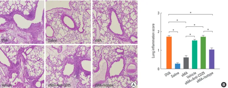
Histological analysis of the lungs from the mice described above revealed that fewer peribronchial and perivascular in- flammatory cells were recruited to the lungs in sMA-treated mice than in OVA-treated mice. However, the inflammatory cells were observed in vehicle- and anti-CD25 mAb-treated mice (Fig. 3A). The inflammation scores were significantly low- er in sMA-treated mice than in OVA-treated mice (0.60±0.07 vs. 1.72±0.07, P<0.01; Fig. 3B). The mean values of the inflam- mation score in vehicle- and anti-CD25 mAb-treated mice are similar to OVA-treated mice (Fig. 3B).
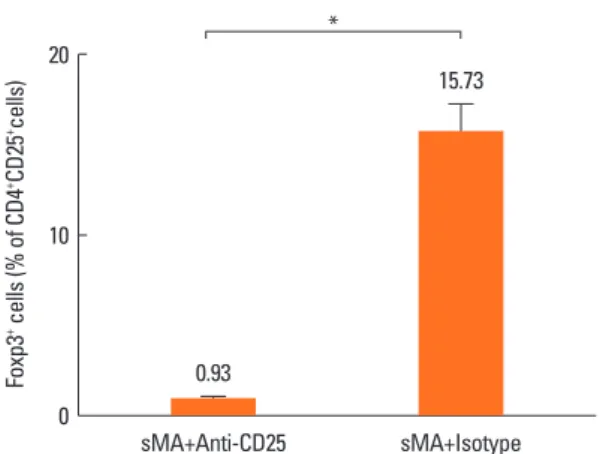
sMA induces the production of Foxp3+ Treg cells
Treg cells were totally depleted from mice treated with anti- CD25 mAb in sMA treated-mice (0.93%±0.09%) compared to mice treated with IgG isotype with sMA (15.73%±1.55%, P=
0.05, respectively, Fig. 4). However, Treg cell numbers were slightly higher in mice treated with sMA (15.05%±0.73%) than in mice treated with OVA (13.57%±0.27%), although the differ- ence was not significant statistically (P=0.121, data not shown).
DISCUSSION
A number of studies have examined the role of mycobacterial cell components, such as peptidoglycan, LAM, and LPS in the development of allergic inflammatory disorders in both humans Fig. 1. Treatment of ovalbumin (OVA)-induced allergic asthmatic mice with syn- thetic mycolic acid (sMA) and effects of anti-CD25 monoclonal antibody (mAb) treatment on the BHR. OVA: mice sensitized with intraperitoneal injections of OVA on Days 0 and 7 and then challenged with aerosolized OVA (1%) on Days 14-16 (a single 30-min challenge per day); Saline: mice sensitized and challenged with saline following the same time schedule described for OVA group; sMA:
mice that were injected intraperitoneally with the sMA on 5 days after sensiti- zation and challenge with OVA; Vehicle: mice were injected intraperitoneally with liposome as a control for sMA on 5 days after sensitization and challenge with OVA; sMA + Anti-CD25: mice received sMA plus the anti-CD25 mAb; sMA + Isotype: mice received sMA and rat IgG (isotype) as a control for anti-CD25 mAb. Bronchial hyperresponsiveness (BHR). *P<0.05; **P<0.01 compared to OVA; †P<0.05; ††P<0.01 compared to sMA + Anti-CD25. The values represent the mean±SEM of the results from 5 mice per group.
Penh (Enhanced pause)
12.5 10.0 7.5 5.0 2.5
0.0 saline 5 10 25 50
Methacholine concentration (mg/mL) OVA
Saline sMA Vehicle sMA+Anti-CD25 sMA+Isotype
** ††
* †
Cell in BAL fluid (×105/mL)
OVA Total cell
Saline sMAVehicle
sMA+Anti-CD25sMA+Isotype 30
20
10
0
*
* ***
*
Cell in BAL fluid (×105/mL)
OVA Eosinophil
Saline sMAVehicle
sMA+Anti-CD25sMA+Isotype 12
10 8 6 4 2 0
*
*
*
**
*
**
Cell in BAL fluid (×105/mL)
OVA Lymphocyte
Saline sMAVehicle
sMA+Anti-CD25sMA+Isotype 1.0
0.8 0.6 0.4 0.2 0
*
*
*
Cell in BAL fluid (×105/mL)
OVA Neutrophil
Saline sMAVehicle
sMA+Anti-CD25sMA+Isotype 10.0
7.5 5.0 2.5 0
* Cell in BAL fluid (×105/mL)
OVA Macrophage
Saline sMAVehicle
sMA+Anti-CD25sMA+Isotype 12.5
10.0 7.5 5.0 2.5 0
*
***
*
Fig. 2. Treatment of OVA-induced allergic asthmatic mice with sMA and effects of anti-CD25 mAb treatment on the differential cell numbers in BAL fluid. The plot legends are as described in Fig. 1. sMA treatment inhibited the number of total cells, macrophages, eosinophils, lymphocytes, and neutrophils in BAL fluid. However, it did not show in vehicle and anti-CD25 mAb treatment. Values represent the mean±SEM of the results from five mice per group. *P<0.05; **P<0.01; ***P=0.05.
B OVA
Vehicle
Saline
sMA+Anti-CD25
sMA
sMA+Isotype A
Lung inflammation score
3
2
1
0
* *
*
*
*
*
OVA Saline sMAVehicle
sMA+Anti-CD25sMA+Isotype
Fig. 3. Treatment of OVA-induced allergic asthmatic mice with sMA and effects of anti-CD25 mAb treatment on the lungs. The lungs were were stained with hema- toxylin and eosin. The plot legends are as described in Fig. 1. (A) Lung pathology. (B) Peribronchial and perivascular lung inflammation score. The values represent the mean±SEM of the results from 5 mice per group. *P<0.01.
and experimental animal models.14,15 MA is a novel cell wall component derived from Mycobacterium tuberculosis and the role of MA in asthma development remains unknown. Thus, the aim of the present study was to examine the role of sMA in an OVA-induced asthmatic mouse model.
Treatment with sMA inhibited allergic inflammatory respons- es, including the development of BHR and infiltration of inflam- matory cells into the airways and lungs. In addition, sMA showed tendency to increase the number of CD4+CD25+Foxp3+ T cells.
These effects were abrogated by an anti-CD25mAb. These find- ings suggest that new sMA might have a novel function as an immunomodulator to control allergic inflammation. This is the first report to the best of our knowledge to identify sMA as a novel modulator of the immune responses that cause asthma.
In previous study, this sMA used for identifying the effect on the biogradation.11 but it was not studied on the allergic diseases, such as asthma. Thus far, a study has been performed on exper- imental mouse model of asthma for identifying effects and mechanisms of MA using natural MA derived from M. tubercu- losis.10 Unlikely, we used artificially synthesized MA based on basic MA-motif. This implies that it is easy to produce in large quantities and can be newly contributing factor for the devel- opment of novel preventive and therapeutic approaches on the allergic diseases including asthma.
Our data showed tendency to increase in the expression of Foxp3+ Treg cells after the treatment of sMA. In addition, the suppressive effects of BHR and allergic inflammation were ab- rogated by an anti-CD25mAb. These suggest that the mecha- nism of sMA may be associated with CD4+CD25+Foxp3+ Treg cells possibly through not only the number but also the function.
Macrophages and dendritic cells are antigen presenting cell and a major cell population that resides in tissues throughout the body, where they play a crucial role as a first-line of defense against invading pathogens. When MA-treated macrophages were instilled into the airways of OVA-sensitized mice , total in- flammatory cell counts and eosinophil counts in the BAL fluid
decreased.10 Also, alveolar macrophage-depletion after sensiti- zation significantly increased the number of eosinophils and the production of IL-4 and IL-5.16 In addition, a mixture of cell wall components derived from BCG, including peptidoglycans, arabinogalactans, and MA, induced the maturation of DCs via TNF-α secreted from themselves.17 Therefore, APCs contribute to MA-related mechanism.
Further studies are needed to refine several aspects of this study. We found that the number of Foxp3+ Tregs in sMA-treat- ed mice was not significantly high compared with that in OVA- treated mice. Therefore, it was not completely confirmed that sMA has affected the development of Tregs. In addition, the an- ti-CD25mAb experiments only implicate Treg cells in the un- derlying mechanisms indirectly. Anti-CD25 mAb is not really sufficient to fully deplete murine Treg cells, and it also depletes activated T cells.18 Further studies are needed to identifiy the mechanism related Treg of sMA, especially focused on the ex- pression of Treg-related cytokine, IL-10 or TGF-β, and Treg cells from regional immune organ in more detail.
MA is expressed in different forms, which may influence its regulatory function and pathogenic behavior.19 Therefore, the novel structure of the sMA used in the present study may play a role in its effects against allergic inflammation.
In conclusion, we have developed a novel sMA molecule that appears to modulate allergic inflammatory responses, which might be related with CD4+CD25+Foxp3+ T cells. This novel compound may provide a new strategy for treating or control- ling asthma.
ACKNOWLEDGMENTS
This study was supported by a grant from the Korea Healthcare Technology R&D Project of the Ministry for Health, Welfare and Family Affairs, Republic of Korea (A084144).
REFERENCES
1. Walker C, Bode E, Boer L, Hansel TT, Blaser K, Virchow JC Jr. Aller- gic and nonallergic asthmatics have distinct patterns of T-cell acti- vation and cytokine production in peripheral blood and bronchoal- veolar lavage. Am Rev Respir Dis 1992;146:109-15.
2. Eder W, Ege MJ, von Mutius E. The asthma epidemic. N Engl J Med 2006;355:2226-35.
3. Hong SJ, Ahn KM, Lee SY, Kim KE. The prevalences of asthma and allergic diseases in Korean children. Korean J Pediatr 2008;51:343- 50.
4. Lee SI. Prevalence of childhood asthma in Korea: international study of asthma and allergies in childhood. Allergy Asthma Immunol Res 2010;2:61-4.
5. Strachan DP. Hay fever, hygiene, and household size. BMJ 1989;
299:1259-60.
6. Riedler J, Braun-Fahrländer C, Eder W, Schreuer M, Waser M, Maisch S, Carr D, Schierl R, Nowak D, von Mutius E; ALEX Study Team. Exposure to farming in early life and development of asth- Foxp3+ cells (% of CD4+CD25+cells)
20
10
0 sMA+Anti-CD25 sMA+Isotype
*
0.93
15.73
Fig. 4. Effect of sMA on the percentage of Foxp3+ Treg cells on the splenocytes.
The Treg cell population was totally depleted by the anti-CD25 mAb. The values represent the mean±SEM of the results from 5 mice. *P =0.05.
ma and allergy: a cross-sectional survey. Lancet 2001;358:1129-33.
7. Ege MJ, Mayer M, Normand AC, Genuneit J, Cookson WO, Braun- Fahrländer C, Heederik D, Piarroux R, von Mutius E; GABRIELA Transregio 22 Study Group. Exposure to environmental microor- ganisms and childhood asthma. N Engl J Med 2011;364:701-9.
8. Korf J, Stoltz A, Verschoor J, De Baetselier P, Grooten J. The Myco- bacterium tuberculosis cell wall component mycolic acid elicits pathogen-associated host innate immune responses. Eur J Immu- nol 2005;35:890-900.
9. Obihara CC, Beyers N, Gie RP, Potter PC, Marais BJ, Lombard CJ, Enarson DA, Kimpen JL. Inverse association between Mycobacte- rium tuberculosis infection and atopic rhinitis in children. Allergy 2005;60:1121-5.
10. Korf JE, Pynaert G, Tournoy K, Boonefaes T, Van Oosterhout A, Ginneberge D, Haegeman A, Verschoor JA, De Baetselier P, Groot- en J. Macrophage reprogramming by mycolic acid promotes a tolerogenic response in experimental asthma. Am J Respir Crit Care Med 2006;174:152-60.
11. Lee M, Kim MK, Kwon MJ, Park BD, Kim MH, Goodfellow M, Lee ST. Effect of the synthesized mycolic acid on the biodegradation of diesel oil by Gordonia nitida strain LE31. J Biosci Bioeng 2005;100:
429-36.
12. Yu J, Jang SO, Kim BJ, Song YH, Kwon JW, Kang MJ, Choi WA, Jung HD, Hong SJ. The effects of Lactobacillus rhamnosus on the pre- vention of asthma in a murine model. Allergy Asthma Immunol Res 2010;2:199-205.
13. Tournoy KG, Kips JC, Schou C, Pauwels RA. Airway eosinophilia is not a requirement for allergen-induced airway hyperresponsive- ness. Clin Exp Allergy 2000;30:79-85.
14. Sayers I, Severn W, Scanga CB, Hudson J, Le Gros G, Harper JL.
Suppression of allergic airway disease using mycobacterial lipogly- cans. J Allergy Clin Immunol 2004;114:302-9.
15. Ito T, Hasegawa A, Hosokawa H, Yamashita M, Motohashi S, Naka T, Okamoto Y, Fujita Y, Ishii Y, Taniguchi M, Yano I, Nakayama T. Hu- man Th1 differentiation induced by lipoarabinomannan/lipoman- nan from Mycobacterium bovis BCG Tokyo-172. Int Immunol 2008;
20:849-60.
16. Bang BR, Chun E, Shim EJ, Lee HS, Lee SY, Cho SH, Min KU, Kim YY, Park HW. Alveolar macrophages modulate allergic inflamma- tion in a murine model of asthma. Exp Mol Med 2011;43:275-80.
17. Tsuji S, Matsumoto M, Takeuchi O, Akira S, Azuma I, Hayashi A, Toyoshima K, Seya T. Maturation of human dendritic cells by cell wall skeleton of Mycobacterium bovis bacillus Calmette-Guérin:
involvement of toll-like receptors. Infect Immun 2000;68:6883-90.
18. Fehérvari Z, Sakaguchi S. Development and function of CD25+CD4+
regulatory T cells. Curr Opin Immunol 2004;16:203-8.
19. Vander Beken S, Al Dulayymi JR, Naessens T, Koza G, Maza-Iglesias M, Rowles R, Theunissen C, De Medts J, Lanckacker E, Baird MS, Grooten J. Molecular structure of the Mycobacterium tuberculosis virulence factor, mycolic acid, determines the elicited inflammato- ry pattern. Eur J Immunol 2011;41:450-60.