INTRODUCTION
Long-term treatment of Parkinson’s disease with dopamine- replacing agents such as L-3,4-dihydroxy-phenylalanine (L- DOPA) is compromised by many side-effects including in- voluntary movements termed L-DOPA-induced dyskinesia, which is one of the most common complications (1, 2). In contrast to L-DOPA, de novo administration of the D2-like dopamine agonist such as bromocryptine or lisuride is asso- ciated with the lower incidence of significant dyskinesia either in patients or 1-methyl-4-phenyl-1,2,3,6-tetrahy- dropyridine (MPTP)-treated monkeys (3-5). However, selec- tive D2 agonist produces severe dyskinesias comparable with that induced by L-DOPA once L-DOPA was administered and elicited dyskinesias (6-8). In 6-hydroxydopamine (6- OHDA)-lesioned hemiparkinsonian rat model acute chal- lenge with dopamine-replacing agents elicits a rotational response contraversive to the lesion. This rotational response shows plasticity and repeated administration of L-DOPA or apomorphine is accompanied by a marked enhancement in
this response (9-11). This phenomenon is known as prim- ing and has pharmacological characteristics similar to L- DOPA-induced dyskinesia seen in MPTP-lesioned primates and men (12). We investigated the change of the rotation response to a selective D2 agonist quinpirole in hemiparkin- sonian rat model with an intermittent administration of apomorphine. Electrophysiological changes associated with priming and the effect of quinpirole on neuronal activity were examined in substantia nigra pars reticulata (SNr) using extracellular single unit recording.
MATERIALS AND METHODS Nigrostriatal lesions
Adult male Sprague-Dawley rats (200-220 g) were anes- thetized with ketamine (150 mg/kg, intraperitoneal, i.p.) and fixed to a stereotaxic frame (David Kopf Instruments, Tujunga, CA, U.S.A.). The skull was exposed, and a burr Jung-Il Lee, Hee Jung Shin, Do-Hyun Nam, Jong-Soo Kim, Seung-Chyul Hong, Hyung Jin Shin, Kwan Park, Whan Eoh, Jong Hyun Kim, Won Yong Lee*
Department of Neurosurgery and Neurology*, Samsung Medical Center, Sungkyunkwan University School of Medicine, Seoul, Korea
Received : 15 February 2001 Accepted : 7 June 2001
Address for correspondence Jung-Il Lee, M.D.
Department of Neurosurgery, Samsung Medical Center, Sungkyunkwan University School of Medicine, 50 Ilwon-dong, Kangnam-gu, Seoul 135-710, Korea
Tel : +82.2-3410-3494, Fax : +82.2-3410-0048 E-mail : jilee@smc.samsung.co.kr
636
Increased Burst Firing in Substantia Nigra Pars Reticulata Neurons and Enhanced Response to Selective D2 Agonist in Hemiparkinsonian Rats After Repeated Administration of Apomorphine
Intermittent administrations of dopaminergic agents in hemiparkinsonian rat enhances the behavioral response to subsequent administration of the drugs.
This phenomenon is known as“priming”and thought as comparable to drug- induced dyskinesia in patients with Parkinson’s disease. We investigated the behavioral and electrophysiological changes in 6-hydroxydopamine (6-OHDA)- lesioned hemiparkinsonian rats after repeated administrations of apomorphine.
Administration of apomorphine (0.32 mg/kg, intraperitoneal, i.p.) twice daily for 6 days enhanced the rotation induced by apomorphine from 341 turns/hour at the beginning to 755 turns/hr at the end. At the same time, the response to selective D2 agonist quinpirole (0.26 mg/kg, i.p.) was also enhanced from 203 to 555 turns/hr. Extracellular single unit recording revealed no significant difference in the basal firing rates of substantia nigra pars reticulata (SNr) neurons between the ipsilateral and contralateral side of the 6-OHDA lesion regardless of the repeated administrations of apomorphine. In SNr of the lesion side, the units with burst firing pattern were found more frequently after repeated administra- tions of apomorphine and the suppressive effect of quinpirole on the firing rate was enhanced. These findings suggest that the increased percentage of the burst units is the important electrophysiological change in the development of enhanced response to selective D2 agonist.
Key Words : Parkinsonian Disorders; Quinpirole; Dyskinesias; Substantia Nigra
hole was drilled. Animals were lesioned by injecting 2.5 L and 2 L of 6-OHDA hydrobromide (Sigma, St. Louis, MO, U.S.A., 3 g free base/ L in 0.2% ascorbate-saline) into 2 sites at the right side nigrostriatal pathway with the follow- ing stereotaxic coordinates respectively: tooth bar 2.4 mm below the interaural line, 4.4 mm posterior and 1.2 mm lateral with respect to the bregma, and 7.8 mm ventral with respect to the dura; tooth bar 3.4 mm above the interaural line, 4.0 mm posterior, and 0.8 mm lateral, and 8 mm ven- tral. Injection was done at a rate of 1 L/min using a Hamil- ton 10 L syringe with 26-gauge needle.
Drug treatment and behavioral analysis
Three weeks after the operation, rats were injected with apomorphine (0.32 mg/kg, i.p.) (Sigma, St. Louis, MO, U.S.A.) and the number of complete 360 degree rotations ipsiversive and contraversive to the lesion were counted over 1 hr using rotometer (Rotorat, Med Associates Inc., Lafayette, IN, U.S.A.) connected to a personal computer. The number of net rotations was obtained by subtracting the number of ipsiversive rotations from that of contraversive rotations.
Only the rats with more than 100 turns of net rotations were used for further experiment. On the following day, quinpirole (0.26 mg/kg, i.p.) (Sigma) was injected and rota- tions were counted. From the third day of the drug treatment, one group of rats (apomorphine treament group) were repeat- edly injected with apomorphine (0.32 mk/kg, i.p.) twice daily (9 a.m. and 5 p.m.) for 6 days. The other group of rats (vehicle treatment group) were injected with sterile water with the same schedule. All the rats were injected with quin- pirole once again on the day following 6 days of drug or vehicle treatment. Rotations were counted at every injections of apomorphine, quinpirole, and sterile water.
In vivo extracellular single unit recording
Extracellular single unit recording in SNr was performed in rats anesthetized with urethane (1.2 g/kg, i.p.) (Sigma) and secured to a stereotaxic frame. Single unit activity in SNr was recorded bilaterally using the following stereotaxic coordinates: 5.4 mm posterior and 2.2 mm lateral with respect to the bregma, and 6.6 mm ventral with respect to the dura.. A glass-coated platinum-iridium microelectrode was introduced using hydraulic microdrive (David Kopf Instruments, Tujunga, CA, U.S.A.) and single unit activity was recorded in the range of 6.6 to 7.8 mm ventral to the dura. When isolation of single unit and continuous record- ing with enough duration was not possible, the second tra- jectory was located 0.3 mm medial from the first one. The exposed tip of the microelectrodes ranged from 5 to 10 m and the electrode impedance ranged from 0.5 to 1.0 M . Raw neural activity was amplified and displayed on a oscil- loscope and stored on computer using a neurophysiological
data acquisition and analysis package (Spike2, Cambridge Electronic Design, Cambridge, England). Spontaneously active neurons with 10 Hz or higher frequency of firing were recorded for at least 5 min. In each animal, one unit from the ipsilateral side of the lesion was finally selected to record response to quinpirole. Increasing doses of quinpirole (0.0325, 0.065, 0.13, 0.26, and 0.52 mg/kg) were given through the tail vein at intervals of 10 min.
Histology
The bottom of the last track was marked by direct cur- rent (+100 A, 30 sec). The rat was killed by pentobarbital overdose, exsanguinated and perfused with warm saline and followed by 10% formalin. The brain was removed and postfixed in formalin overnight. It was sectioned on a freez- ing microtome at a 100 m thickness and stained with hematoxylin/eosin and cresyl violet. The locations of the recorded neurons were reconstructed with reference to the histology and only those located within SNr were used for further analysis.
Data Analysis
Difference in the number of rotations between apomor- phine and vehicle treatment groups was tested by Student t-test for statistical significance. Difference according to repeated drug treatment was tested by repeated measure ANOVA with post hoc adjusted comparisons (Newman- Keuls procedure).
The stored digital record of neural activity was retrieved and analyzed off-line. The numbers of spontaneous action potentials (spikes) were counted for at least 5 min for all recorded units and the mean firing rate was calculated. The mean firing rate over a period from 5 min to 10 min after injection of quinpirole was obtained to evaluate the effect of the drug. The firing patterns of each individual cells were analyzed and classified into the burst and non-burst units by visual inspection, the frequency distribution of the inter- spike intervals (ISI), autocorrelograms and coefficient of variation of at least 500 intervals obtained from the repre- sentative segments of signal. The one-way ANOVA with post hoc adjusted comparisons (Newman-Keuls procedure) and Fisher’s exact test were used to determine the statistical significance of differences in firing rate, coefficient of varia- tion, and incidence of the burst units. Statistical signifi- cance was defined as p value less than 0.05.
RESULTS Behavioral evaluation
Lesioning, behavioral test, drug treatment, and electro-
physiological recording were completed in 20 rats. There were 12 rats in apomorphine treatment group and 8 rats in vehicle treatment group. In apomorphine treatment group, apomorphine-induced rotations increased significantly in proportion to the number of apomorphine injection from 341±28 turns/hr (mean±standard error of mean, SEM) at the beginning to 755±87 turns/hr at the end of 6 days’ treatment (Fig. 1). Also quinpirole-induced rotations in- creased significantly after 6 days of repeated apomorphine
administration (203±26 turns/hr before apomorphine treatment vs. 555±93 turns/hr after apomorphine treat- ment, p<0.01) (Fig. 1). In vehicle treatment group quinpi- role-induced rotations also increased at the end of the test, however, it was not statistically significant (164±34 turns/
hr vs. 266±51 turns/hr, p>0.05).
*
* * *
* * *
* *
*
Number of Contraversive Rotation/Hr
1,000
800
600
400
200
0
-200
1 2 3 4 5 6 7 8 9
Rotation induced by apomorphine (0.32 mg/kg i.p.) Rotation induced by quinpirole (0.26 mg/kg i.p.) Rotation induced by sterile water
Day
Fig. 1. Behavioral hyperkinesia following repeated apomorphine administration in 6-OHDA-lesioned rats. Net 360 degree rota- tions contraversive to the lesion were measured following twice- daily (9 AM and 5 PM) injections of apomorphine or sterile water.
At the beginning and the end of the test, rotations induced by quinpirole were measured. Data are expressed as mean±stan- dard error of mean. (n=12 for apomorphine treatment group, n=8 for vehicle treatment group; *p<0.05 compared to day 1 apomorphine administration, repeated measures ANOVA fol- lowed by Newman-Keuls test; �p<0.05 compared to day 2 quin- pirole administration and day 9 of vehicle treatment group, Stu- dent t-test).
1 sec 1 sec
1 sec
0.1 mV
500 msec
0.1 mV
Raster
Raster Autocorrelogram
Autocorrelogram ISI
ISI
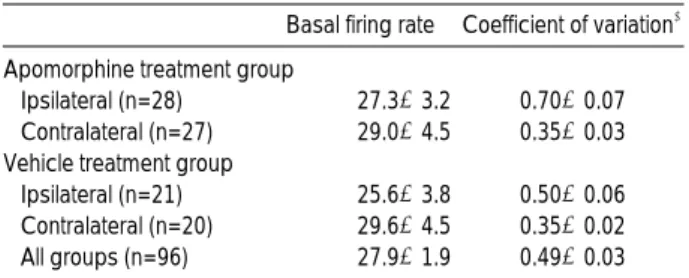
Fig. 2. Single unit recordings in the substantia nigra pars reticu- lata of 6-OHDA-lesioned rats. They could be classified into two types on the basis of their firing pattern. (A) A typical burst unit showed a high frequency firing interrupted by pause, an asym- metric interspike interval frequency distribution, and flat autocor- relogram with a single initial peak. (B) A typical non-burst unit with regular firing pattern showed a symmetric interspike interval frequency distribution and several peaks in autocorrelogram occurring with intervals approximating mean interspike interval.
A
B
msec msec
msec msec
Apomorphine treatment group
Ipsilateral (n=28) 27.3±3.2 0.70±0.07
Contralateral (n=27) 29.0±4.5 0.35±0.03
Vehicle treatment group
Ipsilateral (n=21) 25.6±3.8 0.50±0.06
Contralateral (n=20) 29.6±4.5 0.35±0.02
All groups (n=96) 27.9±1.9 0.49±0.03
Basal firing rate� Coefficient of variation� Table 1.Spontaneous activity of substantia nigra pars reticu- lata units in unilateral 6-OHDA-lesioned rats: firing rates and coefficients of variation*
*The data are the mean±standard error of mean of n cells per group,
�The spontaneous firing rates did not differ among groups: one-way ANOVA, �Ipsilateral SNr had significantly higher coefficient of variation than contralateral side regardless of treatment. Also ipsilateral SNr of apomorphine treatment group had a higher coefficient of variation than that of vehicle treatment group (p<0.05, Newman-Keuls procedure after a significant one-way ANOVA, p<0.05).
Burst 15 (54%) 0 5 (24%) 0 0.93±0.08
Non-burst 13 (46%) 27 (100%) 16 (76%) 20 (100%) 0.38±0.02 Table 2. Incidence of substantia nigra pars reticulata units in unilateral 6-OHDA-lesioned rats with different firing patterns
Type of unit *
Apomorphine treatment group
Ipsilateral Contralateral Ipsilateral Contralateral Vehicle treatment
group Coefficient of variation�
*Ipsilateral SNr had significantly higher incidence of the burst units than contralateral SNr in each treatment group. Ipsilateral SNr of apo- morphine treatment group showed higher incidence of the burst units than that of vehicle treatment group (p<0.05, Fisher’s exact test), �The data are the mean±standard error of mean. Burst units had signifi- cantly higher coefficient of variation than non-burst units (p<0.05, stu- dent t-test).
Electrophysiological recording
Single units isolated in SNr of 20 rats (n=96) were recorded and analyzed. The basal firing rates in the ipsilat- eral and contralateral side of 6-OHDA lesion were 25.6± 3.8 Hz (mean±SEM, n=21) and 29.6±4.5 Hz (n=20),
respectively, in vehicle treatment group and 27.3±3.2 Hz (n=28) and 29.0±4.5 Hz (n=27), respectively, in apomor- phine treatment group (Table 1). The differences in basal firing rates between either treatment groups or hemispheres were not statistically significant, however, coefficient of variation was significantly higher in ipsilateral SNr of apo-
Cells from the rats without previously repeated apomor- phine administration (n=4)
Cells from the rats with previously repeated apomorphine adminis- tration (n=8)
Ratio of firing rate
Quinpirole (mg/kg) 1.4
1.2 1 0.8 0.6 0.4 0.2 0
Baseline 0.0325 0.065 0.13 0.26 0.62
Ratio of firing rate
Quinpirole (mg/kg) 1.2
1 0.8 0.6 0.4 0.2 0
Baseline 0.0325 0.065 0.13 0.26 0.62
Ratio of firing rate
Quinpirole (mg/kg) 3
1.2 1 0.8 0.6 0.4 0.2
0 Baseline 0.0325 0.065 0.13 0.26 0.62
quinpirole 0.0325 mg/kg i.v.
quinpirole 0.065 mg/kg i.v.
quinpirole 0.13 mg/kg i.v.
haloperidol 0.2 mg/kg i.v.
quinpirole 0.26 mg/kg i.v.
haloperidol 0.2 mg/kg i.v.
haloperidol 0.2 mg/kg i.v.
Firing Rate (Hz)
80
60
40
20
0
0 10 20 30 40 50 60 70 80 90 100 min
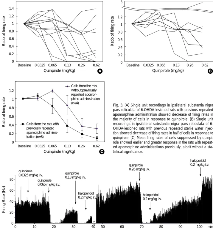
Fig. 3. (A) Single unit recordings in ipsilateral substantia nigra pars reticulata of 6-OHDA lesioned rats with previous repeated apomorphine administration showed decrease of firing rates in the majority of cells in response to quinpirole. (B) Single unit recordings in ipsilateral substantia nigra pars reticulata of 6- OHDA-lesioned rats with previous repeated sterile water injec- tion showed decrease of firing rates in half of cells in response to quinpirole. (C) Mean firing rates of cells suppressed by quinpi- role showed earlier and greater response in the rats with repeat- ed apomorphine administrations previously, albeit without a sta- tistical significance.
Fig. 4. Effect of quinpirole and haloperidol on the single unit firing in substantia nigra pars reticulata of the 6-OHDA-lesioned rat is shown as a histogram. Apomorphine was administered repeatedly for 6 days in this rat. Continuous recordings over 110 min period showed a suppression of firing by relatively low dose of quinpirole that was reversed by haloperidol.
A B
C
morphine treatment group than in contralateral side or vehicle treatment group. Typical burst firing pattern with long-duration high-frequency action potentials separated by a period of low-frequency tonic activity or complete absence of discharge (13) was identified in 20 units exclusively in the ipsilateral side of the 6-OHDA lesion regardless of apo- morphine treatment (Table 2). The other 76 units showed variable degree of irregularity in firing pattern which could not be classified into the burst units. The burst units had autocorrelograms without peaks or with a single initial peak, a very asymmetric ISI frequency distribution, and very high coefficients of variation (0.92±0.09). The non- burst units with typically regular firing had autocorrelo- grams with multiple peaks, a symmetric and narrow range of ISI frequency distribution, and low coefficients of varia- tion (0.42±0.02) (Fig. 2). In ipsilateral SNr, 15 (54%) among 28 units of apomorphine treatment group and 5 (24%) among 21 units of vehicle treatment group showed burst firing pattern. The differences in incidence of burst units between ipsilateral and contralateral side of the lesion or treatment groups were statistically significant. Quinpi- role suppressed 8 (67%) of 12 units (7 burst and 5 non- burst units) in ipsilateral SNr of apomorphine treatment group and 4 (50%) of 8 (3 burst and 5 non-burst units) in ipsilateral SNr of vehicle treatment group (Fig. 3A, B). Five burst and 2 non-burst units responded to quinpirole in apo- morphine treatment group and 2 burst and 2 non-burst units in vehicle treatment group. The suppressive effect of quinpirole was confirmed by spontaneous recovery of firing over time or reversal of suppression by dopamine antagonist haloperidol (0.2 mg/kg, intravenous) (Fig. 4). The units re- sponding to quinpirole showed more sensitive dose-response relation in apomorphine treatment group than in vehicle treatment group with a trial of increasing dose (Fig. 3C), albeit without a statistical significance.
DISCUSSION
The main findings of this study could be summarized as follows: after repeated administrations of apomorphine, (1) the behavioral response to selective D2 agonist was enhanced;
(2) SNr of 6-OHDA-lesioned rats showed increased per- centage of units with bursting activity; (3) selective D2 agonist quinpirole suppressed higher percentage of units more effectively though it could not be confirmed statisti- cally.
It is widely known that dopamine replacement therapy elicits rotational behavior in the 6-OHDA-lesioned rats (14, 15). The direction of this rotation is contraversive to the side of the lesion and is generally considered to repre- sent an antiparkinsonian effect (15). However, following repeated treatment, this response shows a markedly enhanced plasticity (9-11). Though the behavior in rats
shows none of the complex movements that are characteris- tic of dyskinesia in MPTP-treated monkeys or patients with Parkinson’s disease, the enhanced behavioral response to repeated dopamine replacement therapy in 6-OHDA- lesioned rats has pharmacological characteristics similar to L-DOPA-induced dyskinesia seen in MPTP-treated mon- keys and men (12). So it has been suggested that the enhanced response might be the result in changes in neural activity that are similar to those that underlie L-DOPA- induced dyskinesia in man and in the MPTP-treated mon- key. Our result of behavioral test is consistent with those in previous literatures showing progressively increased rota- tion with repeated administrations of apomorphine. In the rats primed with apomorphine selective D2 agonist alone also caused a markedly enhanced response as in drug- induced dyskinesia in men or MPTP-treated monkeys.
According to our results, quinpirole-induced rotation seems to be also enhanced in vehicle treatment group. This can be understood as an combined effect of priming which can elicit an enhanced response even with a single dose (12) and progressive degeneration of dopaminergic neurons after 6- OHDA lesioning (16).
Extracellular single unit recording is a useful method for the measurement of neuronal activity in vivo, and basic information acquired by it is about firing rate and pattern.
In this study, firing pattern was classified mainly by visual analysis and there were units with firing pattern of variable irregularity though they were classified as non-burst units.
When the pattern of unit activity could not be overtly defined by the inspection of the spike train, the digital raster display, ISI histogram, and autocorrel ogram, bursts were identified according to the definition of Legendy and Salcman (17). Burst was defined as at least three consecutive ISIs with a duration shorter than half of the mean ISI and units showing more than 5% of the spikes within bursts were classified as burst units.
The presence of burst units is probably the most constant finding in the spontaneous activity of basal ganglia output nuclei neurons in Parkinson’s disease. They were found in 6-OHDA-lesioned rats (13, 18, 19), in acute monoamine- depleted rats (20), in monkeys after electrolytic lesions of the ventromedial midbrain (21), in monkeys after chronic treatment with haloperidol or reserpine (21), and in MPTP- lesioned monkeys (22-25). Similar finding was reported also in patients with Parkinson’s disease (26). On the other hand, changes in the basal firing rate of output nuclei cells were found inconsistently: there was no differences in mon- keys with ventromedial midbrain lesions or after chronic treatment with haloperidol or reserpine (21), while other study observed an increased basal firing rate of internal pall- idal neurons in MPTP-lesioned monkeys (22, 23). More- over, it has been reported that the changes in basal firing rate of internal pallidal cells of MPTP-lesioned monkeys were inconsistent among animals and behavioral states (24).
′
The reports from experiments done in rat models were also inconsistent. Burbaud et al. (18) showed an increase in the mean firing rate of SNr units in 6-OHDA-lesioned rats, while Weick and Walters (27) reported no difference. Rob- ledo and Feger (20) did not observe an increase in firing rate of SNr neurons in reserpinized rats. Besides, Rohlfs et al.
(19) reported that basal firing rate of SNr in ipsilateral side of 6-OHDA lesion was lower than that in contralateral side.
Therefore, it was suggested that the neuronal firing pattern, rather than the mean firing rate of output nuclei neurons, is modified in parkinsonism (13, 19). Our results also support this hypothesis with a new finding that the percentage of the burst units in SNr ispilateral to 6-OHDA lesion increased in apomorphine treatment group. It suggests that drug treat- ment itself changes the physiology as well as anatomical lesion in parkinsonian rat though we cannot absolutely exclude the possibility that different degree of completeness of the lesion in each animal group may have partly contribut- ed to this finding. Though we did not try to compare the firing rates from parkinsonian rat with that of normal rat, the findings from the contralateral side of the lesion reveals that unilateral lesion or repeated drug treatment does not cause significant changes in firing pattern in contralateral hemisphere without dopamine depletion. It is known that selective D2 agonist suppresses neuronal activity in SNr dif- ferentially depending on the firing pattern, that is, the burst units are more frequently suppressed by quinpirole (28). So it is predictable that more units are suppressed by quinpirole in apomorphine treatment group. One hypothe- sis about bursting activity is that it is originated from the subthalamic nucleus and the change in firing pattern of SNr units is the consequence of striatal denervation and is mediated by the indirect pathway (13, 28). Alternatively, bursting activity could be originated in other structures projecting to the subthalamic nucleus such as the sensori- motor cortex (29) or the intralaminar thalamic nuclei (30).
Our experiment shows that burst units appear after 6- OHDA lesioning and apomorphine treatment exaggerates this tendency with an increasing behavioral response. How- ever, the mechanisms of these changes are still unknown. In addition to the higher percentage of units suppressed by quinpirole after repeated apomorphine injections, our results showed a left-shift of the dose-response curve. It is thought that the increased D2 receptor expression following 6-OHDA lesioning (31) may be exaggerated by apomor- phine treatment. Alternatively, qualitative change in striatal outflow after priming (32) may be accompanied by an increased sensitivity to D2 agonist.
In conclusion, this study suggests that an increased per- centage of burst firing units in SNr reflects a plasticity responding to repeated dopamine replacement in 6- OHDA-lesioned rats and is related with the enhanced behavioral response comparable to drug-induced dyskinesia in man. In addition, the concomitant enhancement of
behavioral and electrophysiological responses to quinpirole with priming suggests that the mechanism acting on D2 dopamine receptor is important in the development of dyskinesia.
ACKNOWLEDGMENT
This work was supported by the Samsung Grant, #SBRI C-A0-019.
REFERENCES
1. Marsden CD, Parkes JD. Success and problems of long-term levo- dopa therapy in Parkinson’s disease. Lancet 1977; 1: 345-9.
2. Obeso JA, Grandas F, Vaamonde J, Luquin MR, Artieda J, Lera G, Rodriguez ME, Martinez-Lage JM. Motor complications associated with chronic levodopa therapy in Parkinson's disease. Neurology 1989; 39: 11-9.
3. Bedard PJ, Di Paolo T, Falardeau P, Boucher R. Chronic treatment with L-DOPA, but not bromocriptine induces dyskinesia in MPTP- parkinsonian monkeys. Correlation with [3H]Spiperone binding.
Brain Res 1986; 379: 294-9.
4. Calne DB, Teychenne PF, Claveria LE, Eastman R, Greenacre JK, Petrie A. Bromocriptine in parkinsonism. Br Med J 1974; 4: 442-4.
5. Lees AJ, Stern GM. Sustained bromocriptine therapy in previously untreated patients with Parkinson’s disease. J Neurol Neurosurg Psychiat 1981; 44: 1020-3.
6. Bedard PJ, Mancilla BG, Blanchett P, Gagnon C, Di Paolo T. Levo- dopa-induced dyskinesia: facts and fancy. What does the MPTP monkey model tell us? Can J Neurol Sci 1992; 19: 134-7.
7. Blanchet PJ, Gomez-Mancilla B, Bedard PJ. DOPA-induced‘peak- dose’dyskinesia: clues implicating D2 receptor-mediated mecha- nisms using dopaminergic agonists in MPTP monkeys. J Neural Transm 1995; 45: 103-12.
8. Pearce RK, Banerji T, Jenner P, Marsden CD. De novo administra- tion of ropinirole and bromocriptine induces less dyskinesia than L- dopa in the MPTP-treated marmoset. Mov Disord 1998; 13: 234- 41.
9. Bevan P. Repeated apomorphine treatment causes behavioural super-sensitivity and dopamine D2 receptor hyposensitivity. Neu- rosci Lett 1983; 35: 185-9.
10. Carey RJ. Naloxone reverses L-DOPA induced overstimulation effects in a Parkinson’s disease animal model analogue. Life Sci 1991; 48: 1303-8.
11. Duty S, Brotchie JM. Enhancement of behavioural response to apo- morphine administration following repeated treatment in the 6- hydroxydopamine-lesioned rat is temporally correlated with a rise in striatal preproenkephalin-B, but not preproenkephalin-A, gene expression. Exp Neurol 1997; 144: 423-32.
12. Henry B, Crossman AR, Brotchie JM. Characterization of enhanced behavioral responses to L-DOPA following repeated administration in the 6-hydroxydopamine-lesioned rat model of Parkinson’s dis-
′
ease. Exp Neurol 1998; 151: 334-42.
13. Murer MG, Riquelme LA, Tseng KY, Pazo JH. Substantia nigra pars reticulata single unit activity in normal and 6-OHDA-lesioned rats: effects of intrastriatal apomorphine and subthalamic lesions.
Synapse 1997; 27: 278-93.
14. Ungerstedt U, Arbuthnott GW. Quantitative recording of rotational behavior in rats after 6-hydroxy-dopamine lesions of the nigrostri- atal dopamine system. Brain Res 1970; 24: 485-93.
15. Ungerstedt U. Postsynaptic supersensitivity after 6-hydroxy-dopamine induced degeneration of the nigro-striatal dopamine system. Acta Physiol Scand Suppl 1971; 367: 69-93.
16. Hong KS, Kim BG, Jeon BS, Yoon BW, Lee KW, Roh JK, Lee SB, Myung HJ. Chronological changes in the rotational behavior in response to apomorphine administration in 6-hydroxydopamine parkinsonian rat. J Korean Neurol Ass 1999; 17: 117-21.
17. Legendy CR, Salcman M. Bursts and recurrences of bursts in the spike trains of spontaneous active striate cortex neurons. J Neuro- physiol 1985; 53: 926-39.
18. Burbaud P, Gross C, Benazzouz A, Coussemacq M, Bioulac B.
Reduction of apomorphine-induced rotational behavior by subtha- lamic lesion in 6-OHDA lesioned rats is associated with a normal- ization of firing rate and discharge pattern of pars reticulata neu- rons. Exp Brain Res 1995; 105: 48-58.
19. Rohlfs A, Nikkhah G, Rosenthal C, Rundfeldt C, Brandis A, Samii M, Loscher W. Hemispheric asymmetries in spontaneous firing characteristics of substantia nigra pars reticulata neurons following a unilateral 6-hydroxydopamine lesion of the rat nigrostriatal path- way. Brain Res 1997; 761: 352-6.
20. Robledo P, Feger J. Acute monoaminergic depletion in the rat poten- tiates the excitatory effect of the subthalamic nucleus in the substan- tia nigra pars reticulata but not in the pallidal complex. J Neural Transm 1991; 86: 115-26.
21. Filion M. Effects of interruption of the nigrostriatal pathway and of dopaminergic agents on the spontaneous activity of globus pallidus neurons in the awake monkey. Brain Res 1979; 178: 425-41.
22. Miller WC, DeLong MR. Parkinsonian symptomatology: an anatom- ical and physiological analysis. Ann NY Acad Sci 1988; 515: 287-
302.
23. Filion M, Tremblay L. Abnormal spontaneous activity of globus pallidus neurons in monkeys with MPTP-induced parkinsonism.
Brain Res 1991; 547: 142-51.
24. Bergman H, Wichmann T, Karmon B, DeLong MR. The primate subthalamic nucleus. II. Neuronal activity in the MPTP model of parkinsonism. J Neurophysiol 1994; 72: 507-20.
25. Wichmann T, Bergman H, Starr PA, Subrmanian T, Watts RL, DeLong MR. Comparison of MPTP-induced changes in sponta- neous neuronal discharge in the internal pallidal segment and in the substantia nigra pars reticulata in primates. Exp Brain Res 1999; 125: 397-409.
26. Hutchison WD, Lozano AM, Tasker RR, Lang AE, Dostorovsky JO. Identification and characterization of neurons with tremor-fre- quency activity in human globus pallidus. Exp Brain Res 1997;
113: 557-63.
27. Weick BG, Walters JR. Effects of D1 and D2 dopamine receptor stimulation on the activity of substantia nigra pars reticulata neu- rons in 6-hydroxydopamine-lesioned rats: D1/D2 coactivation induces potentiated responses. Brain Res 1987; 405: 234-46.
28. Tseng KY, Riquelme LA, Belforte JE, Pazo JH, Murer MG. Sub- stantia nigra pars reticulata units in 6-hydroxydopamine-lesioned rats: responses to striatal D2 dopamine receptor stimulation and subthalamic lesions. Eur J Neurosci 2000; 12: 247-56.
29. Canteras NS, Shammah-Lagnado SJ, Silva BA, Ricardo JA. Soma- tosensory inputs to the subthalamic nucleus: A combined retrograde and antegrade HRP study in the rat. Brain Res 1988; 458: 53-64.
30. Mouroux M, Feger J. Evidence that the parafascicular projection to the subthalamic nucleus is glutamatergic. Neuro Report 1993; 4:
613-5.
31. Gerfen CR, Engber TM, Mahan LC, Susel Z, Chase TN, Monsna FJ, Jr.,Sibley DR. D1 and D2 dopamine receptor-regulated gene expression of striatonigral and striatopallidal neurons. Science 1990;
250: 1429-32.
32. Pollack AE, Turgeon SM, Fink JS. Apomorphine priming alters the responses of striatal outflow pathways to D2 agonist stimulation in 6-hydroxydopamine-lesioned rats. Neuroscience 1997; 79: 79-93.
′