INTRODUCTION
Elbow flexion synergy of stroke survivors may occur reflex- ively or as early stages of voluntary control when spasticity is present. However, when spasticity of the elbow flexors is marked, it may contribute to the typical upper extremity posture in hemiplegia and limit rehabilitation efforts to im- prove function or at least minimize impairments (1). For patients who are ambulatory, walking with the elbow flexed impairs balance and may be cosmetically unacceptable. The patients with spasticity of the elbow flexion are most likely to show the pronation of the forearm, which is not performed by the biceps brachii. Furthermore, a major flexor of the elbow is the brachialis muscle, that has no role in supination (2).
Thus, blocking the brachialis is expected to reduce spasticity of elbow flexion without eliminating the ability to generate supination torques.
Chemodenervation of the musculocutaneous nerve with neurolytic agents is an effective treatment of spasticity of the elbow flexors. The flexion spasticity at the elbow can be treated using injections of neurolytic agents designed to block the musculocutaneous nerve at the level of the axilla or upper arm (3-6). Blocking the main nerve trunk may produce pro- found weakness of the hemiparetic upper extremity (7, 8).
Neurolytic agents may be spilled over the adjacent arteries and have a direct effect on vascular smooth muscles, result- ing in a significant vasoconstriction (9). There might be tem- porary dysesthetic pain over the distribution of the lateral antebrachial cutaneous nerve after neurolysis (8, 10).
To avoid undesirable complications, motor point blocks of the biceps brachii or brachialis muscle may be more satis- factory. For phenol or alcohol neurolysis, precise localization of the motor points of each muscle is necessary to avoid block- ing the sensory or major motor nerve. Previous anatomic studies have defined the motor point as the location where the motor branch entered the muscle belly (11, 12). How- ever, there have been few studies that have investigated the location of the motor points of the biceps brachii and brachialis muscles. The present study, therefore, was conducted to iden- tify the location of the motor points of these muscles relative to anatomic landmarks in order to facilitate the efficacy of motor point block.
MATERIALS AND METHODS
Twenty-three limbs from 12 cadavers were dissected for the study. There were 5 male and 7 female cadavers with an
Byung Kyu Park*, Yong Beom Shin, Hyun-Yoon Ko, Jae Heung Park�, Sun-Yong Baek�
Department of Physical Medicine & Rehabilitation*, Korea University College of Medicine, Seoul;
Department of Rehabilitation Medicine, Department of Anatomy�, Pusan National University School of Medicine, Busan; Department of Rehabilitation Medicine�, HakjangKeunsol Medical Hospital, Busan, Korea
Address for correspondence Yong Beom Shin, M.D.
Department of Rehabilitation Medicine, Pusan National University School of Medicine, Medical Research Institute, Pusan National University Hospital, 1-10 Ami-dong, Seo-gu, Busan 602-739, Korea Tel : +82.51-240-7485, Fax : +82.51-247-7485 E-mail : [email protected]
459 J Korean Med Sci 2007; 22: 459-62
ISSN 1011-8934
Copyright � The Korean Academy of Medical Sciences
Anatomic Motor Point Localization of the Biceps Brachii and Brachialis Muscles
Injection of the neurolytic agents into motor points of the biceps brachii or brachialis muscles is an effective treatment of spasticity of the elbow flexors in many stroke survivors. Accurate localization of the motor points of each muscle is necessary for enhancing the efficacy of motor point blocks. To identify the precise locations of the motor points (terminal nerve endings) of the biceps brachii and brachialis mus- cles in relation to anatomic surface landmarks for motor point blocks, we dissected 23 limbs from 12 cadavers. A reference line was defined as a line connecting the coracoid process with the lateral epicondyle of the humerus. The location of the motor points of the biceps brachii and brachialis muscles was identified in reference to the reference line. The motor point of the biceps brachii muscle was found to be approximately half of the reference line. In the brachialis muscle, the location of the motor point was 70% of the reference line from the coracoid process and 2 cm medi- al to the line. The results are expected to facilitate effective localization of the motor point block of these muscles in selective motor nerve block.
Key Words : Elbow; Flexor; Muscle Spasticity; Nerve Endings; Nerve Block
Received : 5 June 2006 Accepted : 20 October 2006
460 B.K. Park, Y.B. Shin, H.-Y. Ko, et al.
average age at time of death of 66 yr (range, 31 to 87). One limb was unsuitable for the study due to significant contrac- ture of the elbow joint. Each cadaver was placed supine with the elbow extended in the anatomic position.
The skin and subcutaneous tissue were dissected from the elbow crease, exposing the entire biceps brachii. After cut- ting the biceps brachii tendon at the elbow, the muscle was detached from the brachialis. The musculocutaneous nerve was identified and the branches to the brachialis and biceps
brachii were observed. The center of the location where the motor branch entered the muscle belly was designated as the motor point of each muscle.
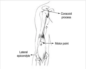
A tape measure was used to form a reference line connect- ing the coracoid process to the lateral epicondyle of the hu- merus (Fig. 1). For the brachialis muscle, the perpendicular line from the reference line to the motor point was measured and was recorded as an X value. The Y value was defined as the distance from the coracoid process to the point where the perpendicular line crossed the reference line. For the biceps brachii muscle, the X value was not measured as the location of the motor point was not fixed in a transverse plane after cutting the muscle at the elbow. The shortest distance bet- ween the coracoid process and the motor point was measured and was defined as the Y value. For both muscles, the Y value was also normalized into a percentage of the total length of the reference line and hence compared across all specimens.
RESULTS
The mean length of the reference line between the coracoid process and the lateral epicondyle of the elbow was 26.76± 1.59 cm. Each head of the biceps muscle was consistently innervated by a single branch of the nerve, respectively. The motor points of the biceps brachii were found to be approx- imately half of the distance from the coracoid process to the lateral epicondyle of the humerus (Fig. 2). The motor point of the short head of the biceps muscle was located 12.91± 1.99 cm (48.24±6.88% of the reference line) distal to the coracoid process. In the long head of the muscle, the location of the motor point from the coracoid process was 14.22± 1.75 cm (53.19±6.21%).
All brachialis muscles were innervated by one or two bran-
Fig. 1. Diagrammatic drawing of measurement of the location of the motor point of the brachialis muscle. The coracoid process and lateral epicondyle of the humerus were identified as reference points. The shortest distance between the reference points was measured and was defined as a reference line. The distance from the reference line to the motor point was recorded as an x value and the distance from the coracoid process to the point where the perpendicular line crossed the reference line was defined as a y value.
Coracoid process
Lateral epicondyle
X Y
Motor point
Normalized ratio
0.8 0.7 0.6 0.5 0.4 0.3 0.2 0.1
00 5 10 15 20 25
Cases
LH
Fig. 2.A plot of the location of the motor points in the biceps brachii muscles. The normalized ratio indicates the ratio of the distance from the coracoid process to the length of the reference line con- necting from the coracoid process to the lateral epicondyle of the humerus. SH, short head; LH, long head.
SH
Normalized ratio
1
0.8
0.6
0.4
0.2
00 1 2 3 4 5
Distance (cm)
MP1
Fig. 3.A plot of the location of the motor points in the brachialis muscle; x axis is the distance from the reference line to the motor point and y axis is the distance from the coracoid process to the point where the perpendicular line crosses the reference line. MP1, the first motor point; MP2, the second motor point.
MP2
Motor Point of the Biceps and Brachialis 461
ches of the musculocutaneous nerve (seventeen with one branch and six with two branches); 7 arms also received a branch from the radial nerve. The first motor point of the brachialis muscle innervated by the musculocutaneous nerve was found to be 2.28±0.74 cm medial to the reference line (x value) and 17.84±1.77 cm (66.73±5.95%) of the reference line distal to the coracoid process (y value) (Fig. 3). The location of the second motor point was 2.33±0.53 cm in the x value and 18.57±1.98 cm (70.09±4.41%) in the y value. The motor point of the brachialis from a branch of the radial nerve was found to be 0.76±0.77 cm in the x value and 18.90± 2.95 cm (70.19±10.61%) in the y value.
DISCUSSION
Our study demonstrated that the motor point of the bra- chialis muscle was found to be approximately 70% of the reference line from the coracoid process to the lateral epicon- dyle of the elbow and 2 cm medial to the line. Buchanan and Erickson (13) performed anatomic dissections on 26 arms from 13 cadavers. They observed that the motor point of the biceps muscle was located at 53% of the length of the humerus and that of the brachialis was found to be distal one third of the humerus. These results are consistent with those of our dissection study. However, they did not document a horizon- tal location (the perpendicular distance from the reference line) of the motor point of the brachialis because they con- sidered that its motor point could be best blocked from the medial side. Recently, Kim et al. (14) also reported similar results with no consideration of the horizontal distance.
The approach from the medial side of the arm is carried out by lifting adjacent neurovascular bundles along with the biceps brachii muscle (5). The brachial artery or the median nerve may be injured if these structures are not adequately lifted ventrally in cases of severe flexion spasticity. We need to predict the depth of the needle insertion to avoid passing through the motor nerve branches of the musculocutaneous nerve. Based on our data, an anterior approach may be con- sidered; the needle is located approximately 2 cm medial to 70% of the reference line from the coracoid process to the lateral epicondyle of the elbow. However, the needle has to pass through the biceps brachii with the potential of minor spilling of the neurolytic agent. Further detailed studies are needed to determine any negative effect of the anterior app- roach on functional status.
The nerve supply to the brachialis muscle has not been clearly understood. Recently, standard testbooks of anatomy des- cribe a contribution from the radial nerve to inferolateral portion of this muscle but do not mention its incidence (15, 16). Ip and Chang (17) reported that the brachialis receives a constant innervation from the radial nerve in eight Chinese cadavers. Mahakkanukrauh and Somsarp (18) investigated the dual innervation of the brachialis muscle in 152 Thai
cadaveric limbs. They found that all brachialis muscles received innervation from the musculocutaneous nerve and the radial nerve provided a dual supply in 81.6% of cases. These results imply that the musculocutaneous nerve or motor point block may not completely paralyze the brachialis muscle. The func- tional significance of the radial branch to brachialis has not been reported. The size of the motor distribution from the radial branch to brachialis may be variable (17, 18).
Blackburn et al. (19) recently reported the radial innerva- tion of the brachialis in 42 UK Caucasian cadaveric limbs.
They found that a radial nerve contribution to the innerva- tion of the brachialis was present in 67% of cases, less than the incidence reported by previous studies. In our study, the dual innervation, occurred in a higher proportion of speci- mens than 30.4%. This may reflect interracial or ethnic dif- ferences in embryological development, or may be due to the small number of specimens. In our study, the motor point of the brachialis from a branch from the radial nerve was locat- ed approximately 1 cm lateral to the motor point from the musculocutaneous nerve innervation. Thus, if a motor point block of the brachialis muscle is not successful, there might be the possibility of a radial contribution which may require more lateral additional block.
According to our study, the motor point of the biceps brachii is likely to be located at approximately the halfway point of the upper arm. The motor point can be easily identified by the use of a surface stimulator on the skin of the mid arm. A needle electrode is then used to more accurately determine the location of the motor points of the muscle. In addition, our data may be helpful in determination of the recording location of the biceps muscle. In standard needle electromyo- graphy, the needle electrode is placed over the bulk of the biceps muscle (20). The lack of knowledge regarding the loca- tion of the motor points of the biceps muscle may lead to an erroneous placement of the needle electrode because the max- imum bulk of the muscle may be found more distally in elbow extension.
In summary, the motor point of the biceps brachii muscle was located at approximately half of the arm. In the brachialis muscle innervated by one or two branches of the musculo- cutaneous nerve, the location of the motor points was likely to be 2 cm medial to distal one third of a reference line from the coracoid process to the lateral condyle of the elbow. Our results may allow precise localization of the motor points and offer more accurate approach. To validate this anatomic work in clinical practice, further clinical studies will be need- ed to compare therapeutic effects of selective motor point block with those of the musculocutaneous nerve block.
REFERENCES
1. Mizrahi EM, Angel RW. Impairment of voluntary movement by spas- ticity. Ann Neurol 1979; 5: 594-5.
462 B.K. Park, Y.B. Shin, H.-Y. Ko, et al.
2. Calais-Germain B. The elbow. In: Anderson S, editor, Anatomy of movement. Seattle: Eastland Press, 1993; 131-46.
3. Keenan MA, Tomas ES, Stone L, Gersten LM. Percutaneous phe- nol block of the musculocutaneous nerve to control elbow flexor spas- ticity. J Hand Surg Am 1990; 15: 340-6.
4. Garland DE, Rhoades ME. Orthopedic management of brain-injured adults. Part II. Clin Orthop Relat Res 1978; 131: 111-22.
5. Keenan MA. Management of the spastic upper extremity in the neu- rologically impaired adult. Clin Orthop Relat Res 1988; 233: 116-25.
6. Khalili AA, Betts HB. Isolated block of musculocutaneous and per- ineal nerves in the management of spasticity with special reference to the use of a nerve stimulator. Anesthesiology 1967; 28: 219-22.
7. Garland DE, Thompson R, Waters RL. Musculocutaneous neurec- tomy for spastic elbow flexion in non-functional upper extremities in adults. J Bone Joint Surg Am 1980; 62: 108-12.
8. Glenn MB. Nerve blocks for the treatment of spasticity. In: Katz RT, editor, Physical medicine and rehabilitation: state of the art reviews.
Philadelphia: Hanley & Belfus 1994; 481-505.
9. Johnson ME, Sill JC, Brown DL, Halsey TJ, Uhl CB. The effect of the neurolytic agent ethanol on cytoplasmic calcium in arterial smooth muscle and endothelium. Reg Anesth 1996; 21: 6-13.
10. Kong KH, Chua KS. Neurolysis of the musculocutaneous nerve with alcohol to treat poststroke elbow flexor spasticity. Arch Phys Med Rehabil 1999; 80: 1234-6.
11. Albert T, Yelnik A, Colle F, Bonan I, Lassau JP. Anatomic motor point localization for partial quadriceps block in spasticity. Arch Phys Med Rehabil 2000; 81: 285-7.
12. Kim HS, Hwang JH, Lee PK, Kwon JY, Oh-Park MY, Kim JM, Chun MH. Localization of the motor nerve branches and motor points of the triceps surae muscles in Korean cadavers. Am J Phys Med Rehabil 2002; 81: 765-9.
13. Buchanan TS, Erickson JC. Selective block of the brachialis motor point. An anatomic investigation of musculocutaneous nerve branch- ing. Reg Anesth 1996; 21: 89-92.
14. Kim JS, Kwon JY, Kang SY, Park JW. Anatomical locations of the motor points of the biceps brachii and brachialis muscles. J Korean Acad Rehabil Med 2004; 28: 592-5.
15. Jenkins DB. Hollinshead’s functional anatomy of the limbs and back.
8th ed. Philadelphia: Saunders; 2002.
16. Johnson D, Ellis H. Upper arm. In: Standring S, Ellis H, Healy JC, Johnson D, Williams A, Collins P, Wigley C, editors, Gray’s anato- my: the anatomical basis of clinical practice. 39th ed. Edinburgh:
Churchill Livingstone 2005; 851-8.
17. Ip MC, Chang KS. A study on the radial supply of the human bra- chialis muscle. Anat Rec 1968; 162: 363-71.
18. Mahakkanukrauh P, Somsarp V. Dual innervation of the brachialis muscle. Clin Anat 2002; 15: 206-9.
19. Blackburn SC, Wood CP, Evans DJ, Watt DJ. Radial nerve contri- bution to brachialis in the UK Caucasian population: position is pre- dictable based on surface landmarks. Clin Anat 2007; 20: 64-7.
20. DeLagi EF, Perotto A, Iazzetti J, Morrison D. Anatomic guide for the electromyographer the limbs. 2nd ed. Springfield (IL): Charles C Thomas; 1981.