165
Received:September 6, 2016, Revised:(1st) November 21, 2016, (2nd) November 28, 2016, Accepted:December 14, 2016
Corresponding to:Wan-Hee Yoo, Division of Rheumatology, Department of Internal Medicine, Chonbuk National University Medical School and Research Institute of Clinical Medicine of Chonbuk National University Hospital, 20 Geonji-ro, Deokjin-gu, Jeonju 54907, Korea.
E-mail:[email protected]
Copyright ⓒ 2017 by The Korean College of Rheumatology. All rights reserved.
This is a Open Access article, which permits unrestricted non-commerical use, distribution, and reproduction in any medium, provided the original work is properly cited.
Case Report
pISSN: 2093-940X, eISSN: 2233-4718
Journal of Rheumatic Diseases Vol. 24, No. 3, June, 2017 https://doi.org/10.4078/jrd.2017.24.3.165
Hypereosinophilic Syndrome Associated with the Onset of Rheumatoid Arthritis: A Case Report
Jae-hee Park1, Won-Seok Lee2, Seoung Ju Park3, Wan-Hee Yoo2
1Department of Internal Medicine, Chonbuk National University Hospital, 2Division of Rheumatology, Department of Internal Medicine, Chonbuk National University Medical School and Research Institute of Clinical Medicine of Chonbuk National University Hospital, 3Division of Pulmonology and Allergy, Department of Internal Medicine, Chonbuk National University Hospital, Jeonju, Korea
Idiopathic hypereosinophilic syndrome (HES) is a disorder characterized by the sustained overproduction of eosinophils and multiple organ damage. Rheumatologic manifestations of HES are infrequent, but persistent eosinophilia is observed in approx- imately 10% to 40% of patients with rheumatoid arthritis (RA). This finding may be a result of the RA itself and is often associated with active disease and the presence of extra-articular features. We describe the case of a 48-year-old man affected by HES who subsequently developed RA. Both HES and RA responded rapidly to the corticosteroid and methotrexate therapy. In this patient, the initiation of RA and HES was related, suggesting a common pathogenetic link between these two diseases. (J Rheum Dis 2017;24:165-168)
Key Words. Hypereosinophilic syndrome, Rheumatoid arthritis
INTRODUCTION
Idiopathic hypereosinophilic syndrome (HES) is a leu- koproliferative disorder characterized by sustained over- production of eosinophils. Although all organ systems can be affected, dermatological involvement is the most common, followed by pulmonary, gastrointestinal, and cardiac involvement [1]. However, HES manifestations are commonly observed in many other medical con- ditions, making the initial diagnosis more difficult to establish. The first step is to rule out other conditions that present with similar symptoms. Though reports of late onset HES in patients with rheumatoid arthritis (RA) have been confirmed in several cases, it is still unknown if HES is related to the drugs used for RA or to the disease itself [2,3].
The patient described in this case was diagnosed with RA during 4 months of follow-up for HES, when he pre- sented with joint symptoms. Our case differs from pre-
vious cases that described the onset of HES in long- standing RA cases. To our knowledge, such case is re- ported for the first time in Korea.
CASE REPORT
In October 2015, a 48-year-old man was admitted with a 3 week history of generalized weakness, cough, and rap- idly progressive dyspnea. He described a nonproductive cough, but denied fever, chest pain, changes in weight, or skin lesions.
On admission, physical examination revealed bilateral lower extremity edema and facial edema. Laboratory ex- amination revealed leukocytosis (peak 29.5×10³/μL [normal 4.8∼10.8×10³/μL]), associated with 21.7%
eosinophils (peak 5.01×10³/μL [normal 0∼0.45×10³/ μL]). Allergy and parasitic helminths (worms) are the most commonly identified causes of eosinophilia. However, the serology tests for parasites and allergic skin prick test
Jae-hee Park et al.
166 J Rheum Dis Vol. 24, No. 3, June, 2017
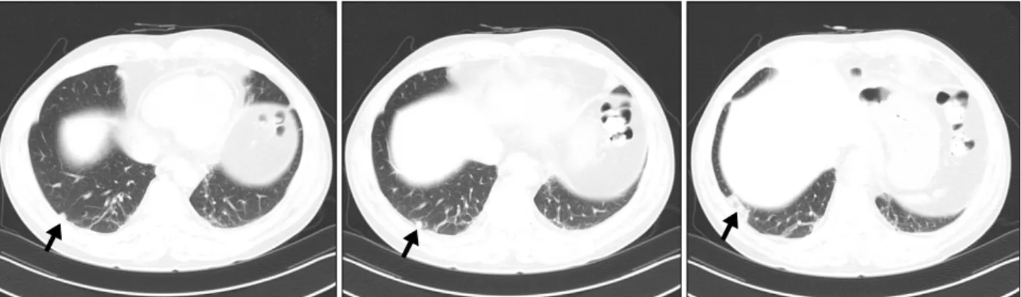
Figure 1. High-resolution computed tomography scan of the lungs reveals subpleural distribution of small hyperdense nodules (black arrow) in the right lower lobe.
Figure 2. Bone scan shows hot spots in both ankles and wrists, a sign of arthritis. Delayed re- gional bone images show in- creased radiotracer uptake in both ankles, both feet, and both wrists.
results of the patient were all negative. Chest X-ray was unremarkable. An extensive workup excluded other caus- es of secondary hypereosinophilia. The results of a bone marrow sample showed normal myeloid blasts, thus rul- ing out eosinophilia caused by myeloproliferative diseases.
A high-resolution computed tomography (HRCT) scan of the lungs revealed subpleural distribution of small hyper- dense nodules in the right lower lobe (Figure 1). We per- formed pulmonary function test and arterial blood gas analysis and found normal results (forced expiratory vol- ume [FEV]1 2.90 L, FEV1/forced vital capacity 70%, PaO2 75.5 mmHg).
Upper gastrointestinal endoscopy showed mild uneven mucosal lesions with swelling, while endoscopic biopsies obtained from the stomach and duodenum revealed chronic gastritis with metaplasia and no evidence of eosi- nophilic infiltration. Electrocardiography and echo- cardiography showed no abnormal findings. The patient’s history did not suggest medication-induced HES. In order to rule out the eosinophilic granulomatous polyangiitis,
we checked the level of C-antineutrophil cytoplasmic an- tibody (ANCA), and P-ANCA. The results were negative.
Consequently, the patient was diagnosed with idio- pathic HES based on the presence of marked blood eosi- nophilia associated with an evidence of eosinophil-in- duced organ damage, in the absence of other causes of hy- pereosinophilia such as allergic, parasitic, and malignant disorders. Following the diagnosis of HES, therapy with oral prednisolone (60 mg/d) and antihistamines was initiated. One month after the initiation of therapy, the pulmonary infiltrations disappeared on HRCT. Two months after the presentation, the patient was doing well on 15 mg/d of prednisolone, but any attempts to lower the dose below 10 mg/d resulted in peripheral eosinophilia recur- rence.
In February 2016, while the patient was on steroid taper- ing therapy (15 mg/d), general edema recurred and he complained of worsening pain in both ankles and wrists.
This prompted an admission for further evaluation, dur- ing which, the recurrence of facial and peripheral edema
Hypereosinophilic Syndrome and Rheumatoid Arthritis
www.jrd.or.kr 167
was also noted. Laboratory examination revealed leuko- cytosis (peak 20.9×10³/μL) and eosinophilia (40.4% eo- sinophils, peak 8.46×10³/μL). The erythrocyte sed- imentation rate (ESR) (70 mm/h [normal, 0∼9 mm/h]) and C-reactive protein level (CRP) (37 mg/dL [normal, 0∼5 mg/dL]) were also elevated. A bone scan performed to further evaluate the ankle and wrist pain revealed hy- peremic uptake in both joints (Figure 2). Rheumatoid fac- tor (RF) level was increased to 78.8 IU/mL and anti-cyclic citrullinated peptide (CCP) antibody level was elevated to 269.2 IU/mL.
A diagnosis of RA was made based on the American College of Rheumatology/European League Against Rheumatism (ACR/EULAR) 2010 RA classification cri- teria [4]. A diagnosis of RA coexisting with HES was made and the patient was started on methotrexate (MTX, 10 mg/wk) and methylprednisolone (30 mg/d). He showed rapid symptomatic improvement after the treat- ment, with complete resolution of the dyspnea and joint symptoms. White cell count was 10.6×10³/μL with 1.6% eosinophils, and the total eosinophil count was 0.5×10³/μL. The ESR and CRP levels returned to the normal range. The patient was subsequently treated with methotrexate (10 mg/wk) and low dose oral predniso- lone (2.5 mg/d) with satisfactory control of the clinical symptoms.
DISCUSSION
HES is a rare and heterogeneous group of disorders de- fined as persistent and marked blood eosinophilia (>1,500 eosinophils/mm3 for more than 6 consecutive months) associated with evidence of eosinophil-induced organ damage, in the absence of other causes of hypereosi- nophilia, such as allergic, parasitic, and malignant dis- orders [1]. The present case did not meet the criteria for persistent eosinophilia, as the time frame was less than 6 months after the initial diagnosis. However, we suspect that the generalized edema and lung abnormalities seen in our patient were clinical features of HES, given the lack of other causes of eosinophilia. Target-organ damage mediated by eosinophils is highly variable among pa- tients, with involvement of skin, heart, lungs, and central and peripheral nervous systems in more than 50% of cas- es [5]. Eosinophilia may be primary or secondary. Cases of eosinophilia in which an underlying cause has been sought but not found fall into the “idiopathic” category. If the condition is chronic and has led to tissue damage, the
term “idiopathic hypereosinophilic syndrome” is used [6]. Secondary eosinophilia is a cytokine-derived (inter- leukin-5) reactive phenomenon. Worldwide, parasitic dis- eases are the most common cause, whereas in developed countries, allergic diseases are the most common cause [7].
Although rheumatologic manifestations of HES are in- frequent, several previous reports showed HES associa- tion with an inflammatory joint disease mimicking RA [3,8-10]. Furthermore, Tay [11] described 10 patients from Singapore with acute polyarthritis and marked hy- pereosinophilia of unknown etiology. He attached the la- bel ‘eosinophilic arthritis’ to this condition. The articular involvement represented soft tissue and synovial fibri- noid degeneration with eosinophilic infiltration. Brogadir et al. [8] reported a case of articular involvement in HES and suggested that this could be one of a multitude of cases of hand deformity resembling rheumatoid arthritis. In our case, we suspected HES-induced arthritis based on the gradual onset of ankle pain, but since the patient also complained about concomitant knee and wrist pain, we evaluated anti-CCP antibody and RF levels and these findings further supported our diagnosis. In a retro- spective study of 45 cases of RA, certain extra-articular manifestations of RA were found to occur more fre- quently in patients with eosinophilia [3]. Furthermore, in a previous study, HES developed during the course of long-standing RA and was directly associated with an ex- acerbation of the arthritic condition [6,12]. The con- version of seronegative into seropositive RA along with the onset of HES during treatment has also been reported [13]. It is, however, not clear whether HES is a con- sequence of the rheumatoid inflammatory process itself or induced by disease modifying anti-rheumatic drugs. In our patient, HES initially occurred in the absence of joint symptoms and RA was diagnosed after the initiation of HES therapy, and in the presence of newly developed joint symptoms and elevated levels of RF and anti-CCP antibody. Therefore, in this case, it is unlikely that HES was a consequence of the drugs used in the treatment of RA. Therefore, although the precise mechanism is un- known, the possibility of a common pathogenetic link be- tween the two diseases was raised, and the common de- nominator, the eosinophils, is believed to play a central role [14].
Because of the rarity of HES, no evidence-based guide- lines address its management. Corticosteroids are first-line treatment, with prednisolone with hydroxyurea or inter- feron alpha as second-line agents. Our patient was treated
Jae-hee Park et al.
168 J Rheum Dis Vol. 24, No. 3, June, 2017
with disease-modifying antirheumatic drugs including MTX, and these drugs proved to be very efficacious both on articular pathology and on the clinical and laboratory manifestations of HES. These data also suggest the com- mon pathogenetic mechanisms in RA and HES.
SUMMARY
This case differs from preceding cases described in the literature because HES did not develop in a patient who was originally treated for RA, but rather the symptoms of RA developed subsequently to HES. The onset of RA and HES was directly related, implying a common pathoge- netic link between these two diseases. This case calls for increased attention to autoimmune diseases such as RA in cases where joint symptoms develop in HES patients.
CONFLICT OF INTEREST
No potential conflict of interest relevant to this article was reported.
REFERENCES
1. Weller PF, Bubley GJ. The idiopathic hypereosinophilic syndrome. Blood 1994;83:2759-79.
2. Chaudhuri K, Dubey S, Zaphiropoulos G. Idiopathic hyper- eosinophilic syndrome in a patient with long-standing rheu- matoid arthritis: a case report. Rheumatology (Oxford) 2002;41:349-50.
3. Winchester RJ, Koffler D, Litwin SD, Kunkel HG.
Observations on the eosinophilia of certain patients with rheumatoid arthritis. Arthritis Rheum 1971;14:650-65.
4. Aletaha D, Neogi T, Silman AJ, Funovits J, Felson DT, Bingham CO 3rd, et al. 2010 Rheumatoid arthritis classi- fication criteria: an American College of Rheumatology/
European League Against Rheumatism collaborative initiative. Arthritis Rheum 2010;62:2569-81.
5. Roufosse FE, Goldman M, Cogan E. Hypereosinophilic syndromes. Orphanet J Rare Dis 2007;2:37.
6. Bain BJ. Eosinophilia--idiopathic or not? N Engl J Med 1999;341:1141-3.
7. Seifert M, Gerth J, Gajda M, Pester F, Pfeifer R, Wolf G.
Eosinophilia--a challenging differential diagnosis. Med Klin (Munich) 2008;103:591-7.
8. Brogadir SP, Goldwein MI, Schumacher HR. A hyper- eosinophilic syndrome mimicking rheumatoid arthritis.
Am J Med 1980;69:799-802.
9. Prattichizzo FA, Bernini L. An idiopathic hypereosinophilic syndrome mimicking seronegative rheumatoid arthritis:
20-year follow-up with clinical and laboratory findings. Clin Exp Rheumatol 1992;10:79-81.
10. Martín-Santos JM, Mulero J, Andréu JL, de Villa LF, Bernaldo-de Quirós L, et al. Arthritis in idiopathic hyper- eosinophilic syndrome. Arthritis Rheum 1988;31:120-5.
11. Tay C. Eosinophilic arthritis. Rheumatology (Oxford) 1999;38:1188-94.
12. Bonanno D, Fedele R, Minciullo P, Quattrocchi P, Ferlazzo B. Idiopathic hypereosinophilic syndrome associated with rheumatoid arthritis. A case report. Reumatismo 2003;
55:181-3.
13. Rosenstein RK, Panush RS, Kramer N, Rosenstein ED.
Hypereosinophilia and seroconversion of rheumatoid arthritis. Clin Rheumatol 2014;33:1685-8.
14. Boomars KA, van Velzen-Blad H, Mulder PG, Koenderman L, Lammers JW, van den Bosch JM. Eosinophil cationic pro- tein and immunoglobulin levels in bronchoalveolar lavage fluid obtained from patients with chronic eosinophilic pneumonia. Eur Respir J 1996;9:2488-93.