기저핵부 뇌실질내 출혈에 대한 수술후 뇌농양으로 오인한 다형성 교아세포종
한양대학교 의과대학 구리병원 신경외과학교실, 해부병리학교실*
정진환·김재민·백광흠·박용욱*·김충현·오석전
= Abstract =
Glioblastoma Misdiagnosed as Brain Abscess after Surgical Evacuation of Spontaneous Basal Ganglia Hemorrhage
Jin Hwan Cheong, M.D., Jae Min Kim, M.D., Koang Hum Bak, M.D., Yong Wook Park, M.D.,* Choong Hyun Kim, M.D., Suck Jun Oh, M.D.
Department of Neurosurgery and Anatomical Pathology,* Hanyang University Kuri Hospital, Kuri, Korea
51-year-old woman presented with sudden severe headache, vomiting, and right hemiparesis at first admission.
Computed tomography(CT) scans revealed an hemorrhagic density at left basal ganglia. Preoperative cerebral angiography showed no vascular lesion. Under the diagnosis of hypertensive intracerebral hemorrhage(ICH), total extirpation of hematoma was done. The postoperative neurological condition improved gradually and discharged without any neurological sequelae. Two months later, she revisited with headache, vomiting and progressive right hemiparesis. CT scans at second admission showed an irregular rim enhanced mass with central low density with surrounding edema at the initial bleeding area. Repeated craniotomy was performed and the mass was partially removed. The histopathological diagnosis of the specimen was confirmed as glioblastoma.
The authors report a glioblastoma, which occurred at initial ICH site and regarded as a brain abscess with literature review.
KEY WORDS:Glioblastoma・Brain abscess・Hypertensive intracerebral hemorrhage.
서 론
대부분의 자발성 두개강내 출혈은 뇌동맥류, 뇌동정맥 기 형, 고혈압 등의 여러 원인에 의해 발생하며, 뇌종양에 의 한 자발성 두개강내 출혈도 약 0.9~14.6%로 보고되고 있 다4)9). 이와 관련 있는 뇌종양으로는 악성 흑색종, 융모 상 피선종 등 두개강내로 전이한 종양에서는 종종 두개강내 출혈을 동반하기도 하지만, 신경교종 특히 다형성 교아세포 종에서는 5%미만으로 드문 경우로 알려져 있다7). 이러한 뇌종양에 의한 두개강내 출혈은 뇌 전산화 단층 촬영(CT) 이나 핵자기 공명영상(MRI), 뇌혈관 조영술로 감별이 되 기도 하지만 간혹 혈종 양이 많아 이러한 진단적 방법으로
도 감별이 되지 않는 경우가 종종 있다. 따라서 정확한 진단 이 이루어지지 않음으로써 수술시 술자를 당황하게 하거나 환자의 치료와 예후 판정을 어렵게 하기도 한다. 최근 본 교 실에서는 기저핵부위 뇌실질내 출혈에 대한 수술후, 뇌농 양으로 오인되었던 다형성 교아세포종 1례를 경험하였기에 문헌 고찰과 함께 보고하는 바이다.
증 례
51세 여자 환자가 갑자기 시작된 심한 두통과 구토를 주 소로 응급실을 통해서 내원하였다. 평소 건강하게 지내던 환자로 가족력상에도 특기할 만한 사항은 없었다. 신경학 적 검사상 의식은 기면 상태였으며, 뇌신경 장애의 증상은
AAAA
없었고, Grade IV의 우측 부전마비의 소견이 관찰되었다.
병적 반사는 관찰되지 않았으며 수막 자극 징후도 없었다.
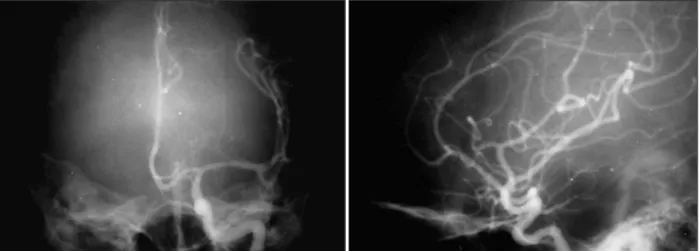
내원 당시의 혈압은 110/70mmHg이었고, 혈당치는 127 mg%이었으며 혈액 검사소견상 혈액 응고기능의 장애는 관 찰되지 않았다. 단순 두개골 X-선 촬영상 이상 소견은 보 이지 않았으며, CT에서 좌측 기저핵 부위와, 전두엽및 측두 엽에 직경 약 4cm의 둥근 모양의 고밀도 종괴가 발견되었 고, 종괴 자체에 의한 주변 뇌실의 압박 소견은 보였으나, 종괴 주위의 부종은 거의 없었다. 조영제 증강 영상에서 혈 관 질환이나 종양을 의심할만한 소견은 발견되지 않았다 (Fig. 1). 뇌혈관 조영술상에서도 종괴에 의한 혈관들의 전 위 소견외에는 특기할 만한 소견이 없었(Fig. 2). 이상과 같 이 CT에서 급성의 혈종에 합당한 소견이었으며 조영제 증 강 영상 및 뇌혈관 조영술에서 특이한 소견이 보이지 않아 고혈압성 뇌출혈 진단하에 수술을 시행하였다. 개두술후 대 뇌 피질을 통한 접근법시 검붉은 빛깔의 혈종의 일부가 대 뇌피질을 뚫고 터져나왔으며, 이 부위를 통해 혈종을 흡입 하였다. 혈종 주위의 혈관은 양극 전기 소작기로 지혈하였으 며, 전반적으로 혈관 벽이 연약하여 미세한 출혈이 많았으나
완전지혈은 가능하였고 정상 뇌실벽을 확인후 수술을 종료 하였다. 술후 10일째 환자는 별다른 신경학적 장애 없이 퇴 원하였다.
환자는 2개월후 다시 두통과 구토, 우측 상지의 부전마 비를 호소하였고, CT 추적검사에서 이전의 뇌출혈과 동일 한 부위에 주변부가 강한 조영 증강의 소견을 보이는 종괴 가 발견되었다(Fig. 3). 환자의 말초혈액 검사상 백혈구는 5950/mm3이었으며 적혈구 침강속도는(ESR) 1mm/hr, C 반응성 단백(CRP)은 0.01mg/dl로 정상소견이었으며, 뇌척 수액 배양검사도 정상이었다. 환자는 1차 수술시 혈종 외 에 특이 소견이 없었기 때문에 뇌농양 의심 하에 일단 보존 적 치료를 시행하였다. 내원 10일째 환자는 갑작스런 의식 저하의 소견을 보였으며, CT 추적 검사에서 종괴의 확장소 견 및 주변부의 부종이 심해진 양상을 나타내었다(Fig. 4).
응급개두술 및 배액술을 시행하였고, 조직검사상 핵의 이형 성증과 지형학적 괴사소견이 관찰되었으며, 다형성 교아세 포종으로 진단되었다(Fig. 5). 술 후 시행한 MRI에서 종양 크기의 심한 증가와 더불어 종양이 뇌실을 통해 소뇌로 전 이된 소견을 보였다(Fig. 6). 환자는 2개월후 사망하였다.
Fig. 1. Left:Pre-enhanced CT scan demonstrating a high density mass lesion without surrounding edema in left basal ganglia.
Right:Post-enhanced CT scan demonstrating no enhancement.
고 찰
뇌종양에 의한 자발성 출혈은 흔히 발견되는 소견은 아 니지만, 전이된 융모 상피선종,악성 흑색종, 폐암에서는 간
혹 볼 수 있는 소견이다. 그러나 원발성 뇌종양에서는 종양 의 출혈이 드물며 신경 교종에서는 그 빈도가 약 10%로 보고되고 있으나 다형성 교아세포종에서는 5%미만으로 드 문 경우로 알려져 있다4)9). 그 임상 양상은 혈종의 위치 및 혈종의 크기, 연령에 따라 다양하게 나타날 수 있으며2)10),
Fig. 2. Antero-posterior(Left) and Lateral(right) view of preoperative left carotid angiogram demonstrating displacement of vascular structure without abnormal staining.
Fig. 3. Follow up post-enhanced CT scan 2 months after initial operation demonstrating thick rim-enhancement with central low density area.
Fig. 4. Ten days after second admission, she was deteriorated to coma and post-enhanced CT scan demonstrated a huge mass with severe shifting of midline structure and surrounding edema.
정확한 신경학적 및 방사선학적 진단이 시행되지 않으면 뇌 졸중으로 오인되는 경우가 빈번하다고 보고되고 있다3).
다형성 교아세포종의 갑작스런 자발성 출혈의 기전에 대 해서는 아직까지 정확하게 밝혀져 있는 것은 없으나, 여러 가지 다양한 원인에 의한 것으로 추정하고 있다2)8). 일반적 으로 뇌종양에 의한 자발성 두개강내 출혈의 원인은 혈관 내 피의 증식으로 인해 혈관이 막히거나, 종양 자체의 증식으로 인한 혈관의 압박, 이로 인한 허혈성 괴사와 종양의 혈관 침 식으로 등으로 출혈을 일으킨다는 가설이 있고2)7)8), Kond- ziolka 등4)은 조직 병리학적인 특성을 1)혈관 벽의 초자화 (hyalinization)와 종양 괴사, 2) 혈관 벽의 변성 및 괴사, 3) 많은 내경이 작은 혈관의 혈전증 때문이라고 보고하였다.
그리고 Linwnicz 등6)은 출혈과 관계된 요인들을 소인 인자 (predisposing factor), 개시 인자 (initiating factor), 악화 인자 (aggravating factor)등으로 분류하였으며 이들 중 혈 관과 모세 혈관의 상태에 해당하는 소인 인자가 중요한 의미 를 가진다고 보고하였다. 신경 교종과 연관된 두개강내 출혈 의 소인 인자가 되는 모세혈관의 구조를 axial, retiform, glo- meruloid 등으로 분류하였으며, 이 중 retiform 형의 모세 혈관이 성상 세포종 및 다형성 교아 세포종의 두개강내 출혈 과 밀접한 관계가 있으며, axial과 glomeruloid 형의 모세 혈관은 종양의 주변부에서 모세 혈관의 망상 조직(network) 을 형성하며 이는 정상적인 뇌조직을 지지하여 출혈을 예방 하는 기능을 하는 것으로 밝혀 졌다6).
대체로 출혈은 천막 상부 종양에 많이 생기는 것으로 알 려져 있으나, Kondziolka 등4)은 소뇌 종양 환자에서도 뇌압 이 높은 경우 뇌압 조절과 출혈과의 관계를 고려해야 한다 고 강조하였다. 또한 뇌혈관 조영술상에서 보이는 종양의
혈관 분포의 정도는 두개강내 출혈과 통계학적인 연관성을 찾아볼 수 없으며, 종양의 위치에 따라서 지주막하 출혈, 뇌 실 내 출혈, 경막하 출혈 등 모든 경우가 발생할 수 있는 것 으로 보고되고 있다1)4)5). 종양이 출혈을 일으킬 때는 갑작 스런 두개강내 혈종의 형성으로 몇시간내에 두개강 내압의 상승으로 인한 신경학적 증상의 악화와 사망을 유발할 수 도 있지만, 무증상 및 느리게 진행하여 감별 진단이 어려울 수 있으며, 본 례와 같이 처음부터 뇌졸중과 같은 증상으로 내원하여, 여러 방사선학적 소견상 뇌종양을 의심할 수 없 었던, 단순한 두개강내 출혈로 증상이 처음 발현된 경우는 정확한 치료및 예후 판정에 혼동을 줄 수도 있다. 그러나 원인 불명의 두개강내 혈종의 경우, 수술시 출혈에 대한 지 혈이 어렵거나 혈종과는 다른 이상 조직의 발견시 수술중 조직 검사를 통한 확진이 필요하며, 방사선학적 검사에서 뇌농양이 의심되나 혈액 검사와 뇌척수액 검사에서 이상 소 견이 보이지 않는 경우는 뇌종양에 의한 출혈을 고려해야 할 것으로 사료된다.
결 론
본 교실에서는 1차 증상의 발현시 단순한 뇌실질내 출혈 로만 진단되었다가, 2차 증상 발현시 뇌농양으로 오인되었 던 다형성 교아세포종 1례를 경험하였기에 문헌 고찰과 함 께 보고하고자 한다.
• 논문접수일:2000년 7월 21일
•심사완료일:2001년 2월 12일
• 책임저자:김 재 민
471-701 경기도 구리시 교문동 249-1 한양대학교 의과대학 구리병원 신경외과학교실 전화:031) 560-2323, 전송:031)560-2327 E-mail:[email protected]
Fig. 5. Microphotograph of specimen demonstrating nuclear angulation, pleomorphism, hyperchromatism, and mi- toses, vascular proliferation with endothelial hyperplasia.
H & E, original magnification ×400.
Fig. 6. Gadolinium enhanced MRI scan demonstrating thick rim-enhancement with central low density area on fourth vetricle wall.
References
1) Andrews BT, Raffel C, Rosegay H:Subarachnoid hemorr- hage from a peripheral intracranial aneurysm associated with malignant glioma. Neurosurgery 17:645-649, 1985 2) Hirano A, Matsui T:Vascular structures in brain tumors.
Human Pathology 6(5):611-621, 1975
3) Kalyanaraman K, Smith BH, Alker GJ:Intracranial tumors of apopletiform onset. NY state J Med 73:2133-2139, 1973 4) 4. Kondziolka D, Bernstein M, Resch L, Tator CH, Fleming
JF, Vanderlinden RG, Schutz H:Significance of hemorrhage into brain tumors:Clinicopathological study. J Neurosurg 67:852-857, 1987
5) Lee SH, Wang KC, Kim JS, Lee SH, Kim HJ, Cho BK, et al:
Spontaneous intracranial hemorrhage caused by brain tumor.
J Korean Neurosurg 21(9):1053-1063 , 1988
6) Liwnicz BH, Wu SZ, Tew JM:The relationship between the capillary structure and hemorrhage in gliomas. J Neurosurg 66:536-541 , 1987
7) Richardson RR, Siqueira EB, Cerullo LJ:Malignant glioma: its initial presentation as intracranial hemorrhage. Acta Neu- rochir 46:77-84, 1979
8) Scatliff JH, Radcliffe WB, Pittman HH, Park CH:Vascular structure of glioblastomas. Am J Roentgenol Radium Ther Nucl Med 105:795-805, 1969
9) Wakai S, Yanakawa K, Manaka S, Takakura K:Spontaneous intracranial hemorrhage caused by brain tumor:its incidence and clinical significance. Neurosurgery 10:437-444, 1982 10) You H, Kim DG, Lee HK, Han DH:A case of malignant
meningioma Presented with intracerebral hemorrhage. J Ko- rean Neurosurg 21(6):706-710, 1992