VOL.23 NO.3 : 038-055 (2010)
四物湯加味方의 항암효과에 대한 실험적 연구
1
동국대학교 대학원 한의학과 한방부인과교실,
2동국대학교 바이오시스템대학 생명과학과 정재중
1, 구선영
2, 임새롬
2, 성정석
2, 김동일
1ABSTRACT
Anti-cancer Effects of Samultang-Gami on HeLa, HepG2, AGS Cells Jae-Joong Jung
1, Sun-Young Goo
2, Sae-Rom Lim
2, Jung-Suk Sung
2, Dong-Il Kim
11
Dept. of OB & GY, Graduate school of Oriental Medicine, Dong-Guk Univ.
2
Dept. of Life Science, Dong-Guk Univ.
Purpose: This study was designed to find out the anti-cancer effects of Samultang-Gami which was composed of Rehmanniae Radix(RR), Angelicae Gigantis Radix(AGR), Cnidii Rhizoma(CR), Paeoniae Radix(PR), Cortex Moutan Radicis(CMR), Hedyotis Diffusa(HD) and Caesalpinia Sappan on HeLa, HepG2 and AGS cells.
Methods: Various cancer cell lines including HeLa, HepG2 and AGS cells, were used. In vitro anti-cancer effects were measured by MTT assay using cancer cell lines treated with various concentrations of 70% ethanol extract of Samultang-Gami . Expression of cell cycle arrest mediators including Bax, Bcl-2, p53 and DARP-1 proteins were measured by Western blot analysis.
Results:
1. Samultang-Gami decreased the viability of HeLa and HepG cells in a dose- dependent manner.
2. AGR, CMR, PR and HD decreased the viability of HeLa, HepG2 and AGS cells.
3. We could observe that the decreased Bax and Bcl-2 expression level and the increased PARP-1 expression level by Samultang-Gami extracts treated in HeLa cells.
4. We could observe that the decreased Bcl-2 expression level and the increased Bax, p53 and PARP-1 expression level by RR extracts treated in HeLa cells.
and also could observe that the reduction of the protein level of Bcl-2, p53 and PARP-1 and the increase of the protein level of Bax by PR in HeLa cells.
5. We could observe that the increased p53 expression level, the decreased PARP-1's that and the unchanged Bax and Bcl-2's that by Samultang-Gami extracts treated in HepG2 cells.
6. We could observe that the reduced Bcl-2 expression level by each of RR extracts and PR extracts in HepG2 cells.
7. The treatment of Samultang-Gami in AGS cells didn't have any effect on the expression level of Bax, Bcl-2, p53 and PARP-1 .
8. We could observe that the increased p53 and PARP-1 expression level by each of CR, RR and PR extracts in AGS cells.
Conclusion: Taken together, we suggest that Samultang-Gami exhibits cytotoxic effects on HeLa, HepG2 and AGS cells, causing apoptosis. The results showed that Samultang-Gami may do so by regulating the expression of specific target molecules that promote efficient apoptotic cell death in a dose-dependent manner.
Key Words: Samultang-Gami , HeLa, HepG2, AGS, Apoptosis, Bax, Bcl-2, p53, PARP-1, anti-cancer.
4 )
교신저자(김동일) : 경기도 고양시 일산동구 식사동 814 동국대학교 일산한방병원 한방여성의학과 전화 : 031-961-9060 이메일 : [email protected]
Ⅰ. 서 론
2008년 통계청 통계결과에 따르면 암 은 우리나라 사망원인 중 28%로 1위를 차지하고 있다. 특히 60대는 암이 전체 사망자의 42.1%를 차지하고, 매년 그 비 중이 증가추세에 있다
1). 미국에서는 심 장병에 이어 암이 두 번째로 흔한 사망 원인이다
2). 그러나 정부와 유관기관에서 의 연구와 과학기술의 발전, 암 조기 진 단의 발전과 암 예방 사업으로 우리나라 의 경우, 암 진단 후 5년 생존율이 1993~
1995년의 41.2%에서 2003~2007년의 57.1%
로 현저히 증가하였다
1).
이렇듯 지난 수십 년간 암을 정복하려 는 노력으로 다양한 화학요법, 수술요법, 방사선 요법 등의 암 치료법이 개발되었 지만, 인구의 노령화와 산업화로 인한 환경파괴 및 오염물질의 증가및 잘못된 식이습관으로 인해 암의 발생억제와 근 원적 치료에 대해서는 임상적 한계에 부 딪히고 있는 실정이다
3). 또한 현재 널리 사용되고 있는 항암제는 대부분 합성 물 질들로 부작용이 심각한 문제로 대두되 고 있으며, 이로 인해 최근에는 부작용 이 적으면서 유효한 천연 항암제의 개발 을 위하여 천연물을 대상으로 예방 및 치료물질의 검색과 개발에 대한 노력이 활발히 전개되고 있다
4,5).
이에 저자는 부작용이 적고 안정적인 암 치료제 개발을 위한 연구의 일환으로 調益營衛, 滋養血의 대표적인 처방인 四 物湯을 기본으로 하되, 方中의 熟地黃을 生地黃으로 대체하고, 牧丹皮, 白花蛇舌 草, 蘇木을 가미한 四物湯加味方을 구성 하여, 생쥐 배아섬유아세포(MEF cells)
에 四物湯加味方 추출액을 처리하여 세 포의 생존율을 조사하고, 인간 자궁경부 암 세포주(HeLa cells), 인간 간암 세포 주(HepG2 cells), 인간 위암 세포주(AGS cells)에 이 처방의 추출액을 처리하여 각 세포의 생존율, Bax, Bcl-2, p53, PARP-1 발현량을 조사하였다. 이러한 일련의 연 구를 통해 유의한 결과를 얻었기에 이 논문을 통하여 발표하고자 한다.
Ⅱ. 연구 방법
1. 재료 및 추출물의 제조
四物湯加味方 구성약물은 生地黃(SJH) 當歸(DK) 白芍藥(BJY) 川芎(CK) 蘇木 (SM) 牧丹皮(MDP) 白花蛇舌草(BSC) 로써 각각 옴니허브(서울, 한국)를 통하 여 구입하여 정선하여 사용하였고 한 첩 의 용량은 Table 1과 같다.
Herbal
Name Scientific Name Amount (g) 生地黃 Rehmanniae
Radix 8
當 歸 Angelicae Gigantis
Radix 4
川 芎 Cnidii Rhizoma 4 白芍藥 Paeoniae Radix 4 牧丹皮 Cortex Moutan
Radicis 3
白花蛇舌草 Hedyotis Diffusa 8 蘇 木 Caesalpinia
Sappan 3
Total amount 34 Table 1. Prescription of Samultang- Gami
fluorescent mounting medium
(DakoCytomation, CA)를 제외한, 4'-6-
diamidino-2-phenylindole(DAPI),[3-(4,5 -dimethylthiazol-2-yl)-2,5-diphenyltetra zolium bromide](MTT), dimethylsulfoxide (DMSO) 및 모든 화학약품은 Sigma(USA) 에서 구입하였다.
에탄올 추출물(ethanol extract)은 수 직으로 환류냉각관을 부착시킨 둥근 플 라스크에 시료의 10배에 해당하는 70%
ethanol을 넣고 70℃의 수욕상에서 3시 간 동안 추출한 후 추출액을 filter paper (Whatman No. 5)로 여과하였다. 남은 잔사를 위와 같은 방법으로 2회 더 추출 하여 모은 각 추출액을 감압농축하여, 동결건조기(freeze dryer, Ilshin Lab Co., LTD, Korea)로 동결건조하여 시료로 사용하였다. 四物湯加味方 추출물 ×1는 100㎎/㎖ 농도로 추출하였으며, 각 실험 에 필요에 따라 희석하여 사용하였다.
2. 실험방법
1) 세포와 세포배양
실험에 사용된 세포주는 MEF cells, HeLa cells, HepG2 cells, AGS cells로 아 메리칸 타입 컬쳐 콜렉션(American Type Culture Collection, USA)에서 분양받았 다. MEF 세포는 10% FBS와 1× penicillin /streptomycin이 함유된 Dulbecco’s modified Eagle’s medium(DMEM)(PAA, Austria) 에서 배양하였다. AGS cells는 10% FBS, 1× penicillin/streptomycin, 1mM Sodium pyruvate, 4.5g/L glucose가 포함된 RPMI1640(Welgene, Korea)배지에서 배 양하였고, HepG2, HeLa cell은 10% FBS, 1× penicillin/streptomycin, 1mM Sodium pyruvate가 포함 된 MEM(Welgene, Korea) 배지에서 배양하였다. 배양 시 온도는 37℃를 유지하고 5% CO
2와 95% 공기를
공급하였다.
2) 세포 활성도 측정
세포활성도(cell viability)는 MTT 분 석법으로 측정하였다. MEF cells는 4.0 × 10
4cells/mL로, HeLa, HepG2, AGS cells 는 각각 5.0 × 10
4cells/mL로 96-well 배 양접시에 분주하고 24시간 동안 배양하 였다.
MEF cells에 24시간 동안 PBS를 처리 한 Control군을 설정하고, 四物湯加味方 개별약물(0.1, 0.5, 1. 5 ㎎/㎖)을 처리한 것과 四物湯加味方(×1000, ×500, ×100,
×50, ×10)을 처리한 것을 각각 실험군으 로 설정하여 개별 실험을 진행하였다.
AGS, HepG2, HeLa cells는 24시간 동 안 PBS를 처리한 대조군, 四物湯加味方 약재 처리한 실험군으로 나누어 四物湯 加味方의 암세포주 손상여부와 정도를 알아보았다.
세포를 PBS(Welgene, Korea)로 2회 세척하고 MTT 용액(5 ㎎/㎖) 10 ㎕첨가 하여 37℃ 배양기에서 3시간 동안 두었다.
배지를 제거하고 DMSO를 200 ㎕첨가했 다. 10분 간 배양기에 둔 후, microplate reader(Molecular Devices)를 이용하여 595 nm에서 흡광도를 측정하였다.
3) Western blot analysis
48시간 동안 四物湯加味方 처리를 한
HeLa cell을 배양하고 PBS로 세척한 다
음, mammalian cell lysis buffer인 RIPA
buffer(50 mM Tris (pH 7.5), 150 mM
NaCl, 0.02% sodium azide, 0.1% sodium
dodecyl sulfate(SDS), 1% Nonidet P-40,
protease inhibitors(1 ug/mL leupeptin, 1
ug/mL aprotinin, and 1 mM PMSF)로
resuspension하고 ice상에서 30분 동안
incubation 한 다음 centrifuge(16200rpm,
4℃, 20분)을 하여 세포파쇄액을 만들었 다. 세포파쇄액의 단백질을 Bradford protein assay를 통해 정량하고, 단백질 을 lane 당 60 ug으로 표준화하고, 10%
SDS-PAGE gel에서 분리한 다음, 이동완 충용액(transfer buffer; 39 mM glycine, 48 mM Tris base, 0.037% SDS, and 20%
methanol)를 사용하여 PVDF membrane 에 옮겼다. Transfer된 membrane을 1%
skim milk로 blocking시킨 다음, Protein detection system인 SNAP i.d를 이용하 여 Bax(primary antibody)(Santa Cruz Biotechnology, Inc, CA, USA) 1:1500으 로 10분씩 2번 반응시키고, 1 × TBST(20 mM Tris-HCl(pH 7.6), 137 mM NaCl, 0.1% Tween-20)로 3회 세척한 다음, mouse anti-rabbit IgG-HRP(secondary antibody 1:1500)(Santa Cruz Biotechnology, Inc, CA, USA)도 10분씩 2회 반응시키고 난 후 1 × TBST (20 mM Tris-HCl(pH 7.6), 137 mM NaCl, 0.1% Tween-20)로 3회 세척 한 다음 enhanced chemilumine-scence (ECL)로 발색시키고 X-ray film으로 감 광하여 시각화하였다. 그 다음, 단백질 band의 농도를 Image Quant software (Amersham Biosciences, UK)를 사용하 여 각각의 四物湯加味方, 川芎, 生地黃, 白芍藥에서의 Bax, Bcl-2, p53, PARP-1 을 정량하였다.
4) Statistical analysis
실험 결과는 Mean±SD값으로 나타내 었으며, 본 실험에서 얻은 결과를 one-way ANOVA Tukey's test(GraphPad Prism) 로 분석하여 p값을 구하였다. 대조군을 각 실험군에 비교하여 p<0.05일 때 유의 성이 있는 것으로 판정하였다.
Ⅲ. 결 과
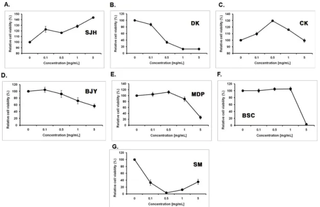
1) 四物湯加味方 개별 약재 처리 농도 에 따른 MEF cells의 생존율 四物湯加味方의 안전성을 확인하기 위 해 먼저 四物湯加味方의 개별 약물 안전 성을 알아보기 위하여 MEF cells에 각 약재의 70% ethanol 추출물을 처리하여, 세포의 활성도를 알아 볼 수 있는 MTT assay를 시행하였다.
生地黃은 농도가 증가함에 따라 농도 의존적으로 cell viability가 증가하는 것 을 보였고 실험군의 모든 농도에서 대조 군에 비해 높은 cell viability를 보였으 며, 川芎 또한 0.1, 0.5, 1㎎/㎖ 농도의 실험 군에서 대조군에 비해 높은 cell viability 를 보였고, 5㎎/㎖ 농도의 실험군에서도 대조군에 비해 cell viability 감소폭이 크 지 않았다. 白芍藥 牧丹皮 白花蛇舌草도 0.1, 0.5, 1㎎/㎖ 농도에서 cell viability가 대조군에 비해 증가하거나 소폭 감소하 였고, 5㎎/㎖ 농도에서는 대조군에 비해 50%이하로 감소하였다. 當歸의 경우 0.5
㎎/㎖ 농도에서부터 cell viability가 급격 히 감소하였고, 蘇木은 결과 확인이 어 려웠다(Fig. 1).
2) 四物湯加味方 처리 농도에 따른 MEF cells의 생존율
四物湯加味方의 안전성을 확인하기 위 해 MEF cells에 四物湯加味方 70% ethanol 추출물 ×1 100㎎/㎖를 기준으로 ×1000,
×500, ×100, ×50, ×10농도로 희석 처리하 여, MTT assay를 시행하였다.
각각의 농도로 24시간 유지 후 확인한
결과 ×1000, ×500 농도에서 안전성이 확
인되었다(Fig. 2).
Fig. 1. The Effects of 7 Medicinal Herbs Extracts on Viability of MEF Cells Determined by MTT Assay.
MEF wild-type cells were treated with various concentrations(0.1, 0.5, 1, 5 ㎎/㎖) of 70%
ethanol extracts of 7 medicinal herbs for 24 hr. Results are expressed as the mean + S.D. of three independent experiments.
Fig. 2. The Dose-dependent Cytotoxicity Effect of Samultang-Gami against MEF Cells Determined by MTT Assay.
MEF cells (4×10⁴cells per well) were treated for 24 hr with various concentrations of
Samultang-Gami
extracts [×1000, ×500, ×100,×50, ×10.(×1=100㎎/㎖)]. Results are expressed as the mean + S.D. of three independent experiments.
2. 四物湯加味方의 암세포 억제효능 1) HeLa, HepG2, AGS cells의 四物湯
加味方 처리 농도에 따른 생존율 四物湯加味方의 항암효과를 알아보기 위하여 암세포주 HeLa, HepG2, AGS cells에 四物湯加味方을 처리하여 세포의 생존율을 알아볼 수 있는 MTT assay를 시행하였다. 四物湯加味方을 ×1000, ×500,
×100, ×50, ×10 농도로 처리한 후 24시간,
48시간, 72시간 후 관찰하였다. HeLa,
HepG2 세포에서는 ×100, ×50, ×10 농도
에서 세포의 생존율이 감소하는 것을 뚜
렷하게 관찰 할 수 있었고, AGS 세포에
서는 ×100, ×50농도에서 생존율이 급격히
감소하는 것을 관찰할 수 있었다(Fig. 3).
Fig. 3. The Effects of Samultang-Gami on The Viability of HeLa, HepG2, AGS Cells Determined by MTT Assay.
Cells were treated with various concentrations (x1000, x500, x100, x50, x10) of
Samultang -Gami
extracts for 24 h, 48 h, 72 h. Results are expressed as the mean + S.D. of three independent experiments.(A) HeLa cells. (B) HepG2 cells. (C) AGS cells.
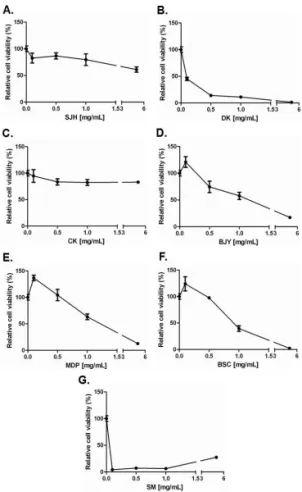
2) HeLa cells의 四物湯加味方 개별 약재 처리 후 생존율
HeLa cells에 대한 四物湯加味方 구성 약재의 활성 억제 효과를 알아보기 위해 MTT assay를 시행한 결과 當歸, 白芍 藥, 牧丹皮, 白花蛇舌草에서 농도가 증가 함에 따라 세포활성도가 감소되는 것을 확인할 수 있었다(Fig. 4).
Fig. 4. The Effects of 7 Medicinal Herbs Extracts on The Viability of HeLa Cells Determined by MTT Assay.
HeLa cells were treated with various concentrations(0.1, 0.5, 1, 5 ㎎/㎖) of 7 Medicinal Herbs Extracts for 24 h, 48 h, 72 h.
This Figures show only 48h. Results are expressed as the mean + S.D. of three independent experiments.
(A) SJH. (B) DK. (C) CK. (D) BJY. (E) MDP. (F) BSC. (G) SM.
3) HepG2 cells의 四物湯加味方 개별 약재 처리 후 생존율
HepG2 cells에 대한 四物湯加味方 구
성 약재의 활성 억제 효과를 알아보기
위해 MTT assay를 시행한 결과 白芍藥,
白花蛇舌草가 농도 의존적으로 활성을
억제하는 것이 관찰되었으나 當歸, 牧丹
皮는 5㎎/㎖의 농도에서만 유의한 생존
율 감소를 보였다(Fig. 5).
Fig. 5. The Effects of 7 Medicinal Herbs Extracts on The Viability of HepG2 Cells Determined by MTT Assay.
HepG2 cells were treated with various concentrations(0.1, 0.5, 1, 5 ㎎/㎖) of 7 Medicinal Herbs Extracts for 24 h, 48 h, 72 h.
This Figures show only 48h. Results are expressed as the mean + S.D. of three independent experiments.
(A) SJH. (B) DK. (C) CK. (D) BJY. (E) MDP. (F) BSC. (G) SM.
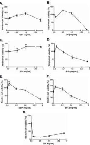
4) AGS cells의 四物湯加味方 개별 약 재 처리 후 생존율
AGS cells에 대한 四物湯加味方 구성 약재의 활성 억제 효과를 알아보기 위해 MTT assay를 시행한 결과 當歸, 牧丹 皮, 白花蛇舌草에서 세포활성도가 감소 되는 것을 확인할 수 있었다(Fig. 6).
Fig. 6. The Effects of 7 Medicinal Herbs Extracts on The Viability of AGS Cells Determined by MTT Assay.
AGS cells were treated with various concentrations(0.1, 0.5, 1, 5 ㎎/㎖) of 7 Medicinal Herbs Extracts for 24 h, 48 h, 72 h.
This Figures show only 48h. Results are expressed as the mean + S.D. of three independent experiments.
(A) SJH. (B) DK. (C) CK. (D) BJY. (E) MDP. (F) BSC. (G) SM
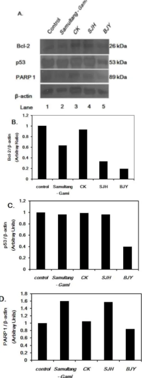
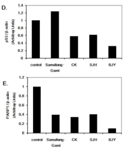
5) 四物湯加味方과 川芎, 生地黃, 白芍 藥이 HeLa cells의 Bax, Bcl-2, p53, PARP-1 발현에 미치는 영향 四物湯加味方을 처리한 실험군이 대조 군에 비해 Bax protein 발현량이 감소되 고, 川芎, 生地黃, 白芍藥에서는 대조군 에 비해 증가하였다(Fig. 7).
anti-apoptosis 작용을 하는 Bcl-2의
발현량을 보면 四物湯加味方 처리한 실
험군에서 대조군에 비해 감소된 수치를
보이고, 특히 개별약재에서는 生地黃, 白 芍藥의 발현량이 매우 감소하였다.
p53에서는 四物湯加味方 처리한 실험 군은 대조군과 비슷한 수치를 보였고, 白芍藥에서만 특이적으로 감소된 수치를 보였다.
PARP-1는 四物湯加味方과 生地黃에 서 대조군에 비해 증가하였다(Fig. 8).
Fig. 7. The Effect of Samultang-Gami extracts on Bax in HeLa cells.
(A) Cells were harvested at 48 h after treatment with various conditions. Cells were then lysed, and the supernatants were subjected to western blot analysis. Aliquots of 60 ug protein extracts were analyzed by 10% SDS-PAGE, transferred to a PVDF membrane and immunoblotted with Bax antibody. Where : Lane 1, untreated cells;
Lane 2, X100
Samultang-Gami
; Lane 3, 1㎎/㎖CK; Lane 4, 1㎎/㎖ SJH; Lane 5, 1㎎/㎖ BJY.
(B) Graphical representation of the data shown in (A)
Fig. 8. The Effects of Samultang-Gami extracts on Bcl-2, p53 and PARP-1 in HeLa cells
(A) Cells were harvested at 48 h after treatment with various conditions. Cells were then lysed, and the supernatants were subjected to western blot analysis. Aliquots of 60 ug protein extracts were analyzed by 10% SDS-PAGE, transferred to a PVDF membrane and immunoblotted with Bax antibody. Where : Lane 1, untreated cells;
Lane 2, X100
Samultang-Gami
; Lane 3, 1㎎/㎖CK; Lane 4, 1㎎/㎖ SJH; Lane 5, 1㎎/㎖ BJY.
The effect of
Gami-Samultang
extracts on Bcl-2(anti-apoptosis protein) (B), p53 (tumor supressor protein) (C) and PARP1(Apoptosis -regulating protein) (D) in HeLa cells.(B), (C), (D) is graphical representation of the data shown in (A).
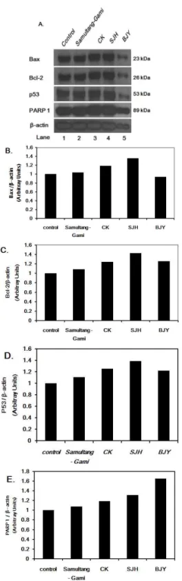
6) 四物湯加味方과 川芎, 生地黃, 白芍 藥이 HepG2 cells의 Bax, Bcl-2, p53, PARP-1 발현에 미치는 영향 Bax 발현량은 대조군에 비해 白芍藥 은 감소되었으나, 그 외 실험군은 큰 차 이를 보이지는 않았다.
Bcl-2 발현량은 대조군에 비해 실험군 모두 감소되었으며, 특히 生地黃, 白芍藥 은 매우 낮은 수치를 보였다.
p53 발현량은 대조군에 비해 四物湯加 味方 처리군에서 증가되었고, 개별 약물 은 대조군에 비해 감소되었다.
PARP-1 발현량은 대조군에 비해 四 物湯加味方, 川芎, 生地黃, 白芍藥 처리 군 모두 감소되었다(Fig. 9).
Fig. 9. The Effect of Samultang-Gami extracts on Bax, Bcl-2, p53 and PARP-1 in HepG2 cells.
(A) Cells were harvested at 48 h after treatment with various conditions. Cells were then lysed, and the supernatants were subjected to western blot analysis. Aliquots of 60 ug protein extracts were analyzed by 10% SDS-PAGE, transferred to a PVDF membrane and immunoblotted with Bax antibody. Where : Lane 1, untreated cells;
Lane 2, X100
Samultang-Gami
; Lane 3, 1㎎/㎖CK; Lane 4, 1㎎/㎖ SJH; Lane 5, 1㎎/㎖ BJY.
The effect of
Samultang-Gami
extracts on Bax(pro-apoptosis) (B), Bcl-2 (anti-apoptosis protein) (C), p53 (tumor supressor protein) (D), PARP 1(Apoptosis-regulating protein) (E) in HepG2 cells.(B), (C), (D), (E) is graphical representation of the data shown in (A).
7) 四物湯加味方과 川芎, 生地黃, 白芍 藥이 AGS cells의 Bax, Bcl-2, p53, PARP-1 발현에 미치는 영향 AGS cells에 대한 실험에서 四物湯加 味方을 처리한 실험군은 모두 대조군과 유의한 차이를 보이지 않았다.
川芎, 生地黃, 白芍藥에 대한 개별 실
험에서는 川芎, 生地黃은 대조군에 비해
Bax와 Bcl-2가 유사하게 증가된 양상을
보였고, 白芍藥은 Bax 발현량이 다소 감
소되었다.
p53, PARP-1에는 3종 약재 모두 대조 군에 비해 증가되었다(Fig. 10).
Fig. 10. The Effect of Samultang-Gami extracts on Bax, Bcl-2, p53 and PARP-1
in AGS cells.
(A) Cells were harvested at 48 h after treatment with various conditions. Cells were then lysed, and the supernatants were subjected to western blot analysis. Aliquots of 60 ug protein extracts were analyzed by 10% SDS-PAGE, transferred to a PVDF membrane and immunoblotted with Bax antibody. Where : Lane 1, untreated cells;
Lane 2, X100
Samultang-Gami
; Lane 3, 1㎎/㎖CK; Lane 4, 1㎎/㎖ SJH; Lane 5, 1㎎/㎖ BJY.
The effect of
Samultang-Gami
extracts on Bax(pro-apoptosis) (B), Bcl-2 (anti-apoptosis protein) (C), p53 (tumor supressor protein) (D), PARP 1(Apoptosis-regulating protein) (E) in AGS cells.(B), (C), (D), (E) is graphical representation of the data shown in (A).