치주염 유발 세균 Aggregatibacter actinomycetemcomitans와 Porphyromonas gingivalis 에 의한 committed osteoclast precursor 분화 증가
박옥진1, 권영각1, 윤철희2, 한승현1*
1서울대학교치의학대학원구강미생물
·
면역학교실2서울대학교농업생명과학대학농생명공학부동물생명공학전공
Received : August 31, 2016 / Revised : September 20, 2016 / Accepted : September 21, 2016
서 론
치주질환은만성염증성질환으로심할경우치조골을파 괴시켜성인치아상실의주된원인이 된다
[11].
유전,
환경 적요인등다양한 원인에의해치주질환이진행 되지만,
치태내병원성세균은치주질환을유발하는핵심요인으로 잘알려져있다[4, 20].
치은열구내에존재하는1,000
여종의 세균 중
Porphyromonas gingivalis, Aggregatibacter actinomycetemcomitans
는대부분의치주질환환자에게서동 정되어강력한치주원인균으로분류되어있다[14, 15].
그람 음성세균인P. gingivalis
와A. actinomycetemcomitans
는 각각만성치주염(Chronic periodontitis)
과국소공격성치주 염(Localized aggressive periodontitis)
발병과밀접하게연관되어있다
[3, 14].
그람음성세균의핵심병독력인자인지질다당체
(Lipopolysaccharide; LPS)
는다양한염증-
매개물 질을유도하여단핵구/
대식세포,
림프구,
다핵구와같은면 역세포를질환부위로몰려오게한다[7, 12].
이들면역세포 들은강한염증반응을일으키고골형성억제및골파괴를야Augmented Osteoclastogenesis from Committed Osteoclast Precursors by Periodontopathic Bacteria Aggregatibacter actinomycetemcomitans and Porphyromonas gingivalis
Ok-Jin Park
1, Yeongkag Kwon
1, Cheol-Heui Yun
2, and Seung Hyun Han
1*
1
Department of Oral Microbiology and Immunology, DRI and BK21 Plus Program, School of Dentistry,
2Department of Agricultural Biotechnology and Research Institute for Agriculture and Life Sciences, Seoul National University, Seoul 08826, Republic of Korea Aggregatibacter actinomycetemcomitans and Porphyromonas gingivalis are gram-negative bacteria frequently found in lesions from patients with periodontitis manifesting alveolar bone loss. Lipopolysac- charides are a major virulence factor of gram-negative bacteria. Bone resorption is known to be regulated by bacteria and their virulence factors. In the present study, we investigated the effects of A. actinomyce- temcomitans and P. gingivalis on bone resorption. Heat-killed A. actinomycetemcomitans (HKAa) and heat- killed P. gingivalis (HKPg) induced bone loss in the femurs of mice after intraperitoneal administration.
HKAa and HKPg augmented the differentiation of committed osteoclast precursors into osteoclasts, while they inhibited the differentiation of bone marrow-derived macrophages into osteoclasts. Concordant with the effects of the heat-killed whole cells, LPS purified from A. actinomycetemcomitans and P. gingivalis also augmented osteoclast differentiation from committed osteoclast precursors but attenuated it from bone marrow-derived macrophages. Taken together, these results suggest that the whole cells and lipopolysac- charides of A. actinomycetemcomitans and P. gingivalis induce the differentiation of committed osteoclast precursors into osteoclasts, potentially contributing to bone resorption in vivo.
Keywords: Aggregatibacter actinomycetemcomitans, Porphyromonas gingivalis, lipopolysaccharide, osteoclast, bone loss
*Corresponding author
Tel : +82-2-880-2310, Fax: +82-2-743-0311 E-mail: [email protected]
© 2016, The Korean Society for Microbiology and Biotechnology
기하게된다
[17].
즉,
치아와잇몸사이의치은열구내병원성 세균의감염은염증반응을유발하고또한과도한골흡수를 야기해치조골소실을유도한다.
골항상성
(Bone homeostasis)
은골흡수를담당하는파골 세포(Osteoclast)
와골형성을담당하는조골세포(Osteoblast)
의활성과분화의적절한균형에의해유지된다
[16].
조혈모세포에서 기원한 파골세포는 핵심 사이토카인인
M-CSF (Macrophage-colony stimulating factor)
와RANKL (Receptor activator of nuclear factor kappa B ligand)
에의해분화된 다핵거대세포(Multinucleated giant cell)
로약해진뼈를파 괴하는역할을수행함으로써골항상성을유지하게한다[1, 8].
하지만,
파골세포에의한골흡수기능이조골세포기능 보다과도하거나,
파골세포의골흡수기능보다조골세포의 기능이과도하여균형이깨지게되면다양한골관련질환 이발생하게된다[2].
세균감염은골수염,
화농성관절염,
치 주질환과같은과도한골파괴를수반하는염증성골질환을 일으킨다[20, 22].
세균은지질단백질, LPS,
리포테이코익산(Lipoteichoic acid)
등과 같은 세균 병독력인자(Pathogen- associated molecular pattern; PAMP)
를가지고있고이들 은숙주의패턴인식수용체들(Pattern-recognition receptor;
PRR)
에의해인지된다[9].
파골세포와조골세포역시이러한패턴인식수용체들을발현하고있어세균과세균병독력인 자는골항상성을조절하게된다
[18, 21].
본연구는 치주질환을 일으키는 대표적 구강세균인
P.
gingivalis
와A. actinomycetemcomitans
를열사멸하여,
쥐의 복강에투여한후대퇴부의해면골을미세단층촬영기(micro- CT)
로촬영/
분석하고이들이단독으로골소실을유발하는지 확인하였다.
또한,
세균에의한골소실과파골세포분화와의 상관관계를 규명하기위하여열사멸한P. gingivalis
와A.
actinomycetemcomitans,
그리고,
이세균들로부터분리한LPS
에의한파골세포분화영향을평가하였다.
재료 및 방법
재료
Brain heart infusion (BHI)
배지는BD Biosciences (USA)
에서 구입하였다. Vitamin K
와hemine
은Sigma- Aldrich Chemical Inc. (USA)
에서 구매하였다. Fetal bovine serum (FBS)
와 α-MEM
은Gibco (USA)
에서, Trypsin-EDTA
와penicillin/streptomycin
은Hyclone (USA)
에서 구매하였다. Recombinant mouse M-CSF
단백질은R&D Systems (USA)
에서, recombinant murine RANKL
은PeproTech (USA)
에서구매하였다.
명시하지않은다른모든 재료들은Sigma-Aldrich Chemical Inc.
에서구매하였다.
미세단층촬영기
모든동물실험은서울대학교동물실험윤리위원회에서승 인을받은후진행하였다
.
생체내에서세균이미치는영향 을확인하기위하여6
주령C57BL/6
수컷쥐의복강에열사 멸된세균또는PBS
를4
일간격으로두차례투여하였고,
실 험시작7
일째 쥐를 희생시켜 오른쪽 대퇴골을 얻어4%
paraformaldehyde
로고정하였다.
고정된대퇴골을미세단 층촬영기(Skyscan 1172 scanner, Skyscan, Belgium)
로촬영하 였다.
촬영된그림을기반으로대퇴골의3
차원영상을얻고,
분석프로그램(SkyScan CT analyzer)
으로뼈의용적비,
뼈 잔기둥의두께,
뼈잔기둥의수,
뼈잔기둥간간격을비교분 석하였다.
열사멸 세균 준비
A. actinomycetemcomitans ATCC43718
과P. gingivalis ATCC49417
은American Type Culture Collection (USA)
에서 구입하였다. P. gingivalis
는BHI
배지에10
µg/ml
의vitamin K
와5
µg/ml
의hemine
을첨가하여37
℃혐기챔버(Whitley DG250 Workstation; Don Whitley Scientific, UK)
배양하 였고A. actinomycetemcomitans
는BHI
배지에서37
℃배 양기에배양하였다.
각세균이mid-log
기에도달하였을때1 × 10
10CFU/ml
로조정하여60
℃에서1
시간동안열처리하였다.
열사멸된A. actinomycetemcomitans (HKAa)
과P. gingivalis (HKPg)
의사멸여부는1.5% agar
가첨가된BHI
배지에사 균을도말하고37
℃에서1
일동안배양하였고세균집락이 관찰되지않는것으로확인하였다(data not shown).
LPS 추출
배양된
P. gingivalis
와A. actinomycetemcomitans
은11,068 × g
로37
℃조건에서원심분리하여배지를모두제거해주고
, PBS
로 세 번세척하였다.
세척된 세균은LPS
extraction kit (iNtRON Biotechnology, Korea)
를사용하여 제조사설명서에따라추출을진행하였다.
세포배양
6
주령의C57BL/6
수컷쥐를오리엔트바이오(Seongnam,
Korea)
에서구매하였고,
쥐의장골에서골수세포를추출하였다
.
추출한골수세포는10% FBS, 100 U/ml penicillin
와100
µg/ml streptomycin
이포함된α-MEM
배지에2 ng/ml M-CSF
를첨가하여37
℃의CO
2배양기에서1
일동안배 양하였고,
기질세포를 제외한 부유세포는20 ng/ml M- CSF
를첨가하여서분화배지를 만든후3
일동안배양하 여 파골전구세포(Bone marrow-derived macrophage;
BMM)
로사용하였다.
그리고BMM
에20 ng/ml M-CSF
와20 ng/ml RANKL
을2
일동안처리해파골세포로운명이 결정된파골전구세포인committed osteoclast precursor
로 사용하였다.
파골세포 분화 및 tartrate-resistant acid phosphatase
(TRAP) 염색
2 × 10
5cells/ml
의BMM
을96-well plate
에well
당200
µl
씩seeding
하고, 20 ng/ml M-CSF
와20 ng/ml RANKL
을첨 가한배지에열사멸된세균또는LPS
를농도별로처리하 여2
일동안배양하였다.
그리고2 × 10⁵ cells/ml
의committed osteoclast precursor
을96-well plate
에well
당200
µl
씩seeding
하고, 20 ng/ml M-CSF
을첨가한배지에열사멸된 세균또는LPS
를농도별로처리하여5
시간또는8
시간동 안배양하였다.
세포는 고정액(26% citrate, 66% acetone, 8% formaldehyde)
으로고정해주고TRAP
염색용액(Sigma- Aldrich Chemical Inc.)
으로염색하였다.
현미경으로관찰시적자색의
TRAP
양성세포중핵이3
개이상인세포를계수하여통계처리하였다
.
통계처리모든실험은
3
번이상반복수행하였으며결과값은 통계 처리하여평균치와표준편차를계산하여나타내었다.
실험 군간의 유의성은 양측 검증법(two-tailed t test)
으로p <
0.05
수준에서유의적차이를검증하였다.
결과 및 고찰
HKAa
와 HKPg에 의한 골흡수능 검사치주염은심할경우치조골소실에의한성인치아상실을일
으키게된다
[11].
그러므로우리는치주염을일으키는대표적세균인
A. actinomycetemcomitans
와P. gingivalis
가골 소실을유발하는지연구해보았다. 6
주령C57BL/6
수컷쥐 의복강에HKAa
와HKPg
를4
일간격으로두차례투여하 였다.
실험수행7
일째에마우스의대퇴부를분리하여,
미세 단층촬영기로촬영하였다.
삼차원그림은HKAa
와HKPg
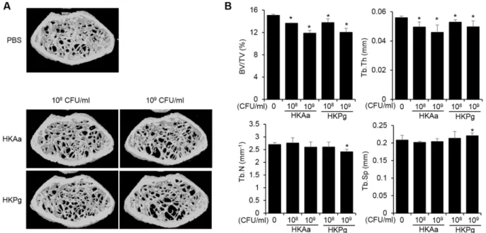
가 투여된마우스의대퇴부해면골이대조군에비해줄어있음 을보여준다(Fig. 1A). HKAa
와HKPg
가투여된마우스는대 조군에비해뼈의용적비와뼈잔기둥두께가유의적으로감 소하였다.
또한10
9CFU/ml
의HKPg
가투여된마우스의뼈 잔기둥수가감소하고,
뼈잔기둥간간격이증가하였다(Fig.
1B).
이결과로HKAa
와HKPg
는골소실을유도함을알수 있고,
이는치주질환쥐모델에서A. actinomycetemcomitans
와P. gingivalis
는골흡수를증가시킨다[5, 23]
는결과와일치 한다.
치주질환쥐모델이치주원인균에의한치조골소실을 평가하는적절한모델이나,
최소한달의시간이소요되는 만큼일주일만에골소실여부가판단가능한치주원인균-
복 강투여마우스모델은이를보완가능할것으로여겨진다.
결 론적으로,
치주원인균인A. actinomycetemcomitans
와P.
gingivalis
는골소실을단독으로유발시킬수있는만큼이Fig. 1. HKAa and HKPg induced bone resorption in vivo. (A) Mice (n = 3 per group) were intraperitoneally administered with HKAa or HKPg, twice with a 4-day interval. At day 7 after the first administration, micro-CT images of the femurs were obtained by the CT analyzer. (B) BV/TV, Tb.Th, Tb.N, and Tb.Sp of the femurs were analyzed. BV/TV = trabecular bone volume; Tb.N = trabecular number;
Tb.Th = trabecular thickness; Tb.Sp = trabecular separation. *p < 0.05.
는치주질환의치조골소실에의한치아상실발생기전을뒷 받침하는결과라하겠다
.
HKAa
와 HKPg의 파골세포분화능력 평가생체외실험에서
A. actinomycetemcomitans
와P. gingivalis
는골소실을유발하였기때문에(Fig. 1),
골소실유발기전을 규명하기위해골흡수를담당하는파골세포의분화에이들 균이어떠한영향을주는지연구하였다.
쥐의장골에서골 수를분리하고, M-CSF
로3
일동안처리하여만든BMM
과BMM
에M-CSF
와RANKL
을2
일간처리하여파골세포로 분화가 운명지어진 파골전구세포(committed osteoclast precursor)
를만들어실험에사용하였다. HKAa
와HKPg
를20 ng/ml
의RANKL
과 함께BMM
에 처리하자, HKAa
와HKPg
처리군은TRAP-
양성다핵세포수가현저히감소하였다
(Fig. 2A, B).
이는HKAa
와HKPg
가RANKL-
매개 파 골세포분화를감소시킴을나타낸다.
그러나, HKAa
와HKPg
를committed osteoclast precursor
에 처리하자, HKPg
와HKAa
처리군은TRAP-
양성다핵세포수가대조군에비해유의적으로 증가하였다
(Fig. 2C, D).
이 결과는HKPg
와HKAa
가파골세포분화를유도할수있음을나타낸다.
이러한결과는
P. gingivalis
가RANKL-
매개파골세포분화를감소시키고
, RANKL
이전처리된파골전구세포의분화를증가시킨다는 보고와 일치한다
[19]. P. gingivalis
와A.
actinomycetemcomitans
가골소실을유도하였기때문에,
파 골세포분화는증가되어져야 하는 것이타당하다.
하지만, committed osteoclast precursor
와는달리RANKL
을만난 적이없는BMM
이세균을만나면파골세포로분화되지않 았고,
이는세균의공통적현상으로보인다.
그이유는대식 세포와파골전구세포의기원이동일하기때문이다[16].
파골 전구세포가세균을만나게되면파골세포로분화되기보다 는대식세포로머물게하여염증반응을더심하게한다[6, 13].
그러나, BMM
이RANKL
에의해비가역적으로운명이 결정된committed osteoclast precursor
는세균에의해파골 세포분화가촉진된다[6, 23].
즉,
치주염병소의BMM
은세 균에의해파골세포로분화되지않고,
대식세포로서남아염 증반응을증가시켜골소실을더욱가속화할수있는환경 을만들고committed osteoclast precursor
의파골세포분화 를증가시킴으로써골소실을증가시키게만드는것으로여 겨진다.
AaLPS
와 PgLPS의 파골세포 분화 유도 능력 평가LPS
는그람음성세균이가지고있는세포외막지질다당Fig. 2. HKAa and HKPg attenuated osteoclast differentiation from BMMs, but induced differentiation of committed osteoclast precursors into osteoclasts. BMMs were treated with 20 ng/ml M-CSF and 20 ng/ml RANKL in the presence of HKAa at MOI of 0.5, 5, or 50 (A) and HKPg at MOI of 0.5, 5, or 50 (B) for 24 h. Committed osteoclast precursors were treated with 20 ng/ml M-CSF in the pres- ence of HKAa at MOI of 0.5, 5, or 50 (C) and HKPg at MOI of 0.5, 5, or 50 (D) for 8 h. The cells were fixed and subjected to TRAP staining.
TRAP-positive multinucleated cells (MNCs) with three or more nuclei were enumerated through microscopic analysis. *p < 0.05.
체로서다양한염증
-
매개물질을유도하는핵심병독력인자 로잘알려져있다[7, 10].
그러므로우리는HKAa
와HKPg
에의한파골세포분화의영향은A. actinomycetemcomitans
와P. gingivalis
의LPS
에의한효과일것이라고가설을세 웠다.
이를 위해 두 세균에서LPS
를 분리하여BMM
과committed osteoclast precursor
의파골세포분화에어떠한 영향을주는지실험하였다.
예측한것처럼, AaLPS
와PgLPS
가처리된
BMM
의TRAP-
양성다핵세포의수가농도의존적으로감소하고
(Fig. 3A, B), AaLPS
와PgLPS
는committed osteoclast precursor
의TRAP-
양성다핵세포수를증가시킴 을관찰하였다(Fig. 3C, D).
이결과는HKAa
와HKPg
와동 일하게, AaLPS
와PgLPS
가RANKL-
매개파골세포분화를 감소시키고committed osteoclast precursor
의파골세포분 화를 유도함을 보여준다.
우리의 결과는Escherichia coli
O55:B5 LPS
가BMM
의 파골세포분화를 감소시키고,
committed osteoclast precursor
의파골세포분화를증가시 킨다는연구결과와일치한다[24].
결과적으로그람음성세균 에의해조절되는파골세포분화능력은LPS
때문에나타나 는 것이라 여겨진다.
추가적으로 세균과LPS
에 의한committed osteoclast precursor
의파골세포분화결과가생 체외실험결과와일치함을확인할수있었다.
이는생체외골소실을예측하기위해
committed osteoclast precursor
를 활용한분화연구진행이적절하다는것을말해준다.
요 약
치주질환은만성염증성질환으로치조골소실을일으켜 성인치아상실을유발하는요인중하나이다
.
그람음성세균인Aggregatibacter actinomycetemcomitans
와Porphyromonas gingivalis
는치주질환환자의병소에서쉽게동정된다.
지질 다당체(Lipopolysaccharide; LPS)
는그람음성세균의핵심 독력인자로알려져있다.
이러한세균과LPS
는파골세포에 의한골소실을조절하는요인중하나이다.
그러므로본연 구에서는동물모델을활용하여A. actinomycetemcomitans
와P. gingivalis
의의한골소실여부를확인하고,
기전규명 을 위하여A. actinomycetemcomitans, P. gingivalis, A.
actinomycetemcomitans
와P. gingivalis
에서분리한LPS
에 의한 파골세포분화 영향을 연구하였다.
열사멸한A.
actinomycetemcomitans (HKAa)
와열사멸한P. gingivalis
(HKPg)
가복강으로투여된쥐의대퇴골은대조군에비해감소된골량을보여주었다