들쭉 추출물의 노인성 황반변성증 예방 효과
김선미*·김혜주·손미원·정세영*,#
동아제약(주)연구본부, *경희대학교 약학대학
(Received August 10, 2012; Revised October 29, 2012; Accepted October 30, 2012)
Protective Effect of Vaccinium uliginosum L. Extract on Age-related Macular Degeneration
Sun Mi Kim*, Hye Ju Kim, Miwon Son and Se Young Choung*,#
Research Center, Dong-A Pharm. Co., Ltd., Gyeonggi-Do 446-905, Korea
*College of Pharmacy, Kyung Hee University, Seoul 130-701, Korea
Abstract — Age-related macular degeneration (AMD) is the leading cause of vision loss and blindness among the elderly.
In this study, extract of Vaccinium uliginosum L. that has potent antioxidant activity was evaluated as effective preventive supplement for AMD using AMD animal model induced by high fat diet and UV A irradiation. Treatment with VU extract protected photoreceptor cells of retina and blocked the accumulation of lipofuscin pigment-granules induced by high fat diet and UV A light irradiation. This study suggests that VU extract may be a useful agent for prevention of progress of AMD.
Keywords □ Vaccinium uliginosum L., Age-related macular degeneration, lipofuscin
진달래과(Ericaceae)에 속하는 낙엽소관목인 들쭉나무 (Vaccinium uliginosum L.)는 주로 고산이나 고원에서 자라며 북 아메리카, 유럽, 아시아의 아한대지방에서 분포하고 있다. 계통적 으로 들쭉과 가까운 Bilberry(Vaccinium myrtillus)나 Blueberry (Vaccinium corymbosum or Vaccinium angustifolium)에 관한 연구는 많이 진행되어 왔으나, 그에 비해 들쭉나무 열매에 관한 연구는 드문 실정이다. 들쭉나무의 열매(들쭉)는 8~9월에 검은 자줏빛으로 익으며 하얀 가루로 덮여 있으며, 효능은 모세혈관 강화작용, 혈당저하, 대장암예방, 피로회복, 정혈작용, 정력증강, 해열작용 등에 좋다고 하여 민간요법으로 널리 사용되어 오고 있 으며, 중요한 생리적 활성물질인 안토시아닌류(anthocyanins)와 플라보놀류(flavonols)가 다량 포함되어 있고, 이로 인해 강한 항 산화능이 있는 것으로 보고되어 있다. 이러한 항산화물질에 의 한 노인성 황반변성증(AMD)의 개선 효과는 이미 보고된 바 있 어, 들쭉 추출물에 의한 노인성 황반변성증 개선 효과를 미리 예 측해 볼 수 있다.1)
노인성 황반병성증(AMD)은 여러 가지 요인에 의해 황반부에 변성이 생겨 시력장애가 일어나는 질환이다. AMD의 위험인자
로 나이,2-4)흡연,5,6)인종,7,8)유전자,9,10)고지방 식이 및 콜레스 테롤11,12)염증성분(complement factor H)13-15)식습관,16,17)태양 광18)등 다양하게 제시되어 왔다. 그 중 나이와 흡연은 30년 동 안 꾸준히 AMD의 위험인자로 보고되고 있으며,19-22) 우리나라 65세 이상 인구의 16.5%가 황반변성을 가지고 있어 노인인구의 실명의 주요 원인이 되고 있다.23)황반변성 중 습성황반변성의 경우 anti-VEGF(anti-vascular endothelial growth factor) 치료 제가 개발되어 사용 중이나, 고가의 치료비와 유리체강내 투여 를 여러 번 반복해야 하는 단점이 있으며, 좀 더 유병률이 높은 건성황반변성의 경우 적절한 치료제가 없어 황반변성을 억제하 는 대체 약물의 개발이 시급한 실정이다. 눈은 빛에 노출되어 있 으며 망막에 불포화지방산24)이 많이 포함되어 있고 산소용존율25) 이 높으며 빛을 받으면 ROS를 발생하는 lipofuscin26,27)이 존재 하기 때문에 다른 기관에 비해 산화 stress를 많이 받는다. 다행 히 눈에는 항산화제가 존재하여 보호를 받을 수 있지만 잘못된 생활습관이나 노화로 인해 항산화제가 부족하게 되면 활성산소, 산화된 lipofuscin 등으로부터 공격을 받아 AMD 같은 질병이 발 병하게 되므로 AMD 예방을 위해서는 산화 stress를 저해할 수 있어야 한다. 따라서, 강한 항산화능을 보이는 들쭉추출물은 AMD 를 예방하거나 지연시키는 데 도움이 될 것으로 사료된다.
본 연구에서는 들쭉 추출물이 노인성 황반변성증을 예방하는 천연물 의약품 또는 건강기능식품의 새로운 소재로서 가능성을
#본 논문에 관한 문의는 저자에게로 (전화) 02-961-9198 (팩스) 02-966-3885 (E-mail) [email protected]
종설
알아 보고, 노인성 황반변성증 발생억제에 대한 효과를 과학적 으로 입증하고 더 나아가 그 기전을 밝히고자 하였다.
실험방법
실험동물
수컷 C57BL/6는 Harlan Laboratories(Indianapolis, IN, USA)로부터 구매하였으며, 노화가 진행되도록 11개월령까지 사 육한 후 실험에 사용하였다. 식이와 물은 자유로이 섭취하도록 하였으며, 22±1oC의 온도에서 12시간 간격의 day/night cycle의 조건에서 사육하였다. 동물실험은 경희대학교 Animal Care and Use Guidelines에 의해 행해졌고, 모든 protocol은 경희대학교의 Institutional Animal Care and Use Committee(승인번호: KHP- 2009-01-9)에 의해 승인되었다.
실험재료 및 시약
3-(4,5-dimethylthiazol-yl)-2,5-diphenyltetrazolium bromide, paraformaldehyde powder 95%는 Sigma Chemical Co.(St.
Louis, USA)로부터 구매하였다. 실험에서 양성 대조군으로 사용 한 아프리칸 메리골드(Tagetes erecta L.)에서 추출한 82% 루테 인(lutein)은 Green Chem(Attibele, India)으로부터 구입하였다.
들쭉 추출물 제조
건조시킨 잘 익은 들쭉(Vaccinium uligisosum L.)을 그라인더 로 분쇄한 후 열수 추출(90oC, 16시간, 2회)하고 여과하였다. 여 과 후 얻은 추출물은 동결건조하여 파우더로 만든 후 사용하기 전까지 -40oC에 보관하였다.
고지방사료와 시료 투여 및 UVA 조사
마우스를 평균체중과 분산이 균일하도록 군을 나누고, 각 군 당 6마리씩 7군으로 하였으며, 모든 마우스는 순화기간 동안 정 상사료(#38057; Cargill Agri Purina, Inc., Korea)를 섭취하였다.
7일간의 순화기간이 끝난 후 normal 군을 제외한 모든 마우스의 눈에 lipofuscin을 축적시키기 위해 normal 군을 제외한 모든 마우스는 고지방사료(#D12492; Research diets Inc., New Brunswick, NJ, USA)로 식이를 변경해 주었으며, normal군은 실 험이 종료될 때까지 정상사료를 식이 하였다. 실험시작 4주 후 부터 각 실험물질을 saline에 녹여 4주 동안 경구투여하였다. UV A 조사는 실험종료 2주 전부터 3 J/cm2까지 매일 일정한 시간에 조사하였다(intensity: 1.5~1.25 mW/cm2, time: 35~40 min).
광학현미경과 투과전자현미경 관찰 및 정량적 평가
실험 종료 후 적출된 마우스의 눈은 2% glutaraldehyde, 2%
paraformaldehyde 혼합물에 12시간 동안 4oC에 보관하여 고정
시켰다. 고정된 눈 조직에서 각막, lens, 과도한 근육이나 지방조 직을 제거한 후 망막, 맥락막, 공막이 포함된 부분(posterior segment)을 4분의 1 크기로 잘라 실험에 사용하였다. 잘려진 조 직은 1% osmium tetroxide에서 1시간 동안 후고정시키고 PBS 로 washing하여 고정액을 제거하였다. 그리고 ethanol series(50, 70, 80, 90, 95, 100% ethanol)를 이용하여 조직을 탈수시킨 후 eponate 12 resin로 embed하였다. 그리고 나서 광학현미경 검경 을 위한 조직은 1 µm로 semi-thin section되었고 투과전자현미 경 검경을 위한 조직은 60 nm로 thin section하였다. UV A 조 사에 의한 photoreceptor 손상에 대한 들쭉의 효능을 관찰하기 위해 section(1 µm)된 조직은 toluidine blue 염색한 후 슬라이드 를 제작하여 광학현미경으로 관찰하였고 stereo investigator software(MBF Bioscience, Williston, VT, U.S.A)를 사용하여 핵 의 수를 세고 outer nuclear layer(ONL)의 두께를 측정하였다.
Thin section(60 nm)된 눈 조직은 4% uranyl acetate와 lead citrate로 염색한 후 투과전자현미경(TEM)을 사용하여 RPE 세 포에서 lipofuscin 축적 정도를 관찰하여 들쭉 추출물의 lipofuscin 축적 저해능을 확인할 수 있었다.
통계학적 분석
모든 data는 spreadsheet(Excel 2007; Microsoft, Redmond, WA)에서 수집하였고 SPSS software(PASW statistic 18; SPSS Inc., Chicago, IL)를 이용하여 분석하였고 모든 통계시험에서 p value가 p 0.05일 때 유의적인 차이가 있는 것으로 처리하였다.
실험결과
들쭉 추출물의 망막조직 손상 보호 효과
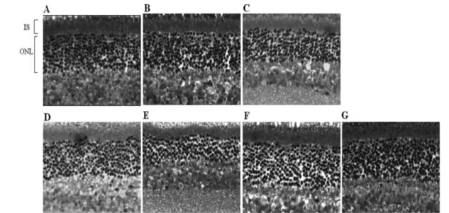
들쭉 추출물 섭취가 고지방식이 및 UV A 조사로 인한 망막손 상을 보호할 수 있는지 알아보기 위해 각 군에서 photoreceptor 세포의 핵으로 구성된 outer nuclear layer(ONL)을 중심으로 망 막의 구조를 관찰하였다(Fig. 1). 그 결과, 정상 식이를 하고 UV A 조사되지 않은 군(SD group)과 고지방식이하고 UV A 조사되 지 않은 군(HD group)의 inner segment(IS)와 ONL 사이 경계 선은 직선으로 유지되어 망막 조직이 변성되지 않은 반면, 고지 방식이와 UV A 조사를 동시에 받은 NC group, lutein 50 mg/
kg 섭취한 군(Lutein 50 group) 그리고 들쭉 추출물 50, 100 mg/
kg 섭취한 군(VU50, VU100)의 경계선은 곡선 형태를 띄는 것으 로 관찰되어 망막 조직의 변성이 나타나는 것을 알 수 있었다 (Fig. 1A though F). 그러나, 들쭉 추출물 200 mg/kg 섭취한 군 (VU200)의 경계선은 조직 손상 없는 직선으로 관찰되어(Fig.
1G), 들쭉 추출물 고용량 투여 시 고지방식이와 UV A 조사에 의한 세포손상으로부터 망막을 보호하는 효과가 있음을 알 수 있었다.
들쭉 추출물의 photoreceptor 세포 보호 효과
망막조직 가로 길이 100 µm 내에 있는 정상적인 핵의 수를 비 교한 결과 고지방식이와 UV A 조사한 군(NC)의 photoreceptor 핵의 수는 SD group의 핵의 수 보다 27.7% 줄어들어 유의적인
감소를 보여, 고지방식이와 UV A 조사가 병용되었을 때 photoreceptor 세포가 손상됨을 확인하였다. 고지방식이와 UV A 조사에 의한 망막 photoreceptor 세포의 손상은 들쭉 추출물 50, 100, 200 mg/kg 섭취에 의해서 각각 21.5, 51.3, 76.7% 보호되었 고, lutein 50 mg/kg 섭취한 군(Lutein 50)에서의 보호능은 28.9%로 들쭉 50 mg/kg 섭취한 군(VU 50)과 비슷한 효능을 보 였다(Fig. 2).
들쭉 추출물의 외과립층(ONL) 보호 효과
각군 마우스의 ONL 두께를 일정한 위치에서 측정한 결과, HD group은 SD group에 비해 ONL의 두께가 68.7% 유의적인 증가 를 보여, 고지방사료 식이 시 망막 내에 지방이 축적되어 ONL 의 두께가 두꺼워지는 현상을 관찰하였다. 또한 NC group의 ONL 두께는 SD group의 ONL 두께와 유의적인 차를 보이지 않아 고 지방 식이로 두꺼워진 ONL이 UV A 조사로 인한 photoreceptor 의 손상으로 그 두께가 얇아지는 현상을 관찰하였다. UV A의 조 사에 의해서 얇아진 ONL의 두께는 들쭉 추출물 50, 100, 200 mg/kg 섭취에 의해서 각각 7.2, 52.2 47.7% 증가되어 있었고, 들 쭉 추출물 100 mg/kg, 200 mg/kg 섭취한 군(VU 100, VU 200) 에서의 차이는 유의적이었다. Lutein 50 group에서의 ONL 두께 는 NC group에 비해 17.7% 증가되는 것을 관찰할 수 있었다 (Fig. 2).
UV A로 인한 lipofuscin 축적에 대한 억제 효과
RPE 세포 내에 lipofuscin 축적도를 조사하기 위해서 RPE 세 포를 TEM을 이용하여 관찰하였다(Fig. 3). SD, HD groups에서 Fig. 1− Light microscopic analysis of outer retina from C57BL6 mice. Photomicrographs show outer retina from a mouse of SD group (A), HD group (B), NC group (C), Lutein 50 group (D), VU 50 group (E), VU 100 group (F), VU200 group (G). Staining is toluidin blue (original magnification, 200) HD: high fat diet, NC: negative control, VU: berry extract of Vaccinium uliginosum L., IS: inner segment ONL:
outer nulear layer.
Fig. 2− Morphologic evaluation by quantitative histology. Histogram show the average number of photoreceptor nuclei per 100µm of ONL (A) and ONL thickness measured from the central retinas of mice (B). Means±SEM of four experiments; #: P<0.005 when compared with SD,
*,**,***: P<0.05, P<0.005, P<0.001 when compared with NC. HD: high fat diet, NC: negative control, VU: berry extract of Vaccinium uliginosum L.
는 lipofuscin이 응집되어 생긴 dense body가 관찰되지 않았으나, NC group의 RPE세포 관찰 시 아래 부분에 불규칙적인 dense bodies가 6개 관찰되었다(Fig. 3A though C). 고지방식이와 UV A 조사에 의해서 유도된 dense bodies가 Lutein 50 group에서 는 1개, VU 50 group에서 1개 발견되었고, VU 200 group에서 는 발견되지 않아(Fig. 3D though F), 들쭉 추출물의 섭취로 lipofuscin 과립의 축적이 현저하게 저해됨을 확인하였다.
고 찰
AMD(age-related macular degeneration)는 RPE(retinal pigment epithelial) 세포의 사멸에 따른 photoreceptor 세포의 2 차적 사멸에 의해 시력을 상실하게 되는 질병이며, 따라서 AMD 로 인한 시력상실을 예방하기 위해서는 RPE 세포를 보호하는 것이 중요하다. 본 연구에서는 들쭉 추출물의 RPE 세포의 보호 효과를 연구하였고, 이를 동물실험을 통하여 알아보고자 하였다.
In vivo 동물실험에서 mouse AMD의 biomarker는 RPE 세포 내에 축적된 lipofuscin 과립이다.28)본 연구에서는 C57/BL6 mice에 고지방사료를 식이 하였고 UV A를 조사하여 AMD 동 물 model을 만들었다.29,30) UV A 조사는 최대 흡광 파장이 315 nm에서 345 nm인 lamp를 사용하였다. UV 빛은 lens에 의해서 다량 흡수되지만 photosensitizer의 반응에 의해 간접적으로 영 향을 끼칠 수 있다.31) AMD가 고지방 식이와 관련이 있다는 것 은 아직까지 논란의 여지가 많지만 지질이 RPE 세포막에 밀집될 수 있고 lipofuscin 축적 시 지질이 substrate로서 작용하기 때문
에 UV A 조사에 의한 망막 손상을 극대화 시킬 수 있다고 판단
하였다.32-35)기대한 것과 같이 transmission electron microscopy
분석에서 수많은 lipofuscin 과립이 고지방 식이와 UV A 조사에 의해서 현저히 증가하였다. 그리고 AMD 유도된 mice에 들쭉 추 출물과 positive control로서 lutein을 경구투여하였을 경우 lipofuscin 과립의 축적이 줄었다. 또한 광학현미경을 이용한 photoreceptor의 관찰 결과는 RPE 세포 내 lipofucin 축적저해 효능과 일치하는 양상을 나타내었다. 이는 RPE 세포 내 lipofuscin 의 축적에 의해 photoreceptor 세포가 손상되었고 들쭉 추출물 이 RPE 세포 내 lipofuscin 축적을 저해하였다. 그러므로, 최종 적으로 photoreceptor 세포의 손상을 예방하는 것을 입증하는 결 과이다.
또한 들쭉은 임신, 수유부에서 많은 양 섭취 시 안전하지 않을 가능성이 있으며, 곰팡이균에 감염된 열매로 섭취하였을 때 독 성(메스꺼움, 구토, 중독, 시각장애 등)이 보고된 바 있으나, 그 외 안전성을 우려할 만한 정보는 현재까지 보고된 바 없어, 안전 한 노인성 황반변성증의 예방 및 치료물질로서 개발이 가능할 것 으로 사료된다.34)
결 론
본 연구를 통하여 들쭉 추출물은 AMD 유도한 동물모델을 대 상으로 한 실험에서 RPE 세포 내 lipofuscin의 축적을 감소시키 며, photoreceptor 세포의 손상을 효과적으로 예방함을 확인하였 다. 그러므로, 들쭉 추출물은 노인성 황반변성증에 예방 및 치료 Fig. 3− Transmission electron microscopic analysis of RPE from C57BL/6 mice. Photomicrographs show RPE from a mouse of Normal group (A), HD group (B), NC group (C), Lutein 50 group (D), VU 50 group (E), VU 200 group (F). White arrows indicate the irregularly shaped lipofuscin pigment granules, which are distinct from the larger oval melanosomes (Bars, 2.0µm). HD: high fat diet, NC:
negative control, VU: berry extract of Vaccinium uliginosum L.
물질로서 가능성이 있으며, 노인 인구의 눈건강을 증진시키는 역 할을 할 수 있을 것으로 사료된다.
참고문헌
1) Michikawa, T., Ishida, S., Nishiwaki, Y., Kikuchi, Y., Tsuboi, T., Hosoda, K., Ishigami, A., Iwasawa, S., Nakano, M. and Takebayashi, T. : Serum antioxidants and age-related macular degeneration among older Japanese. Asia Pac. J. Clin. Nutr. 18, 1 (2009).
2) Klein, R., Klein, B. E. and Linton, K. L. : Prevalence of age- related maculopathy. The Beaver Dam Eye Study. Ophthalmology 99, 933 (1992).
3) Friedman, D. S., Katz, J., Bressler, N. M., Rahmani, B. and Tielsch, J. M. : Racial differences in the prevalence of age- related macular degeneration: the Baltimore Eye Survey.
Ophthalmology 106, 1049 (1999).
4) VanNewkirk, M. R., Nanjan, M. B., Wang, J. J., Mitchell, P., Taylor, H. R. and McCarty, C. A. : The prevalence of age- related maculopathy: the visual impairment project. Ophthalmology 107, 1593 (2000).
5) Delcourt, C., Diaz, J. L., Ponton-Sanchez, A. and Papoz, L. : Smoking and age-related macular degeneration. The POLA Study. Pathologies Oculaires Liees a l'Age. Arch. Ophthalmol.
116, 1031 (1998).
6) Khan, J. C., Thurlby, D. A., Shahid, H., Clayton, D. G., Yates, J. R., Bradley, M., Moore, A. T. and Bird, A. C. : Smoking and age related macular degeneration: the number of pack years of cigarette smoking is a major determinant of risk for both geographic atrophy and choroidal neovascularisation. Br. J.
Ophthalmol. 90, 75 (2006).
7) Klein, R., Klein, B. E., Jensen, S. C., Mares-Perlman, J. A., Cruickshanks, K. J. and Palta, M. : Age-related maculopathy in a multiracial United States population: the National Health and Nutrition Examination Survey III. Ophthalmology 106, 1056 (1999).
8) Chachat, A. P., Hyman, L., Leske, M. C., Connell, A. M. and Wu, S. Y. : Features of age-related macular degeneration in a black population. The Barbados Eye Study Group. Arch.
Ophthalmol. 113, 728 (1995).
9) Haddad, S., Chen, C. A., Santangelo, S. L. and Seddon, J. M. : The genetics of age-related macular degeneration: a review of progress to date. Surv. Ophthalmol. 51, 316 (2006).
10) Scholl, H. P., Fleckenstein, M., Charbel Issa, P., Keilhauer, C., Holz, F. G. and Weber, B. H. : An update on the genetics of age-related macular degeneration. Mol. Vis. 13, 196 (2007).
11) Tomany, S. C., Wang, J. J., Van Leeuwen, R., Klein, R., Mitchell, P., Vingerling, J. R., Klein, B. E., Smith, W. and De Jong, P. T. : Risk factors for incident age-related macular
degeneration: pooled findings from 3 continents. Ophthalmology 111, 1280 (2004).
12) Cho, E., Hung, S., Willett, W. C., Spiegelman, D., Rimm, E. B., Seddon, J. M., Colditz, G. A. and Hankinson, S. E. : Prospective study of dietary fat and the risk of age-related macular degeneration. Am. J. Clin. Nutr. 73, 209 (2001).
13) Okamoto, H., Umeda, S., Obazawa, M., Minami, M., Noda, T., Mizota, A., Honda, M., Tanaka, M., Koyama, R., Takagi, I., Sakamoto, Y., Saito, Y., Miyake, Y. and Iwata, T. : Complement factor H polymorphisms in Japanese population with age- related macular degeneration. Mol. Vis. 12, 156 (2006).
14) Klein, R. J., Zeiss, C., Chew, E. Y., Tsai, J. Y., Sackler, R. S., Haynes, C., Henning, A. K., SanGiovanni, J. P., Mane, S. M., Mayne, S. T., Bracken, M. B., Ferris, F. L., Ott, J., Barnstable, C. and Hoh, J. : Complement factor H polymorphism in age- related macular degeneration. Science 308, 385 (2005).
15) Souied, E. H., Leveziel, N., Richard, F., Dragon-Durey, M. A., Coscas, G., Soubrane, G., Benlian, P. and Fremeaux-Bacchi, V. : Y402H complement factor H polymorphism associated with exudative age-related macular degeneration in the French population. Mol. Vis. 11, 1135 (2005).
16) Flood, V., Smith, W., Wang, J. J., Manzi, F., Webb, K. and Mitchell, P. : Dietary antioxidant intake and incidence of early age-related maculopathy: the Blue Mountains Eye Study.
Ophthalmology 109, 2272 (2002).
17) Van Leeuwen, R., Boekhoorn, S., Vingerling, J. R., Witteman, J. C., Klaver, C. C., Hofman, A. and de Jong, P. T. : Dietary intake of antioxidants and risk of age-related macular degeneration.
JAMA 294, 3101 (2005).
18) Taylor, H. R., West, S., Munoz, B., Rosenthal, F. S., Bressler, S. B. and Bressler, N. M. : The long-term effects of visible light on the eye. Arch. Ophthalmol. 110, 99 (1992).
19) Klein, R., Peto, T., Bird, A. and Vannewkirk, M. R. : The epidemiology of age-related macular degeneration. Am. J.
Ophthalmol. 137, 486 (2004).
20) Bird, A. C., Bressler, N. M., Bressler, S. B., Chisholm, I. H., Coscas, G., Davis, M. D., de Jong, P. T., Klaver, C. C., Klein, B. E. and Klein, R. : An international classification and grading system for age-related maculopathy and age-related macular degeneration. The International ARM Epidemiological Study Group. Surv. Ophthalmol. 39, 367 (1995).
21) Mitchell, P., Smith, W., Attebo, K. and Wang, J. J. : Prevalence of age-related maculopathy in Australia. The Blue Mountains Eye Study. Ophthalmology 102, 1450 (1995).
22) Thornton, J., Edwards, R., Mitchell, P., Harrison, R. A., Buchan, I. and Kelly, S. P. : Smoking and age-related macular degeneration: a review of association. Eye (Lond) 19, 935 (2005).
23) 보건복지부. 질병관리본부. 국민건강통계. 국민건강영양조사 제5기
1차년도 (2010)
24) Fliesler, S. J. and Anderson, R. E. : Chemistry and metabolism of lipids in the vertebrate retina. Prog. Lipid. Res. 22, 79 (1983).
25) Ahmed, J., Braun, R. D., Dunn, R. Jr. and Linsenmeier, R. A. : Oxygen distribution in the macaque retina. Invest. Ophthalmol.
Vis. Sci. 34, 516 (1993).
26) Sparrow, J. R. and Boulton, M. : RPE lipofuscin and its role in retinal pathobiology. Exp. Eye. Res. 80, 595 (2005).
27) Sparrow, J. R., Vollmer-Snarr, H. R., Zhou, J., Jang, Y. P., Jockusch, S., Itagaki, Y. and Nakanishi, K. : A2E-epoxides damage DNA in retinal pigment epithelial cells. Vitamin E and other antioxidants inhibit A2E-epoxide formation. J. Biol.
Chem. 278, 18207 (2003).
28) Mishima, H. and Kondo, K. : Extrusion of lysosomal bodies from apical mouse retinal pigment epithelium. Albrecht Von Graefes Arch. Klin Exp. Ophthalmol. 216, 209 (1981).
29) Young, R. W. : Solar radiation and age-related macular degeneration. Surv. Ophthalmol. 32, 252 (1988).
30) Tomany, S. C., Cruickshanks, K. J., Klein, R., Klein, B. E. and
Knudtson, M. D. : Sunlight and the 10-year incidence of age- related maculopathy: the Beaver Dam Eye Study. Arch.
Ophthalmol. 122, 750 (2004).
31) Beatty, S., Koh, H., Phil, M., Henson, D. and Boulton, M. : The role of oxidative stress in the pathogenesis of age-related macular degeneration. Surv. Ophthalmol. 45, 115 (2000).
32) Jones, C. A., Huberman, E., Cunningham, M. L. and Peak, M. J. : Mutagenesis and cytotoxicity in human epithelial cells by far- and near-ultraviolet radiations: action spectra. Radiat.
Res. 110, 244 (1987).
33) Cousins, S. W., Espinosa-Heidmann, D. G., Alexandridou, A., Sall, J., Dubovy, S. and Csaky, K. : The role of aging, high fat diet and blue light exposure in an experimental mouse model for basal laminar deposit formation. Exp. Eye. Res. 75, 543 (2002).
34) Brunk, U. T. and Terman, A. : Lipofuscin: mechanisms of age- related accumulation and influence on cell function. Free Radic.
Biol. Med. 33, 611 (2002).
35) Natural Medicine Comprehensive Database. : bog bilberry monograph.