ISSN 0378-6471 (Print)⋅ISSN 2092-9374 (Online)
https://doi.org/10.3341/jkos.2017.58.9.1036
Original Article
단안 삼출성 나이관련황반변성 환자의 무증상 반대안의 외망막층 두께 변화
Change of Outer Retinal Thickness in Fellow Eyes of Patients with Unilateral Age-related Macular Degeneration
정현교1⋅김성수2⋅박현주1
Hyun Kyo Jeong, MD1, Sung Soo Kim, MD, PhD2, Hyun Ju Park, MD1
실로암안과병원1, 연세대학교 의과대학 안과학교실2 Siloam Eye Hospital1, Seoul, Korea
Department of Ophthalmology, Yonsei University College of Medicine2, Seoul, Korea
Purpose: To compare the outer retinal thickness in normal fellow eyes of patients with unilateral age-related macular degener- ation (AMD) and normal control eyes.
Methods: We retrospectively reviewed the medical records of 60 patients with unilateral exudative AMD including polypoidal choroidal vasculopathy and 60 normal controls. Spectralis optical coherence tomography was performed in the normal fellow eyes of patients with unilateral AMD and in the normal group. The thicknesses between the retinal pigment epithelium (RPE) line and the cone outer segment tips (COST) line, between the COST line and the photoreceptor inner segment/outer segment (IS/OS) line, and between the IS/OS line and the external limiting membrane (ELM) line were measured at the fovea in both groups.
Results: The thickness between the RPE and COST lines was 32.4 ± 3.0 μm in normal fellow eyes of patients with unilateral AMD and 35.3 ± 3.5 μm in the normal group (p < 0.001). Total retinal thickness, thicknesses between the COST and the IS/OS lines and the IS/OS and the ELM lines in fellow eyes were not significantly different from those of normal eyes (p = 0.126, 0.615, 0.874). There was no significant difference in total retinal thickness or each outer retinal thickness measured in normal fellow eyes between patients with neovascular AMD and polypoidal choroidal vasculopathy.
Conclusions: The thickness between the RPE and the COST lines was thinner in the fellow eyes of patients with unilateral AMD than in the normal eyes. We suggest that less thickness between the RPE and COST lines might indicate a greater risk of AMD.
J Korean Ophthalmol Soc 2017;58(9):1036-1041
Keywords: Age-related macular degeneration, Cone outer segment tips, Outer retinal thickness, Retinal pigment epithelium
■Received: 2017. 6. 22. ■ Revised: 2017. 8. 9.
■Accepted: 2017. 9. 1.
■Address reprint requests to Hyun Ju Park, MD
Siloam Eye Hospital, #181 Deungchon-ro, Gangseo-gu, Seoul 07668, Korea
Tel: 82-2-2650-0880, Fax: 82-2-2650-0725 E-mail: [email protected]
*Conflicts of Interest: The authors have no conflicts to disclose.
ⓒ2017 The Korean Ophthalmological Society
This is an Open Access article distributed under the terms of the Creative Commons Attribution Non-Commercial License (http://creativecommons.org/licenses/by-nc/3.0/) which permits unrestricted non-commercial use, distribution, and reproduction in any medium, provided the original work is properly cited.
단안의 삼출성 나이관련황반변성 환자에서는 현재 임상 적인 특이 이상 소견이 관찰되지 않는 반대안에도 추후 새 로운 신생혈관이 발생할 위험이 양안 모두 나이관련황반변 성에 이환되지 않은 환자에 비해 더 높음이 알려져 있다.1-3 단안의 나이관련황반변성 환자에서 2년 안에 12-22%, 4년 안에 37%, 5년 안에 22-38.7%로 반대안에 새로운 신생혈 관이 발생할 가능성이 있다고 보고되었다.4-7
나이관련황반변성은 주로 외망막층과 그 관련 구조들인 망막색소상피층, 광수용체, 맥락막의 손상으로부터 시작된 다.8,9 Acton et al10은 초기 나이관련황반변성에서 빛간섭단
Figure. 1. Thickness measurements of each layer in the outer retina in normal eye (A) and in fellow eye of patients with unilateral
age-related macular degeneration (B). Thickness between the retinal pigment epithelium (RPE) line (outer border) and the cone outer segment tips (COST) line (inner border), the COST line (inner border) and the photoreceptor inner segment/outer segment (IS/OS) line (center of the line), the IS/OS line (center of the line) and the external limiting membrane (ELM) line (center of the line) and the total retinal thickness were measured in both groups.층촬영을 통한 외망막층의 두께를 분석한 결과, 광수용체 외분절이 얇아지고 망막색소상피층이 두꺼워지는 구조적 변화와 함께 시야 결손 등의 기능적 손상이 동반되기 시작 한다고 하였다. 단안 삼출성 나이관련황반변성 환자의 반 대안에서 새로운 맥락막신생혈관의 발생 가능성이 높다는 점으로 미루어, 임상적으로 특이 망막 이상 소견이 나타나 지 않은 반대안에도 외망막층을 중심으로 한 구조적 변화 가 시작되고 있을 가능성이 있으나 이와 관련한 국내 연구 결과는 보고된 적이 없다. 따라서 저자들은 빛간섭단층촬 영을 이용하여 단안 삼출성 나이관련황반변성 환자의 임상 적 정상 소견인 반대안의 외망막층을 양안 모두 임상적 정 상 소견인 눈과 비교해 보고자 하였다.
대상과 방법
의무기록을 후향적으로 조사하여 2016년 1월부터 2016년 12월까지 연세대학교 세브란스병원 안과에 내원하여 양안의 스펙트럼영역 빛간섭단층촬영기인 Spectralis® OCT (Heidelberg Engineering, Heidelberg, Germany)를 이용한 검사를 시행받
은 환자 중 단안의 삼출성 나이관련황반변성을 진단받은 환자 60명의 안저 검사상 망막의 이상이 발견되지 않은 반 대안 60안과 양안 모두 망막 이상 소견이 발견되지 않은 환 자들 60명의 60안을 대조군으로 하였다. 환자군 60안 중 신 생혈관성 나이관련황반변성은 44안, 결절맥락막혈관병증 은 16안이었다. 환자군 선택 시 비삼출성 나이관련황반변 성 및 망막혈관종성증식으로 진단한 경우는 제외하였다. 또한 환자군의 망막 이상 소견이 없는 반대안 및 대조군의 양안에서 63 μm 이상의 드루젠 및 황반의 색소 이상이 있 는 경우도 제외하였다. 두 군에서 모두 녹내장, 약시, 포도 막염, -6디옵터 미만의 고도 근시, 위수정체안, 조절되지 않 는 당뇨 혹은 고혈압을 동반한 환자도 제외하였다. 모든 환 자에서 IOL Master® (Carl Zeiss, Jena, Germany)를 이용하 여 안축장을 측정하였다.
모든 환자는 산동 상태에서 빛간섭단층촬영을 시행하였으 며, 부피 스캔 방식으로 황반부 20°×20°의 범위를 촬영한 후 빛간섭단층촬영 내의 caliper를 이용하여 중심와에서 전체 망 막 두께 및 각 망막층 사이의 두께를 측정하였다. 각 망막층 사이의 두께는 망막색소상피층(retinal pigment epithelium,
A B
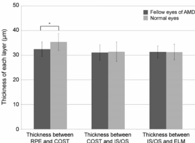
Figure 2. Thickness of each layer at the fovea in fellow eyes of
patients with unilateral age-related macular degeneration (AMD) and normal eyes. Error bar is mean ± standard deviation. RPE = retinal pigment epithelium, COST = cone outer segment tips, IS/OS = photoreceptor inner segment/out- er segment, ELM = external limiting membrane. *p-value <0.05.
RPE)의 외측 경계와 원추세포외절끝(cone outer segment tips, COST)의 내측 경계 사이의 두께(RPE-COST), 원추세 포외절끝의 내측 경계와 광수용체내외절경계(photoreceptor inner segment/outer segment, IS/OS) 중심 사이의 두께 (COST-IS/OS), 그리고 광수용체내외절경계 중심과 외경계 막(external limiting membrane, ELM) 중심 사이 두께 (IS/OS-ELM)를 각각 측정하였다. 빛간섭단층촬영의 해상 도의 한계로 인해 망막색소상피층과 원추세포외절끝은 환 자에 따라 명확히 구분되지 않는 경우들이 있었기 때문에, 망막색소상피층은 외측 경계를, 원추세포외절끝은 내측 경 계를 기준으로 하여 각 층간의 두께를 측정하였으며, 이는 두 군의 모든 환자에서 동일하게 적용하였다(Fig. 1). 두 명 의 독립된 측정자가 각자 상기에 명시한 방법대로 빛간섭 단층촬영상 각 망막층 사이의 두께를 측정하였으며, 측정 자들에게 피험자의 나이, 성별 및 환자군과 대조군 여부 등 의 임상적 자료는 제공하지 않았다. 본 연구는 헬싱키 선언 을 준수하여 연세대학교 의료원 임상연구윤리 심의위원회 의 승인 아래 진행되었다.
통계적 분석은 SPSS version 24.0 (IBM Corp., Armonk, NY, USA)을 사용하였다. 환자군과 대조군 사이, 환자군 내 에서 신생혈관성 나이관련황반변성과 결절맥락막혈관병증 환자들 사이의 측정값 간의 유의한 차이가 있는지를 알아 보기 위해 연속형 자료는 Student t-test를 사용하였고 범주 형 자료는 chi-square test를 사용하여 비교하였다. 모든 통 계적 유의성은 p값 0.05 미만으로 판정하였다. 또한 측정자 간의 재현성을 확인하기 위해 급외상관계수를 분석하였다.
결 과
단안 나이관련황반변성 환자군 총 60명 중 남성이 25명, 여성이 35명이며 평균 연령은 69.6 ± 6.7세, 대조군 총 60 명 중 남성 27명, 여성 33명이며 평균 연령은 68.8 ± 6.8세 였고, 두 군 간 성별 및 연령에서 유의한 차이는 없었다 (p=0.713, 0.518). 단안 나이관련황반변성 환자군의 평균 안 축장은 23.47 ± 1.05 mm, 대조군은 23.46 ± 0.91 mm로 두 군 간 유의한 차이를 보이지 않았다(p=0.936). 단안 나이관 련황반변성 환자의 반대안과 대조군의 중심와에서 측정한 전체 망막두께는 각각 231.6 ± 15.1 μm 및 227.1 ± 16.6 μm 로 통계적으로 유의한 차이는 없었다(p=0.126). 평균 망막 색소상피층과 원추세포외절끝 사이의 두께(RPE-COST)는 단 안 나이관련황반변성 환자군의 반대안에서 32.4 ± 3.0 μm, 대조군에서 35.3 ± 3.5 μm로 유의한 차이를 보였다(p<0.001).
원추세포외절끝과 광수용체내외절경계 사이 두께(COST-IS/OS) 는 환자군에서 31.1 ± 3.2 μm, 대조군에서 31.4 ± 4.0 μm로
측정되었고, 광수용체내외절경계와 외경계막 사이 두께 (IS/OS-ELM)는 각각 31.3 ± 2.5 μm, 31.3 ± 3.2 μm로 측정되었 으며, 두 군 간의 유의한 차이는 없었다(p=0.615, 0.874) (Fig. 2).
두 그룹의 중심와에서 측정한 전체 망막두께에 대한 각 망막층 사이 두께의 상대적인 비율도 비교하였다. 전체 망 막두께에 대한 상대적인 망막색소상피층과 원추세포외절 끝 사이 두께(RPE-COST)의 비율은 단안 나이관련황반변성 환자의 반대안에서 14.1 ± 1.6%, 대조군에서 15.6 ± 1.9%로 유의한 차이를 보였다(p<0.001). 전체 망막두께에 대한 원추세 포외절끝과 광수용체내외절경계 사이의 두께(COST-IS/OS)의 비율은 환자군에서 13.5 ± 1.5%, 대조군에서 13.9 ± 2.0%였으 며, 광수용체내외절경계와 외경계막 사이 두께(IS/OS-ELM) 의 비율은 각각 13.6 ± 1.3%, 13.8 ± 1.8%였고, 두 군 간 유 의한 차이는 발견되지 않았다(p=0.179, 0.374).
환자군 중 신생혈관성 나이관련황반변성 44안과 결절맥 락막혈관병증 16안을 다시 나누어 무증상 반대안의 각 망 막층 사이 두께를 비교하였다. 신생혈관성 나이관련황반변 성 환자들 중 남성은 20명, 여성은 24명이며 평균 연령은 68.9 ± 6.8세, 결절맥락막혈관병증 환자들 중 남성은 5명, 여성은 11명이며 평균 연령은 71.7 ± 6.1세로, 두 그룹 사이 의 성별과 연령에 유의한 차이는 없었다(p=0.324, 0.149). 신 생혈관성 나이관련황반변성 및 결절맥락막혈관병증 환자들 의 반대안의 중심와에서 측정한 전체 망막두께는 각각 230.1
± 15.5 μm, 235.6 ± 13.8 μm였고, 망막색소상피층과 원추세 포외절끝 사이 두께(RPE-COST)는 각각 32.1 ± 2.9 μm, 33.3 ± 3.2 μm였다(p=0.211, 0.175). 원추세포외절끝과 광
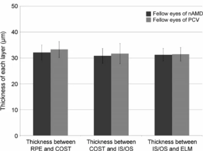
Figure 3. Thickness of each layer at the fovea in fellow eyes of
patients with unilateral neovascular age-related macular de- generation (nAMD) and polypoidal choroidal vasculopathy.Error bar is mean ± standard deviation. PCV = polypoidal choroidal vasculopathy; RPE = retinal pigment epithelium;
COST = cone outer segment tips; IS/OS = photoreceptor inner segment/outer segment; ELM = external limiting membrane.
수용체내외절경계 사이의 두께(COST-IS/OS)는 신생혈관 성 나이관련황반변성 환자군에서 30.8 ± 2.9 μm, 결절맥락 막혈관병증 환자군에서 31.8 ± 3.9 μm로 측정되었고, 광수 용체내외절경계와 외경계막 사이 두께(IS/OS-ELM)은 각각 31.3 ± 2.4 μm, 31.4 ± 2.7 μm로 측정되었다(p=0.323, 0.846).
신생혈관성 나이관련황반변성과 결절맥락막혈관병증 환자들 의 반대안에서 측정한 전체 망막두께 및 각 망막층 사이의 두께는 모두 유의한 차이를 보이지 않았다(Fig. 3).
전체망막두께 및 망막색소상피층과 원추세포외절끝 사 이 두께(RPE-COST), 원추세포외절끝과 광수용체내외절경 계 사이 두께(COST-IS/OS), 광수용체내외절경계와 외경계 막 사이의 두께(IS/OS-ELM)에 대한 두 독립된 측정자 간 의 급외상관계수는 각각 0.91, 0.83, 0.86, 0.86으로 두 측정 자 간의 각 망막층 사이 두께 측정 결과는 신뢰할 만한 재 현성을 보였다.
고 찰
나이가 들수록 원추세포초(cone sheath)가 위축되며 짧아 지고 이로 인해 망막색소상피층과 원추세포외절끝 사이의 두께가 얇아질 수 있다는 연구가 보고되었다.11 망막색소상 피층과 원추세포외절끝 사이의 조직은 망막색소상피세포, 브루크막, 원추세포초 등으로 이루어져 있으며, 나이가 들 수록 망막색소상피세포와 브루크막에는 축적물이 쌓이면 서 그 두께가 증가한다.12,13 이로 인해 망막색소상피층을 통 한 대사 물질들의 이동에 장애가 생기고, 그 결과로 영양
공급의 부족 및 허혈로 인해 원추세포 광수용체의 기능 손 상이 발생할 수 있다.14 한편, 정상 망막 소견의 노인에 비 해 단안 나이관련황반변성 환자의 반대안의 망막색소상피 세포에 리포푸신이 더 많이 축적된다는 점으로 보아,15 단 안 나이관련황반변성 환자의 반대안의 망막색소상피층의 두께는 정상 망막 소견을 가진 노인의 눈에 비해 더 두꺼울 가능성이 높다.11 그럼에도 불구하고 망막색소상피층과 원 추세포외절끝 사이의 두께가 더 얇게 측정되었다는 것은, 이를 구성하는 다른 요소인 원추세포초 등의 구조들이 오 히려 더 얇아졌을 가능성을 의미하며, 나이 증가에 따른 구 조 변화와 마찬가지로 원추세포 광수용체의 기능 손상 및 원추세포초의 위축 등을 그 원인으로 생각해 볼 수 있다. 하지만 현재까지 나이 증가 및 나이관련황반변성 발생 시 작 전후로 원추세포초가 어떠한 구조의 변화를 보이는지 명확하게 실험적으로 증명된 바가 없어 이에 관한 추가적 인 연구가 요구된다.
Kenmochi et al11은 cirrus 빛간섭단층촬영장비를 이용하 여 단안 나이관련황반변성 환자의 정상 소견을 보이는 반 대안과 단안의 기타 다른 망막 질환을 가진 환자의 정상 소 견 반대안의 망막층 사이 두께를 비교하여, 망막색소상피층과 원추세포외절끝 사이의 두께가 단안 나이관련황반변성 환자 의 반대안에서 더 얇게 측정되었음을 보고하였으며 이는 저 자들의 연구와 상통하는 결과이다. 하지만 Kenmochi et al11의 연구에서 대조군의 대부분은 단안에 다양한 망막 질환을 가진 환자들이었기 때문에, 임상적으로 정상 소견으로 보 이는 반대안에도 망막층에 여러 잠재적인 변화의 가능성이 있음을 한계로 제시하였으나, 저자들은 양안 망막 모두 임 상적으로 정상 소견을 보이는 환자들의 단안을 대조군으로 정하여 연구를 진행하였다. 또한 spectralis 빛간섭단층촬영 의 경우 cirrus 빛간섭단층촬영을 포함한 다른 종류의 빛간 섭단층촬영 장비에 비해 정상안에서 망막 두께를 측정함에 있어 더 높은 반복성을 보인다는 점 등을 고려할 때 이는 본 연구의 장점이라 할 수 있다.16,17
본 연구의 한계점은, 비록 뚜렷하게 초기 나이관련황반 변성 관련 소견이 있는 환자는 연구에서 제외하였으나, 환 자군의 반대안에도 작은 드루젠 등 잠재적인 초기 나이관 련황반변성의 변화를 내포하고 있을 가능성을 완전히 배제 할 수는 없다는 점이다. 그리고 환자군에서 나이관련황반 변성에 이환된 눈에 과거에 항혈관내피세포성장인자 치료 를 시행한 여부 및 종류, 횟수, 기간 등은 환자마다 차이가 있었다. 비록 MARINA와 ANCHOR 연구 결과 라니비주맙 으로 치료받은 환자의 반대안에 새로운 신생혈관이 생기는 발생률은 sham 또는 광역학치료와 유의한 차이가 없음을 보고하였으나,18 국내 연구에서는 24개월 이상의 경과 관찰
결과 베바시주맙으로 치료한 환자들에서 반대안의 맥락막 신생혈관 발생률이 라니비주맙 치료군에 비해 높게 나타났 다는 보고도 있으므로,19 이환된 눈에 과거에 시행하였던 항혈관내피세포성장인자 치료가 반대안에 미칠 수 있는 영 향을 완전히 배제할 수는 없겠다. 또한 현재 빛간섭단층촬 영상 각 망막층을 자동적으로 구분해주는 소프트웨어가 개 발되었으나 이에 외망막층을 이루는 미세 구조들은 포함되 지 않은 상태로, 본 연구에서 보고자 한 외망막층 내 각각 의 망막 두께는 caliper를 이용해 측정자가 수기로 측정할 수 밖에 없었으며, 빛간섭단층촬영 해상도의 한계로 인해 망막색소상피층은 외측 경계, 원추세포외절끝은 내측 경계 를 기준으로 하여 망막층 간 두께를 측정하였다. 망막색소 상피층 외측 경계부터 원추세포외절끝 내측 경계 사이에는 브루크막, 망막색소상피세포, 원추세포초, 광수용체 외절의 일부 등의 구조들이 포함될 수 있음을 고려할 때, 그 두께 의 변화에는 이러한 여러 구조들의 변화가 모두 원인이 될 수 있고, 어느 한 구조가 두께 변화의 주 원인이라고 명확 히 밝히기 어렵다. 이는 추후 빛간섭단층촬영 장비의 해상 도가 개선되고 또한 이를 통해 외망막층 내 자동 구분이 가 능해지면, 두 층을 더 명확히 구분하여 측정할 수 있게 되 어 오차의 가능성을 줄일 수 있을 것이다. 또한 실험적으로 외망막층 구조의 변화를 증명할 수 있는 연구도 더 필요하 다고 본다.
저자들은 빛간섭단층촬영을 이용해 단안 삼출성 나이관 련황반변성 환자들의 임상적으로 망막의 정상 소견을 보이 는 반대안과 양안 모두 임상적으로 망막의 정상 소견을 보 이는 환자들에서 외망막층에 속하는 각 구조들 사이의 두 께를 측정하여 비교하였다. 그 결과 망막색소상피층과 원 추세포외절끝 사이의 두께는 단안 나이관련황반변성 환자 의 무증상 반대안에서 정상안인 대조군에 비해 더 얇게 측 정되었다. 단안 나이관련황반변성 환자의 반대안에서 맥락 막 신생혈관의 발생 가능성이 더 높다는 점을 고려하면, 본 연구 결과를 통해 망막색소상피층과 원추세포외절끝 사이 의 두께가 더 얇을수록 나이관련황반변성이 발생할 위험성 도 더 높을 가능성이 있다고 본다. 상기 환자들의 장기적인 경과 관찰을 통해 망막색소상피층과 원추세포외절끝 사이 의 두께가 얇게 측정된 환자들에서 실제로 대조군에 비해 신생혈관이 유의하게 높게 발생하는지에 관한 연구 및 이 에 동반되는 망막 구조의 변화를 비교하기 위한 실험적 연 구도 더 필요할 것으로 생각한다.
REFERENCES
1) Submacular Surgery Trials Research Group, Solomon SD, Jefferys JL, et al. Incident choroidal neovascularization in fellow eyes of
patients with unilateral subfoveal choroidal neovascularization secondary to age-related macular degeneration: SST report No. 20 from the Submacular Surgery Trials Research Group. Arch Ophthalmol 2007;125:1323-30.
2) Five-year follow-up of fellow eyes of patients with age-related macular degeneration and unilateral extrafoveal choroidal neovas- cularization. Macular Photocoagulation Study Group. Arch Ophthalmol 1993;111:1189-99.
3) Risk factors for choroidal neovascularization in the second eye of patients with juxtafoveal or subfoveal choroidal neovascularization secondary to age-related macular degeneration. Macular Photocoa- gulation Study Group. Arch Ophthalmol 1997;115:741-7.
4) Klein R, Klein BE, Jensen SC, Meuer SM. The five-year incidence and progression of age-related maculopathy: the Beaver Dam Eye Study. Ophthalmology 1997;104:7-21.
5) Klein R, Knudtson MD, Klein BE. Statin use and the five-year in- cidence and progression of age-related macular degeneration. Am J Ophthalmol 2007;144:1-6.
6) Mukesh BN, Dimitrov PN, Leikin S, et al. Five-year incidence of age-related maculopathy: the Visual Impairment Project. Ophthalmology 2004;111:1176-82.
7) van Leeuwen R, Klaver CC, Vingerling JR, et al. The risk and natu- ral course of age-related maculopathy: follow-up at 6 1/2 years in the Rotterdam study. Arch Ophthalmol 2003;121:519-26.
8) Dunaief JL, Dentchev T, Ying GS, Milam AH. The role of apopto- sis in age-related macular degeneration. Arch Ophthalmol 2002;
120:1435-42.
9) Hageman GS, Luthert PJ, Victor Chong NH, et al. An integrated hypothesis that considers drusen as biomarkers of immune-medi- ated processes at the RPE-Bruch's membrane interface in aging and age-related macular degeneration. Prog Retin Eye Res 2001;20:
705-32.
10) Acton JH, Smith RT, Hood DC, Greenstein VC. Relationship be- tween retinal layer thickness and the visual field in early age-re- lated macular degeneration. Invest Ophthalmol Vis Sci 2012;53:
7618-24.
11) Kenmochi J, Ito Y, Terasaki H. Changes of outer retinal thickness with increasing age in normal eyes and in normal fellow eyes of pa- tients with unilateral age-related macular degeneration. Retina 2017;37:47-52.
12) Ramrattan RS, van der Schaft TL, Mooy CM, et al. Morphometric analysis of Bruch's membrane, the choriocapillaris, and the cho- roid in aging. Invest Ophthalmol Vis Sci 1994;35:2857-64.
13) Friedman E, Ts'o MO. The retinal pigment epithelium. II. Histologic changes associated with age. Arch Ophthalmol 1968; 79:315-20.
14) Curcio CA, Owsley C, Jackson GR. Spare the rods, save the cones in aging and age-related maculopathy. Invest Ophthalmol Vis Sci 2000;41:2015-8.
15) Sarks SH. Ageing and degeneration in the macular region: a clin- ico-pathological study. Br J Ophthalmol 1976;60:324-41.
16) Pinilla I, Garcia-Martin E, Fernandez-Larripa S, et al. Reproducibility and repeatability of Cirrus and Spectralis Fourier-domain optical co- herence tomography of healthy and epiretinal membrane eyes.
Retina 2013;33:1448-55.
17) Hafner J, Prager S, Lammer J, et al. Comparison of ganglion cell inner plexiform layer thickness by Cirrus and Spectralis optical co- herence tomography in diabetic macular edema. Retina 2017 Apr 3. doi: 10.1097/IAE.0000000000001631. [Epub ahead of print]
= 국문초록 =
단안 삼출성 나이관련황반변성 환자의 무증상 반대안의 외망막층 두께 변화
목적: 단안 삼출성 나이관련황반변성 환자의 무증상 반대안과 정상안 대조군의 외망막층의 두께 변화를 비교하고자 하였다.
대상과 방법: 결절맥락막혈관병증을 포함한 단안 삼출성 나이관련황반변성 환자 60명과 정상안 대조군 60명의 의무기록을 후향적으 로 조사하였다. 환자군의 무증상 반대안과 정상안 대조군에서 Spectralis 빛간섭단층촬영을 시행하여 중심와에서 전체 망막 두께 및 망막색소상피층, 원추세포외절끝, 광수용체내외절경계, 외경계막 사이의 두께를 각각 측정하였다.
결과: 망막색소상피층과 원추세포외절끝 사이의 두께는 단안 삼출성 나이관련황반변성 환자의 무증상 반대안에서 32.4 ± 3.0 μm, 대조군에서 35.3 ± 3.5 μm로 유의한 차이를 보였다(p<0.001). 전체 망막두께 및 원추세포외절끝과 광수용체내외절경계 사이의 두께, 광수용체내외절경계와 외경계막 사이 두께는 두 군 간 유의한 차이를 보이지 않았다(p=0.126, 0.615, 0.874). 환자군 중 신생혈관성 나이관련황반변성 및 결절맥락막혈관병증 환자들 사이의 무증상 반대안에서 측정한 총 망막두께 및 각 외망막층 사이 두께는 모두 유의한 차이가 없었다.
결론: 단안 삼출성 나이관련황반변성 환자의 무증상 반대안은 정상안에 비해 망막색소상피층과 원추세포외절끝 사이의 두께가 더 얇았다. 망막색소상피층과 원추세포외절끝 간의 두께가 더 얇을수록 나이관련황반변성이 발생할 위험성이 더 높을 가능성이 있다고 본다.
<대한안과학회지 2017;58(9):1036-1041>
18) Barbazetto IA, Saroj N, Shapiro H, et al. Incidence of new choroi- dal neovascularization in fellow eyes of patients treated in the MARINA and ANCHOR trials. Am J Ophthalmol 2010;149:939- 46.e1.
19) Lee JJ, Lew YJ, Cho SW, Kim JW. Incidence of new choroidal neo- vascularization in fellow eyes of patients treated for age-related macular degeneration. J Korean Ophthalmol Soc 2013;54:1534-39.