such as muscle wasting are common in COPD, with a high prevalence of co-morbidities such as cardiovascular disease, anxiety, and depression. COPD patients can experience exac- erbations, which are acute symptom worsenings requiring a treatment change. These events are often triggered by infec- tions
5and can result in hospitalisation or death. The natural history of COPD varies greatly between individuals. Although generally believed to be a progressive condition, there is plenty of evidence that some patients can remain relatively stable for long periods of time, even years
6,7. On the other hand, some patients display a very rapid rate of lung function decline, which has been associated with current smoking, emphysema, and exacerbations
7. The multidimensional na- ture of COPD and variation between individuals makes it a difficult condition to manage
8. Treatment approaches need to be tailored to the specific disease components present in an individual.
The typical pathological changes in COPD include (1) mu- cus gland hyperplasia and goblet cell metaplasia in the bron- chial epithelium leading to mucus overproduction, (2) airway inflammation, and (3) parenchymal destruction causing em- physematous lesions with reduced ability for gas exchange
9-11. Small airway disease (SAD) is a recognized feature of COPD
9-11and has been characterized by pathology, imaging, and physiologi- cal studies. The small airways are <2 mm diameter, and there is a dramatic increase in small airway resistance in COPD pa- tients compared to controls
12.
This article focuses on SAD in COPD. The evidence for the importance of SAD in COPD is reviewed, encompassing stud-
Introduction
Chronic obstructive pulmonary disease (COPD) is char- acterized by persistent respiratory symptoms and airflow limitation caused by exposure to noxious particles or gases
1. The most common cause of COPD is long-term exposure to cigarette smoke. The global burden of COPD is enormous, with approximately 400 million cases worldwide
2,3. COPD is a major cause of mortality worldwide, accounting for approxi- mately 3 million deaths per year
3,4.
COPD patients commonly suffer with dyspnoea, cough, and sputum production. This is associated with reduced exercise performance and fatigue. Extra-pulmonary complications
Small Airway Disease in Patients with Chronic Obstructive Pulmonary Disease
Dave Singh, M.D.
University of Manchester, University Hospital of South Manchester, Manchester, UK
Small airway disease (SAD) has been recognized for many years as a central feature of chronic obstructive pulmonary disease (COPD). Histopathology studies have shown that the narrowing and destruction of small airways in COPD combined with inflammatory cell infiltration in the submucosa increases the severity of the disease. SAD is present in the early stages of COPD and becomes more widespread over time as the disease progresses to more severe COPD. The development of inhalers containing extra-fine particles allows the small airways to be pharmacologically targeted. Recent clinical trials have shown the efficacy of extra-fine triple therapy that targets the small airways in patients with COPD.
This article reviews the importance and treatment of SAD in COPD.
Keywords: Particle Size; Therapeutics; Pulmonary Disease, Chronic Obstructive
Address for correspondence: Dave Singh, M.D.
Medicines Evaluation Unit, University of Manchester, University Hospital of South Manchester, Langley Building, Southmoor Road, Manchester M23 9QZ, UK
Phone: 44-1619464073, Fax: 44-1619461459 E-mail: [email protected]
Received: Jul. 4, 2017 Revised: Jul. 6, 2017 Accepted: Jul. 7, 2017 Published online: Sep. 1, 2017
cc It is identical to the Creative Commons Attribution Non-Commercial License (http://creativecommons.org/licenses/by-nc/4.0/).
Copyright © 2017
The Korean Academy of Tuberculosis and Respiratory Diseases.
All rights reserved.
ies that have used a range of different measurement tools to assess the nature and extent of SAD. The pharmacological targeting of SAD is also considered, using inhaled extrafine particles to optimise delivery to this important anatomical site of disease pathophysiology.
Why Are the Small Airways Damaged in Smokers?
The terminal bronchioles serve as conducting airways, lead- ing to respiratory bronchioles containing multiple alveolar openings that facilitate gas exchange. The small airways (<2 mm) are terminal or respiratory bronchioles and can first appear at the fourth generation of airway branching
12. The branching of the airway tree means that there is an increase in the cross sectional luminal area of each successive airway gen- eration. This reduces airflow velocity, which in healthy lungs allows more time for gas diffusion in the alveoli. However, a decrease in airflow velocity in the peripheral lung regions can increased the exposure to particle matter within the inspired air, including the harmful components of cigarette smoke and pollution. Solid particles diffuse at a slower rate than gases;
this increases the contact time for particulate matter with the small airways. These physical factors mean that the small air- ways have a potentially high exposure to the harmful compo- nents in cigarette smoke.
Evidence for SAD in COPD
Much of our knowledge concerning SAD in COPD has come from studying lung tissue obtained from sources such as post-mortems, lung cancer surgical resections, lung volume reduction surgery and lung transplantation programmes.
Some of the key knowledge concerning small airway resis- tance was published approximately 50 years ago, using direct assessment catheter techniques. Non-invasive lung physiol- ogy studies have also contributed information but have often suffered from an inability to robustly determine small airway resistance or the extent of small airway dysfunction. However, recent technical advances have improved the ability of non- invasive lung function testing to detect SAD. Lung imaging studies have also shown potential in recent years to measure SAD. Information regarding SAD in COPD gathered by these different methods is now reviewed.
1. Direct assessment of small airway resistance
In 1967, Macklem and Mead
13reported the direct measure- ment of small airway resistance by catheterisation of post- mortem lungs. They reported that <20% of the total lower airway resistance was attributed to small airways (<2 mm
diameter). Soon afterwards, Hogg et al.
11reported similar find- ings for the contribution of the small airways to total airway resistance in normal lungs, but also that there was a 4- to 40- fold increase in small airway resistance in patents with em- physema. Mucus plugging plus narrowing and obliteration of the small airways were the key pathological features that were associated with this large increase in resistance. Over 20 years later, catherisation during bronchoscopy in order to measure pressure and calculate small airway resistance confirmed the minor contribution of the peripheral airways to the total air- way resistance in healthy subjects, and that peripheral airways resistance is greatly increased in COPD patients
14.
The increase in small airway resistance in COPD could be explained by airway narrowing or airway obliteration. Accord- ing to Poiseuille’s law, airway resistance is proportional to the airway radius to the fourth power; therefore, when the radius is reduced by half, there is a 16-fold increase in resistance. The airways can be regarded as a parallel circuit arrangement, where total resistance for each airway generation is calculated using the formula; 1/R
T=1/R
1+1/R
2+1/R
3+1/R
4, etc. Obliteration of half of the airways would double airway resistance. These mathematical considerations indicate that the observed in- crease in small airway resistance in COPD (>4-fold) must be predominantly due to the overall reduction in the diameter of the small airways
12.
2. Pathology studies
Hogg et al.
11described the key histopathological features of SAD in 1968, showing airway narrowing and obliteration in addition to mucus plugging. In 2004, Hogg et al.
9further de- scribed the “nature of small airway disease in COPD,” report- ing that the number of inflammatory cells in the small airways increases with disease severity. Other researchers have docu- mented increased submucosal inflammatory cell numbers in COPD compared to control small airways
15,16. The cell types involved in small airway inflammation include neutrophils, macrophages, and lymphocytes, with a prominent role for CD8 lymphocytes.
The introduction of micro-computed tomography (micro-
CT) imaging has enabled identification of terminal bronchioles
in lung tissue specimens. This has allowed small airway oblit-
eration to be accurately quantified, in addition to the measure-
ment of the diameter and cross-sectional lumen area of small
airways. McDonough et al.
10showed a reduction of 89% in the
absolute number of terminal bronchioles in COPD patients
with forced expiratory volume in 1 second (FEV
1) <30% pre-
dicted compared to controls, while the cross-sectional lumen
area was reduced by 99.7%. Narrowing of the lumen of the re-
maining terminal bronchioles was observed. Small airway nar-
rowing and destruction was present in lung regions without vis-
ible evidence of emphysema. It has been proposed that small
airway narrowing and destruction precedes the development
of emphysema, and that SAD spreads distally to cause centri- lobular emphysema
10,12. This model associates the processes of SAD and emphysema. Small airway collapse may occur during exhalation in patients with emphysema because of the destruc- tion of structural components that support the small airways.
This is potentially a viscous circle, as SAD causes emphysema, which itself can further impair small airway function.
3. Lung physiology
The measurement of FEV
1by spirometry is not specific for SAD with the larger airways contributing substantially to the expired volume. Mid-expiratory flow rates have been used to detect SAD but can suffer from a high degree of variability making it less useful for follow up measurements
17, for exam- ple when measuring the effect of a therapeutic intervention.
Impulse oscillometry (IOS) offers a more specific measure- ment of small airway disease. IOS uses sound waves of vari- ous frequencies to assess respiratory resistance and reactance during tidal breathing
18. Resistance arises because of friction or air turbulence. Reactance is the energy storage capacity due to the lung’s elastic properties. Total airway resistance is increased and reactance is more negative in COPD patients compared to healthy controls
19. Small airway resistance can be measured by R5–R20, which subtracts large airway resis- tance from total airway resistance; it has been shown that 74%
of COPD patients have SAD using this method, and that the severity of SAD and symptoms using the COPD assessment test (CAT) score were significantly associated
20. SAD can cause gas-trapping on expiration
19,21, leading to hyperinflation;
this study also reported that SAD and the degree of hyperinfla- tion were significantly associated
20.
Small airway closure can impede low frequency IOS sound waves from reaching the lung periphery
19. Small airway clo-
sure during expiration results in “choke points” that cause expiratory flow limitation (EFL). Within breath analysis of IOS measurements allows EFL to be detected by the change in reactance during tidal breathing, using the difference between inspiratory and expiratory reactance measurements at 5 Hz (∆X5). Different ∆X5 thresholds have been used to classify COPD patients with EFL, with 0.28 kPa/L/sec proposed as a threshold with high sensitivity and specificity
22, while another study used ∆X5 ≥0.1 kPa/L/sec
19. Despite the different thresh- olds used, these studies have provided evidence that higher
∆ X5 values are associated with greater hyperinflation and symptoms. A recent study, using the 0.28 kPa/L/sec threshold, reported that 37.4% of COPD patients had EFL, which was as- sociated with more severe airflow obstruction (mean FEV
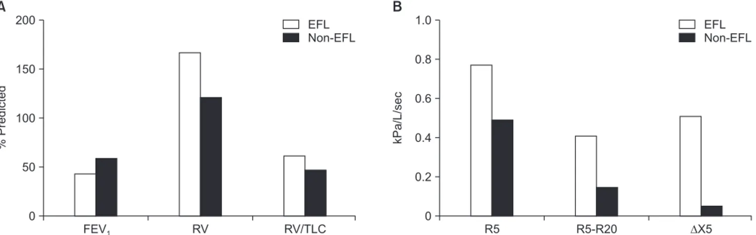
1% predicted, 42 vs. 59; p<0.0001), greater hyperinflation (residual volume medians, 166 vs. 121% predicted; p<0.0001), reduced exercise performance, and increased small airway impair- ment measured by R5–R20 (Figure 1)
23. Overall, these IOS studies have shown that EFL is a common finding in COPD, and contributes to hyperinflation which itself is known to be associated with greater symptoms.
4. Imaging studies
Computed tomography (CT) imaging in clinical practice and research has been used to define the presence and severi- ty of emphysema. The objective quantification of emphysema has used a definition of the percentage of voxels <-950 Houn- sfield Units (HU) on an inspiratory CT scan
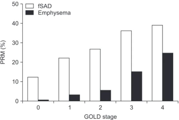
24. Gas trapping may occur due to SAD or emphysema and has been defined as the percentage of voxels <-856 HU on an expiratory CT scan. Parametric response mapping (PRM) matches the CT images from inspiratory and expiratory scans to differentiate gas trapping due to emphysema and SAD. PRM shows that
FEV1 200
150
100
50
0
EFL Non-EFL
%Predicted
RV RV/TLC R5 R5-R20 X5
1.0
0.8
0.6
0.4
0.2
0
EFL Non-EFL
kPa/L/sec
A B
Figure 1. Lung function measurements in chronic obstructive pulmonary disease patients with and without expiratory flow limitation (EFL)
23.
(A) EFL patients have worse airflow obstruction and more hyperinflation measured by residual volume (RV) and total lung capacity (TLC). (B)
EFL patients have more impulse oscillometry evidence of small airway disease (R5 and R5–R20). All differences between groups in panels (A)
and (B) are statistically significant (p<0.05). FEV
1: forced expiratory volume in 1 second.
SAD is the dominant cause of gas trapping in mild to moder- ate COPD, with emphysema gaining more prominence in very severe COPD (Figure 2)
24. This observation supports the micro-CT findings of McDonough et al.
10which suggested that SAD precedes the development of emphysema.
CT imaging lacks the resolution to assess small airway wall thickness. COPD CT imaging studies have focused on the larger airways, measuring sub-segmental bronchial wall thickness
25. Using this method, Ostridge et al.
26reported no relationship between the levels of matrix metalloproteinases (MMPs) in bronchoalveolar lavage and bronchial wall thick- ness, and some significant associations with emphysema severity. The ratio of mean lung density in expiration com- pared to inspiration was used as a marker of SAD (with higher values indicating more gas trapping due to SAD), and showed the strongest relationships with MMP levels. MMPs cause proteolysis that contributes to emphysema, and these findings further connect SAD with the pathogenesis of emphysema.
Pharmacological Treatment of COPD: An Overview
The Global Initiative for Obstructive Lung Disease (GOLD) advocates an individualized approach for the pharmacologi- cal management of COPD patients
1. The management aims are to alleviate symptoms and reduce the future risk of exac- erbations, disease progression, and mortality. GOLD recom- mends an assessment based on symptoms and exacerbation history (to predict future exacerbation risk) to categorise patients into groups (A–D) which have distinct pharmacologi- cal treatment recommendations for initial and follow up treat-
ment. COPD patients categorized into C and D groups have a high risk of future exacerbations, while B and D groups have a high burden of symptoms.
Bronchodilators are the cornerstone of COPD treatment, with long acting bronchodilators commonly used for main- tenance treatment. Long acting-acting β2 agonists (LABAs) and long-acting muscarinic antagonists (LAMAs) are bron- chodilators that can be administered as monotherapies, or as together in combination inhalers (LAMA/LABA combina- tions)
27. Long acting bronchodilators improve lung function and symptoms, and also reduce exacerbation rates. Inhaled corticosteroids (ICS) are anti-inflammatory drugs that are usually administered as part of an ICS/LABA combination;
this combination provides significant benefits compared to monotherapies for clinical outcomes including lung function, symptoms, and exacerbations
28.
COPD patients categorised into the GOLD A and B groups are recommended to receive bronchodilator treatment as initial therapy. LAMA/LABA combinations are an option for more symptomatic patients (GOLD B), as these dual bronchodilator combinations cause greater improvements in lung function and symptoms compared to long acting bronchodilator monotherapies
8,27,29. For patients categorized as GOLD C or D, long acting bronchodilator monotherapy or LABA/LAMA combination treatment are recommended for initial treatment. There is also the option to use ICS/LABA combinations to reduce exacerbation frequency. However, many patients treated with bronchodilators or an ICS/LABA combination require a step up in treatment due to persisting symptoms and/or exacerbations. The use of triple therapy (ICS plus LABA plus LAMA) is common in this situation.
Pharmacological Targeting of the Small Airways in COPD
The size of inhaled particles determines their fate within the respiratory tract, with larger particles being deposited in the oropharynx, trachea, and upper bronchial tree, while smaller particles can reach the distal airways
30. Hydrofluoroalkane propellants in pressurized metered dose inhalers have al- lowed solution formulations to be manufactured that ensure that a greater proportion of particles are deposited within the lungs rather than the oropharynx
30. Additionally, the ex- trafine fraction can be increased in order to achieve greater delivery to the small airways. Modulite technology (Chiesi Farmaceutici SpA, Parma, Italy) has been used to develop an extrafine ICS/LABA combination containing beclomethasone dipropionate and formoterol fumuorate (BDP/FF), and more recently the extrafine triple combination of BDP/FF plus gly- copyronnium bromide (BDP/FF/GB).
0 1 2 3 4
50
40
30
20
10
0
fSAD Emphysema
PRM(%)
GOLD stage