Tuberc Respir Dis 2012;72:8-14
CopyrightⒸ2012. The Korean Academy of Tuberculosis and Respiratory Diseases. All rights reserved.
Contributors of the Severity of Airflow Limitation in COPD Patients
Yoonki Hong, M.D.1, Eun Jin Chae, M.D.2, Joon Beom Seo, M.D.2, Ji-Hyun Lee, M.D.3, Eun-Kyung Kim, M.D.3, Young Kyung Lee, M.D.4, Tae-Hyung Kim, M.D.5, Woo Jin Kim, M.D.6, Jin Hwa Lee, M.D.7, Sang-Min Lee, M.D.8, Sangyeub Lee, M.D.9, Seong Yong Lim, M.D.10, Tae Rim Shin, M.D.11, Ho Il Yoon, M.D.12, Seung Soo Sheen, M.D.13, Seung Won Ra, M.D.14, Jae Seung Lee, M.D.1, Jin Won Huh, M.D.1, Sang-Do Lee, M.D.1, Yeon-Mok Oh, M.D.1
1Department of Pulmonary and Critical Care Medicine and Clinical Research Center for Chronic Obstructive Airway Diseases, and 2Department of Radiology, Asan Medical Center, University of Ulsan College of Medicine, Seoul, 3Division of Pulmonary and Critical Care Medicine, Department of Internal Medicine, Bundang CHA Hospital, CHA University College of Medicine, Seongnam, 4Department of Radiology, East-West Neo Medical Center, Kyunghee University School of Medicine, Seoul, 5Division of Pulmonology, Department of Internal Medicine, Hanyang University Guri Hospital, Hanyang University College of Medicine, Guri, 6Department of Internal Medicine, College of Medicine, Kangwon National University, Chuncheon, 7Department of Internal Medicine, Ewha Womans University Mokdong Hospital, Ewha Womans University College of Medicine, 8Division of Pulmonary and Critical Care Medicine, Department of Internal Medicine, Seoul National University College of Medicine, 9Division of Respiratory and Critical Care Medicine, Department of Internal Medicine, Korea University Anam Hospital, Korea University College of Medicine, 10Division of Pulmonary and Critical Care Medicine, Department of Medicine, Kangbuk Samsung Hospital, Sungkyunkwan University School of Medicine, 11Department of Internal Medicine, Kangnam Sacred Heart Hospital, Hallym University College of Medicine, Seoul, 12Department of Internal Medicine, Seoul National University Bundang Hospital, Seoul National University College of Medicine, Seongnam, 13Department of Pulmonary and Critical Care Medicine, Ajou University School of Medicine, Suwon, 14Department of Internal Medicine, Ulsan University Hospital, University of Ulsan College of Medicine, Ulsan, Korea
Background: Although airway obstruction in chronic obstructive pulmonary disease (COPD) is due to pathologic processes in both the airways and the lung parenchyma, the contribution of these processes, as well as other factors, have not yet been evaluated quantitatively. We therefore quantitatively evaluated the factors contributing to airflow limitation in patients with COPD.
Methods: The 213 COPD patients were aged >45 years, had smoked >10 pack-years of cigarettes, and had a post-bronchodilator forced expiratory volume in one second (FEV1)/forced vital capacity (FVC) <0.7. All patients were evaluated by medical interviews, physical examination, spirometry, bronchodilator reversibility tests, lung volume, and 6-minute walk tests. In addition, volumetric computed tomography (CT) was performed to evaluate airway wall thickness, emphysema severity, and mean lung density ratio at full expiration and inspiration. Multiple linear regression analysis was performed to identify the variables independently associated with FEV1 - the index of the severity of airflow limitation.
Results: Multiple linear regression analysis showed that CT measurements of mean lung density ratio (standardized coefficient β=−0.46; p<0.001), emphysema severity (volume fraction of the lung less than −950 HU at full inspiration; β=−0.24; p<0.001), and airway wall thickness (mean wall area %; β=−0.19, p=0.001), as well as current smoking status (β=−0.14; p=0.009) were independent contributors to FEV1.
Conclusion: Mean lung density ratio, emphysema severity, and airway wall thickness evaluated by volumetric CT and smoking status could independently contribute to the severity of airflow limitation in patients with COPD.
Key Words: Pulmonary Disease, Chronic Obstructive; Forced Expiratory Volumes; Tomography, X-Ray Computed
Address for correspondence: Yeon-Mok Oh, M.D.
Department of Pulmonary and Critical Care Medicine, and Clinical Research Center for Chronic Obstructive Airway Diseases, Asan Medical Center, University of Ulsan College of Medicine, 86, Asanbyeongwon-gil, Songpa-gu, Seoul 138-736, Korea Phone: 82-2-3010-3136, Fax: 82-2-3010-6968, E-mail: [email protected]
Received: Jul. 23, 2011 Revised: Aug. 5, 2011 Accepted: Sep. 26, 2011
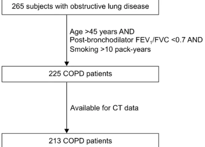
Figure 1. Selection of the study subjects from all subjects with obstructive lung disease. COPD: chronic obstructive pulmonary disease; CT: computed tomography.
Introduction
Chronic obstructive pulmonary disease (COPD) is characterized by airflow limitation with poor reversi- bility and progression1. This airflow limitation is due to pathologic processes in both the airways and lung pa- renchyma2. The relative proportion of these two proc- esses can vary considerably between individuals with the same degree of airflow limitation3.
Recent advances in multi-channel computed tomog- raphy (CT) scanning has allowed the quantitative assess- ment of both the airway and parenchymal processes.
CT measurements of the extent of emphysema, airway thickening, and air trapping, as well as exercise capacity and body mass index (BMI) have been found to corre- late with the severity of airflow limitation in COPD pa- tients4-8. However, the contribution of each factor to the severity of airflow limitation has not yet been evaluated quantitatively. We therefore evaluated factors con- tributing the severity of airflow limitation in COPD pa- tients quantitatively.
Materials and Methods
1. SubjectsThe 213 COPD patients aged >45 years, who had smoked >10 pack-years of cigarettes, and had a
post-bronchodilator forced expiratory volume in one second (FEV1)/forced vital capacity (FVC) <0.7, but did not have bronchiectasis or sequelae of pulmonary tuberculosis were analyzed in this study.
The 213 patients were selected from a group of pa- tients with obstructive lung disease (OLD), including those with COPD, asthma and overlap syndrome (Figure 1). The 265 stable patients with OLD were recruited from the pulmonary clinics of 11 hospitals in South Korea from June 2005 to October 2008. The inclusion criteria for patients with OLD have been described else- where9.
All patients were evaluated by medical interviews, physical examinations, spirometry, bronchodilator rever- sibility tests, and lung volume and six-minute walk tests. In addition, volumetric CT was performed to eval- uate airway wall thickness, emphysema severity, and mean lung density ratio at full expiration and inspira- tion. Multiple linear regression analysis was performed to identify the variables independently associated with FEV1, the index of the severity of airflow limitation.
Our Institutional Review Board approved analyses of the clinical and imaging data. Individual informed writ- ten consent was obtained from all patients.
2. Pulmonary function tests
Spirometry was performed using a Vmax 22 (Sensor- Medics, Yorba Linda, CA, USA) or a PFDX (MedGra- phics, St Paul, MN, USA). To assess post-bronchodilator FEV1 increases, spirometry was performed pre-broncho- dilation and 15 minutes after inhalation of four separate doses of salbutamol 100μg through a metered-dose in- haler (MDI) with a spacer. The post-bronchodilator FEV1 increase was expressed as the percent of the pre- dicted normal value. Lung volumes, including total lung capacity (TLC), vital capacity (VC), functional residual capacity (FRC), and residual volume (RV), were meas- ured by body plethysmography (V6200, SensorMedics or PFDX). Diffusing capacity for carbon monoxide (DLco) was measured by the single-breath method using a Vmax229D (Sensor-Medics) or a Masterlab Body (Jaeger AB, Würtsburg, Germany). All pulmonary func-
tion tests were performed as recommended by the American Thoracic Society (ATS)/European Respiratory Society (ERS)10-12.
3. Computed tomography
Volumetric CT scans were performed on all patients using the Somatom Sensation 16 (Siemens Medical Solutions, Forchheim, Germany), GE Lightspeed Ultra (General Electric Healthcare, Milwaukee, WI, USA), and Philips Brilliance 16 (Philips Medical Systems, Best, Netherlands) 16-slice multi-detector CT (MDCT) scan- ners. Patients were scanned during suspended full in- spiration and expiration in the supine position without respiratory gating. Before CT scans, patients were taught how to inhale and exhale and practiced doing so under the guidance of trained nurses. The CT param- eters were: 16×0.75 mm collimation, 100 eff. mAs, and 140 kVp for the Somatom Sensation 16; 16×0.625 mm, 300 mAs, 140 kVp, Pitch 0.938, and 0.5 sec/rot for the GE Lightspeed; and 16×0.75 mm, 133 mAs, 140 kVp, Pitch 1, and 0.75 sec/rot for the Philips 16. Acquired data were reconstructed using a standard algorithm with thicknesses of 0.625∼0.8 mm and increments of 0.625
∼0.8 mm. Each CT scanner was calibrated for water using a standard water phantom monthly and after ma- jor maintenance, and for air daily. All screening scans were performed within 24 hours after calibration. Image data were stored in the Digital Imaging and Communi- cations in Medicine (DICOM) format. Using in-house software, images of the whole lung were extracted auto- matically and the attenuation coefficient of each pixel was measured and calculated. The cutoff between nor- mal lung density and a low-attenuation area (LAA) was defined as −950 HU. From the CT data, the volume fraction of the lung less than −950 HU (V950) and the mean lung density (MLD) were calculated automatically.
The ratio of MLD on expiration and inspiration was calculated. The airway dimensions, wall area (WA), lu- men area (LA) and wall area percent (WA%; ie, WA/
(WA+LA)×100), were measured near the origin of two segmental bronchus (the right apical and left apico-pos- terior) selected by a consensus reading of two radio-
logists. The software automatically detects the airway lumen and the inner and outer boundaries of the airway wall using a full-width-half-maximum (FWHM) meth- od13. The mean value of each segmental bronchus was used for analysis8.
4. Statistical analysis
To investigate contributors associated with airflow limitation, FEV1 was used as the dependent variable in univariate and multivariate analyses. Selected independ- ent variables were BMI, six-minute walk distance (6MWD), smoking status (current or ex-smokers, smok- ing pack-years), the pulmonary function parameters TLC, FRC, RV/TLC, and DLco and the CT measurements inspiratory V950, WA%, and MLD ratio. Multiple linear regression analysis was performed to identify which var- iables were independently associated with FEV1. All statistical analyses were performed using the SPSS stat- istical package (SPSS version 12.0, SPSS Inc, Chicago, IL) and p-values <0.05 were considered significant.
Results
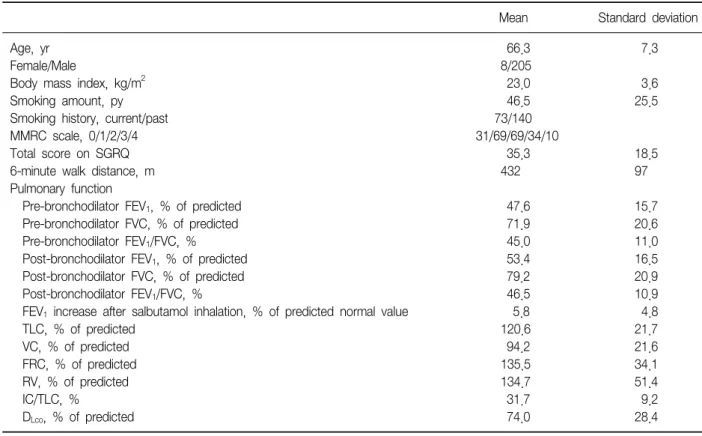
Of the 213 patients, 205 (96.2%) were men. Mean pa- tient age was 66.3±7.3 years, mean smoking history was 46.5±25.5 pack-years, and mean FEV1 was 47.6±
15.7% predicted (Tables 1, 2).
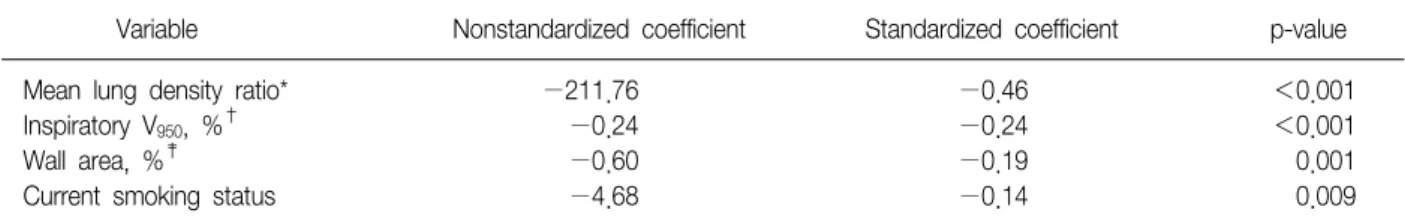
Multiple linear regression analysis showed that a sig- nificant regression model for FEV1 comprised volumetric CT measurements of MLD ratio (standardized coefficient β=−0.46; p<0.001), inspiratory V950 (%, standard- ized coefficient β=−0.24; p<0.001), WA% (standar- dized coefficient β=−0.19; p=0.001) and current smoking status (standardized coefficient β=−0.14;
p=0.009) (Table 3). The model did not include BMI, to- tal lung capacity, 6 MWD, or the other variables ana- lyzed (p≥0.10).
Multiple linear regression analysis showed that ad- justed r2 was 0.381 for volumetric CT measurements of MLD ratio, inspiratory V950 and WA%. And it showed 0.398 for CT measurements and current smoking status (Table 4).
Table 1. Characteristics of subjects
Mean Standard deviation
Age, yr 66.3 7.3
Female/Male 8/205
Body mass index, kg/m2 23.0 3.6
Smoking amount, py 46.5 25.5
Smoking history, current/past 73/140
MMRC scale, 0/1/2/3/4 31/69/69/34/10
Total score on SGRQ 35.3 18.5
6-minute walk distance, m 432 97
Pulmonary function
Pre-bronchodilator FEV1, % of predicted 47.6 15.7
Pre-bronchodilator FVC, % of predicted 71.9 20.6
Pre-bronchodilator FEV1/FVC, % 45.0 11.0
Post-bronchodilator FEV1, % of predicted 53.4 16.5
Post-bronchodilator FVC, % of predicted 79.2 20.9
Post-bronchodilator FEV1/FVC, % 46.5 10.9
FEV1 increase after salbutamol inhalation, % of predicted normal value 5.8 4.8
TLC, % of predicted 120.6 21.7
VC, % of predicted 94.2 21.6
FRC, % of predicted 135.5 34.1
RV, % of predicted 134.7 51.4
IC/TLC, % 31.7 9.2
DLco, % of predicted 74.0 28.4
Py: pack year; FEV1: forced expiratory volume in 1 second; FVC: forced vital capacity; IC: inspiratory capacity; TLC: total lung capacity;
VC: vital capacity; FRC: functional residual capacity; RV: residual volume; DLco: diffusion capacity for carbon monoxide; MMRC scale:
modified medical research council dyspnea scale; SGRQ: St George's respiratory questionnaire.
Table 2. Volumetric CT data in 213 patients with COPD Volumetric computed
tomography Mean Standard
deviation
Inspiratory V950, %* 24.5 15.7
Inspiratory mean lung density −888.6 23.1
Expiratory V950, %* 14.4 14.4
Expiratory mean lung density −841.2 41.8
Mean lung density ratio† 0.947 0.034
Wall area, %*,‡ 66.1 4.8
*Volume fraction of the lung below −950 HU. †Mean lung den- sity ratio at full expiration and inspiration. ‡Wall area/(wall area
+lumen area)×100.
Discussion
We have shown here that mean lung density ratio, inspiratory V950, and wall area %, as evaluated by volu- metric CT, as well as current smoking status were sig- nificant contributors of FEV1 in COPD patients. This
finding indicates that the severity of airflow limitation could be, in part, determined by imaging measurements of the airway and emphysema.
Structural changes, as assessed by CT scanning, have been reported to correlate with the severity of airflow limitation7,14-16. In these previous reports, the structural changes included extent of emphysema, as evaluated by low attenuation area (equivalent to V950 in this study), and abnormalities of large airway walls, as evaluated by wall thickness (equivalent to wall area % in this study).
In addition, comparison of CT scans at expiration and inspiration was reported to reflect small airway dis- eases17,18. To assess the extent of small airway disease in 34 COPD patients, we developed a CT air-trapping index (mean lung density ratio) by comparing the mean lung density seen on CT scans at full expiration and in- spiration8. The results presented here also show that mean lung density ratio is the most significant contrib-
Table 3. Multiple stepwise linear regression model for the severity of airflow obstruction (FEV1)
Variable Nonstandardized coefficient Standardized coefficient p-value
Mean lung density ratio* −211.76 −0.46 <0.001
Inspiratory V950, %† −0.24 −0.24 <0.001
Wall area, %‡ −0.60 −0.19 0.001
Current smoking status −4.68 −0.14 0.009
*Mean lung density ratio of full expiration and inspiration. †Volume fraction of the lung less than −950 HU. ‡Mean wall area, wall area/(wall area+lumen area)×100.
Table 4. Models for studying factors contributing to the severity of airflow obstruction (FEV1), as determined by multiple linear regression
Variables in the 5 different models Adjusted r2
Mean lung density ratio*, Inspiratory V950 (%)†, Wall area (%)‡ 0.381
Mean lung density ratio, Inspiratory V950 (%), Wall area (%), current smoking status 0.398
*Mean lung density ratio at full expiration and inspiration. †Volume fraction of the lung below −950 HU. ‡Wall area (%), wall area/(wall area+lumen area)×100.
utor of the severity of airflow limitation in these patients. This finding is consistent with results showing the importance of small airway disease in the patho- genesis of COPD6,19. Although obstruction of the smaller airways has been found to correlate with the severity of airflow limitation7, to date there have been no accu- rate CT-based measurements of small airway lesions in COPD patients. We have previously shown that mean lung density ratio correlated with the physiologic air-trapping index (vital capacity−FVC) and FEV18
. The results presented here validate the importance of mean lung density ratio, although it has not yet been con- firmed as directly reflecting small airway disease.
Emphysema severity and airway wall thickness, as determined by CT scans, have also been found to corre- late with FEV120-23
. Our findings confirm these results, in that we found that emphysema severity and large air- way wall thickness, as measured by CT scan, could con- tribute airflow limitation. Cigarette smoking, the major risk factor for COPD, causes abnormalities by inducing inflammation in the lung parenchyma and airways.
Indeed, we found that current smoking status was an independent contributor of FEV1 in COPD patients, whereas smoking history, as assessed by pack-years,
was not. Although the amount of previous smoking is related to the severity of COPD, it may not be an in- dependent predictor of FEV1 after correction for the morphologic changes of the airway and emphysema that are thought to result from smoking.
Our multiple linear regression analysis did not in- clude BMI, total lung capacity or 6 MWD, indicating that the contributions of these factors to FEV1 may not be independent but may be indirectly influenced by the morphologic changes measured by CT scans.
Although we evaluated FEV1 as the dependent varia- ble for contributing the severity of airflow limitation, it has been suggested that the severity of airflow limitation be evaluated by post-bronchodilator FEV1 in COPD pa- tients24. Repeat multiple linear regression analysis using post-bronchodilator FEV1 as the dependent variable re- sulted in a similar regression model, with mean lung density ratio (standardized coefficient β=−0.44; p<
0.001), inspiratory V950 (%, standardized coefficient β=
−0.30; p<0.001), wall area% (standardized coefficient β=−0.16; p=0.003) and current smoking status (stan- dardized coefficient β=−0.15; p=0.006) being in- dependent contributors of post-bronchodilator FEV1. This study had several limitations. First, the mean
lung ratio measured by volumetric CT may not com- pletely reflect small airway disease. We have not shown a direct correlation between mean lung ratio and the severity of small airway disease in COPD patients. A di- rect correlation needs to be assessed by the pathologic or pathophysiologic evaluation of small airway disease, in accordance with changes in mean lung density.
Another limitation was that our study subjects were predominantly male smokers, either present or past.
The high proportion of males in our study may be due to the high prevalence of male smokers and very low prevalence of female smokers in Korea. As there may be gender differences in COPD25,26, further inves- tigations are warranted.
In conclusion, we found that mean lung density ratio, emphysema severity, and airway wall thickness, as measured by volumetric CT, as well as smoking status, could contribute the severity of the airflow limitation in patients with COPD.
Acknowledgements
This study was supported by a grant of the Korea Healthcare Technology R&D Project, Ministry for Health, Welfare and Family Affairs, Republic of Korea (A102065).
References
1. Rabe KF, Hurd S, Anzueto A, Barnes PJ, Buist SA, Calverley P, et al. Global strategy for the diagnosis, management, and prevention of chronic obstructive pulmonary disease: GOLD executive summary. Am J Respir Crit Care Med 2007;176:532-55.
2. Hogg JC. Pathophysiology of airflow limitation in chronic obstructive pulmonary disease. Lancet 2004;
364:709-21.
3. Celli BR. Roger S. Mitchell lecture. Chronic obstructive pulmonary disease phenotypes and their clinical relevance. Proc Am Thorac Soc 2006;3:461-5.
4. Cerveri I, Dore R, Corsico A, Zoia MC, Pellegrino R, Brusasco V, et al. Assessment of emphysema in COPD:
a functional and radiologic study. Chest 2004;125:
1714-8.
5. Reilly J. Using computed tomographic scanning to ad-
vance understanding of chronic obstructive pulmonary disease. Proc Am Thorac Soc 2006;3:450-5.
6. Nakano Y, Wong JC, de Jong PA, Buzatu L, Nagao T, Coxson HO, et al. The prediction of small airway di- mensions using computed tomography. Am J Respir Crit Care Med 2005;171:142-6.
7. Hasegawa M, Nasuhara Y, Onodera Y, Makita H, Nagai K, Fuke S, et al. Airflow limitation and airway di- mensions in chronic obstructive pulmonary disease.
Am J Respir Crit Care Med 2006;173:1309-15.
8. Lee YK, Oh YM, Lee JH, Kim EK, Lee JH, Kim N, et al. Quantitative assessment of emphysema, air trap- ping, and airway thickening on computed tomo- graphy. Lung 2008;186:157-65.
9. Kim WJ, Oh YM, Sung J, Kim TH, Huh JW, Jung H, et al. Lung function response to 12-week treatment with combined inhalation of long-acting beta2 agonist and glucocorticoid according to ADRB2 polymorphism in patients with chronic obstructive pulmonary disease.
Lung 2008;186:381-6.
10. Macintyre N, Crapo RO, Viegi G, Johnson DC, van der Grinten CP, Brusasco V, et al. Standardisation of the single-breath determination of carbon monoxide up- take in the lung. Eur Respir J 2005;26:720-35.
11. Miller MR, Hankinson J, Brusasco V, Burgos F, Casaburi R, Coates A, et al. Standardisation of spirometry. Eur Respir J 2005;26:319-38.
12. Wanger J, Clausen JL, Coates A, Pedersen OF, Brusasco V, Burgos F, et al. Standardisation of the measurement of lung volumes. Eur Respir J 2005;26:511-22.
13. Wood SA, Zerhouni EA, Hoford JD, Hoffman EA, Mitzner W. Measurement of three-dimensional lung tree structures by using computed tomography. J Appl Physiol 1995;79:1687-97.
14. Nakano Y, Muro S, Sakai H, Hirai T, Chin K, Tsukino M, et al. Computed tomographic measurements of air- way dimensions and emphysema in smokers. Correla- tion with lung function. Am J Respir Crit Care Med 2000;162:1102-8.
15. Orlandi I, Moroni C, Camiciottoli G, Bartolucci M, Belli G, Villari N, et al. Spirometric-gated computed tomog- raphy quantitative evaluation of lung emphysema in chronic obstructive pulmonary disease: a comparison of 3 techniques. J Comput Assist Tomogr 2004;28:
437-42.
16. Washko GR, Criner GJ, Mohsenifar Z, Sciurba FC, Sharafkhaneh A, Make BJ, et al. Computed tomo- graphic-based quantification of emphysema and corre- lation to pulmonary function and mechanics. COPD
2008;5:177-86.
17. Bakhtavar K, Sedighi N, Moradi Z. Inspiratory and ex- piratory high-resolution computed tomography (HRCT) in patients with chemical warfare agents exposure.
Inhal Toxicol 2008;20:507-11.
18. Lucidarme O, Grenier PA, Cadi M, Mourey-Gerosa I, Benali K, Cluzel P. Evaluation of air trapping at CT:
comparison of continuous-versus suspended-expiration CT techniques. Radiology 2000;216:768-72.
19. Hogg JC, Chu F, Utokaparch S, Woods R, Elliott WM, Buzatu L, et al. The nature of small-airway obstruction in chronic obstructive pulmonary disease. N Engl J Med 2004;350:2645-53.
20. Gevenois PA, De Vuyst P, Sy M, Scillia P, Chaminade L, de Maertelaer V, et al. Pulmonary emphysema:
quantitative CT during expiration. Radiology 1996;199:
825-9.
21. Omori H, Nakashima R, Otsuka N, Mishima Y, Tomiguchi S, Narimatsu A, et al. Emphysema detected by lung cancer screening with low-dose spiral CT:
prevalence, and correlation with smoking habits and pulmonary function in Japanese male subjects. Respir-
ology 2006;11:205-10.
22. Orlandi I, Moroni C, Camiciottoli G, Bartolucci M, Pistolesi M, Villari N, et al. Chronic obstructive pulmo- nary disease: thin-section CT measurement of airway wall thickness and lung attenuation. Radiology 2005;
234:604-10.
23. Berger P, Perot V, Desbarats P, Tunon-de-Lara JM, Marthan R, Laurent F. Airway wall thickness in ciga- rette smokers: quantitative thin-section CT assessment.
Radiology 2005;235:1055-64.
24. Global Initiative for Chronic Obstructive Lung Disease (GOLD). Golobal strategy for the diagnosis, manage- ment, and prevention of chronic obstructive pulmonary disease. NHLBI/WHO workshop report. [place un- known]: GOLD; 2009.
25. Dransfield MT, Washko GR, Foreman MG, Estepar RS, Reilly J, Bailey WC. Gender differences in the severity of CT emphysema in COPD. Chest 2007;132:464-70.
26. Martinez FJ, Curtis JL, Sciurba F, Mumford J, Giardino ND, Weinmann G, et al. Sex differences in severe pul- monary emphysema. Am J Respir Crit Care Med 2007;
176:243-52.