Angiosarcoma is an uncommon tumor and it account- ing for approximately 1% of all soft tissue tumors.
Angiosarcoma is a neoplasm that arises from endothelial cells and it usually originates in the small blood vessels.
It can affect virtually any organ, but it most commonly develops in the skin and deep soft tissues, liver, spleen, heart and breast (1). We present here a rare case of pri- mary angiosarcoma of the chest wall that was associated with chronic empyema and hypervascular pulmonary metastases. To the best of our knowledge, there has been no case report on the computed tomography (CT) and positron emission tomography-computed tomogra- phy (PET-CT) features of a patient with angiosarcoma of the chest wall with hypervascular pulmonary metas-
tases.
Case Report
A 66-year-old man presented with chest tightness, pain and a palpable mass that he’d had for several months. He showed mild dyspnea at the time of admis- sion. He had a 30-year history of chronic tuberculous empyema. A chest radiograph showed a large mass in the left lower chest wall and multiple ill-defined pul- monary nodules in both lungs (Fig. 1A). The axial CT scans were obtained with using 16-channel multidetec- tor CT after the intravenous administration of contrast media. The contrast-enhanced chest CT demonstrated a well-defined lobulated mass that measured 11.5 × 9 cm in diameter with mild heterogeneous enhancement, and it arose in the left chest wall. The mean density of the solid portion of the mass was 31 Hounsfield units (HUs) on the precontrast scan and 43 HUs on the postcontrast scan with non-enhancing low density areas. This mass was associated with calcified thickened parietal pleura,
J Korean Soc Radiol 2011;64:45-48
─ 45 ─
Angiosarcoma of the Chest Wall associated with Chronic Empyema and Pulmonary Metastasis: A Case Report1
Dong Won Kim, M.D., Ki Nam Lee, M.D., Sang Yun Lee, M.D., Mee Sook Roh, M.D.
21
Department of Radiology, College of Medicine, Dong-A University
2Deparment of Pathology
, College of Medicine, Dong-A University
Received September 13, 2010; Accepted October 21, 2010Address reprint requests to : Ki-Nam Lee, M.D., Department of Radiology, College of Medicine, Dong-A University, 1, 3-ga, Dongdaesin- dong, Seo-gu, Busan 602-103, Korea.
Tel. 82-51-240-5365 Fax. 82-51-253-4931 E-mail: [email protected]
Angiosarcoma of the chest wall is a very rare tumor and it is difficult to radiological- ly differentiate this tumor from other malignant tumors. Chronic tuberculous empye- ma is a predisposing factor that has been associated with angiosarcoma. We report here on a case of a 66-year-old man with angiosarcoma that arose in the chest wall.
Computed tomography (CT) demonstrated a heterogeneous enhancing mass in the chest wall with calcified pleural thickening and multiple pulmonary nodules with the halo sign, which all indicated the presence of sarcoma with hypervascular metastases.
Index words : Hemangiosarcoma Empyema
Tuberculosus Thoracic Wall
Chronic Disease
Dong Won Kim,
et al : Angiosarcoma of the Chest Wall associated with Chronic Empyema and Pulmonary Metastasis
─ 46 ─
A B
C D
E F
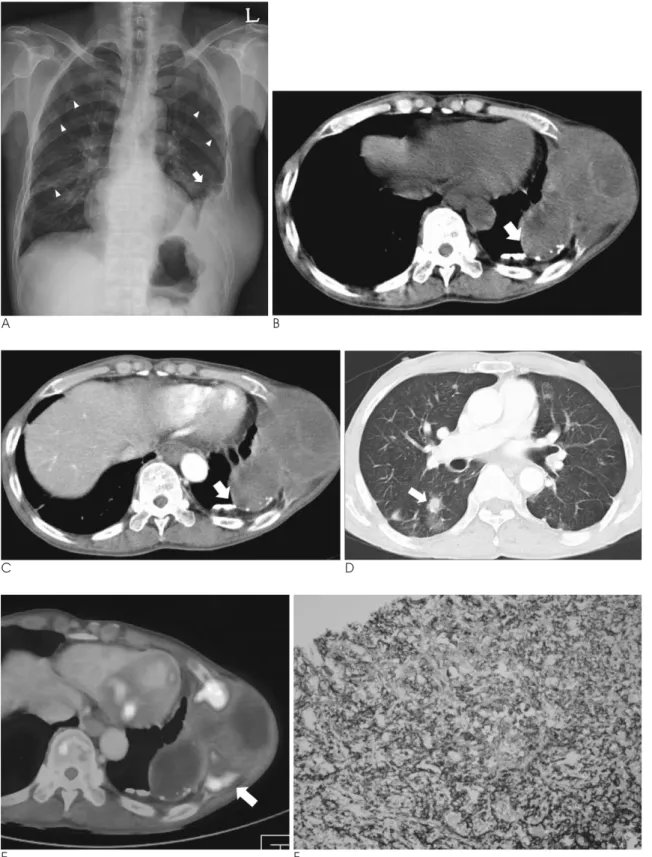
Fig. 1. A 66-year-old man with angiosarcoma of the chest wall and pulmonary metastases.
A. The chest radiograph shows an extrapulmonary mass (arrow) in the left lower chest wall and multiple ill-defined pulmonary nod- ules (arrowheads) in both lungs.
B, C. The axial CT images before (B) and after contrast injection (C) display a lobulated mass associated with calcified thickened pleu- ra, proliferated extrapleural fatty tissue (arrow) and destroyed adjacent multiple ribs, and the mass is extending into the chest wall. - D. The lung setting image at the level of the bronchus intermedius shows multiple pulmonary nodules each with a halo sign (arrow) in the right lung.
E. The PET-CT images show multiple focal areas of increased uptake in the peripheral portion of the tumor (arrow).
F. Immunohistochemically, the tumor cells are strongly positive for CD31 ( 100).
proliferated extrapleural fatty tissue and multiple de- stroyed adjacent ribs, and the tumor extended through the diaphragm with invasion of the superior portion of the spleen (Figs. 1B, C). The lung setting image showed multiple small nodules each with a surrounding halo of ground glass opacity in both lungs (Fig. 1D). PET-CT was performed after 10.3 mCi F-18 fluoro-deoxy-glucose (FDG) administration to identify the metastatic sites.
Multiple focal areas of increased uptake were seen in the peripheral portion of the mass, the sternum, ribs and the vertebral bodies (Fig. 1E). The maximum standard uptake value (maxSUV) of the solid portion of the mass was 4.55.
Surgery was not an option because of the extent of the disease. Careful biopsies were taken from the left lateral portion of the chest wall tumor and the pulmonary nod- ules. The microscopic examinations revealed the same results that the tumor was poorly differentiated, it pro- duced no definite blood vessels and it had the appear- ance of dense clumps of irregular anaplastic cells.
Immunohistochemically, the tumor cells were strongly positive for CD31 (Fig. 1F) and vimentin, while they were negative for cytokeratin, desmin, CD56, TTF-1 and smooth muscle actin. Vascular tumor can be highly suspected when the immunohistochemical staining for cytokeratin is negative and that for vimentin is positive.
CD31 is a highly specific, sensitive endothelial marker that reacts rarely and only weakly with nonvascular tu- mors. So, these were characteristic and specific findings of angiosarcoma. The pathologic diagnosis was angiosar- coma, which was in accordance with the radiologic find- ings.
The patient underwent chemoradiation treatment. A follow-up CT scan obtained six months after the diagno- sis showed newly developed bony metastases and pro- gression of the primary mass and the pulmonary metas- tases.
Discussion
Primary sarcomas of the thorax are rare. Angioarcoma is a sarcoma with a predominantly intrathoracic location such as the cardiac and mediastinal sarcomas rather than having a chest wall location (2). Angiosarcomas originating from the pleura and chest wall are extremely rare.
The pathogenesis and etiology of angiosarcoma of the chest wall are not clear. It may be caused by a long- standing inflammatory process, chronic stimulation of
mesothelial cells or the action of oncogenic substances contained in the pleura. The predisposing factors that have been implicated include trauma, chronic lym- phedema, radiation, foreign bodies, thorium, viral infec- tion and chronic empyema (3). The histopathologic diag- noses of the reported cases associated with chronic empyema have been malignant lymphoma, squamous cell carcinoma, mesothelioma, malignant fibrous histio- cytoma, liposarcoma and angiosarcoma. The mean du- ration of chronic empyema before the diagnosis of ma- lignancy has been reported to be about 25 years (4).
Such a predisposing factor was present in our patient.
Primary malignant chest wall tumors typically mani- fest as large, palpable, rapidly growing masses. The rare empyema-associated malignant tumors that need to be considered in the differential diagnosis of angiosarcoma include lymphoma, malignant fibrous histiocytoma and squamous cell carcinoma. However, not much informa- tion has appeared in the literature with regard to the specific radiologic manifestations of these tumors. The imaging features of many malignant chest wall tumors are nonspecific (5). The specific feature of chronic empyema-associated lymphoma is a symmetric growth pattern of a mass at the margin of the chronic empyema (6). The reported CT findings of angiosarcoma reveal a nonspecific soft tissue chest wall mass with attenuation similar to that of muscle or a circumferential lobular pleural mass with various degrees of enhancement after the intravenous injection of contrast medium (7, 8). In our case, CT demonstrated a large well-defined lobulat- ed mass with mild heterogeneous enhancement around the calcified thickened parietal pleura, which suggested the occurrence of malignancy in the background of chronic empyema. CT also demonstrated multiple small nodules each with a surrounding halo of ground glass opacity in both lungs. This appearance is usually associ- ated with hemorrhagic nodules. Many infectious, neo- plastic and inflammatory processes cause this halo sign (9). However, if this presents together with hypervascu- lar tumor such as angiosarcoma, choriosarcoma, os- teosarcoma and melanoma, then multiple pulmonary nodules with a halo of ground glass opacity suggest the presence of lung metastases. Angiosarcoma may be dis- tinguished from other tumors by a history of chronic empyema along with hypervascular metastasis.
PET with 18F-FDG may be used for the staging of an- giosarcoma. Some reports have shown focal, intense ac- cumulation of FDG in angiosarcomas of the chest wall, pleura, breast and liver, with an SUV up to 7.5 (7, 10). In
J Korean Soc Radiol 2011;64:45-48
─ 47 ─
our case, PET-CT demonstrated multiple focal areas of increased uptake in the peripheral portion of the chest wall mass, with no evidence that the pulmonary nod- ules were hypermetabolic. We suggest that the pul- monary nodules showed no significant uptake of FDG because the size of the solid portion of the nodules was less than 7 mm.
We have described the CT and PET features of an- giosarcoma and pulmonary metastasis. A mass in the chest wall that is associated with chronic empyema and multiple pulmonary nodules with the halo sign suggests the presence of angiosarcoma with hypervascular metastases.
References
1. Patel AM, Ryu JH. Angiosarcoma in the lung. Chest 1993;103:
1531-1535
2. Gladish GW, Sabloff BM, Munden RF, Truong MT, Erasmus JJ, Chasen MH. Primary thoracic sarcomas. Radiographics 2002;22:
621-637
3. Aozasa K, Naka N, Tomita Y, Ohsawa M, Kanno H, Uchida A, et al. Angiosarcoma developing from chronic pyothorax. Mod Pathol 1994;7:906-911
4. Minami M, Kawauchi N, Yoshikawa K, Itai Y, Kokubo T, Iguchi M, et al. Malignancy associated with chronic empyema: radiologic assessment. Radiology 1991;178:417-423
5. Tateishi U, Gladish GW, Kusumoto M, Hasegawa T, Yokoyama R, Tsuchiya R, et al. Chest wall tumors: radiologic findings and pathologic correlation: part 2. Malignant tumors. Radiographics 2003;23:1491-1508
6. Ueda T, Andreas C, Itami J, Miyakawa K, Fujimoto H, Ito H, et al.
Pyothorax-associated lymphoma: imaging findings. AJR Am J
Roentgenol 2010;194:76-847. Del Frate C, Mortele K, Zanardi R, Hunsaker AR, Nikpoor N, Cibas ES, et al. Pseudomesotheliomatous angiosarcoma of the chest wall and pleura. J Thorac Imaging 2003;18:200-203
8. Sugita R, Takezawa M, Itinohasama R. Primary angiosarcoma of the chest wall: CT and MR findings. Radiat Med 2002;20:101-103 9. Pinto PS. The CT Halo Sign. Radiology 2004;230:109-110
10. Maeda T, Tateishi U, Hasegawa T, Ojima H, Arai Y, Sugimura K.
Primary hepatic angiosarcoma on coregistered FDG PET and CT images. AJR Am J Roentgenol 2007;188:1615-1617
Dong Won Kim, et al : Angiosarcoma of the Chest Wall associated with Chronic Empyema and Pulmonary Metastasis
─ 48 ─
대한영상의학회지 2011;64:45-48
만성 농흉과 연관하여 발생한 흉벽의 혈관육종과 폐전이1
1
동아의대병원 영상의학과
2