• 검색 결과가 없습니다.

Massive Necrotizing Fasciitis of the Chest Wall: A Very Rare Case Report of a Closed Thoracostomy Complication

N/A
N/A
Protected

Academic year: 2021

Share "Massive Necrotizing Fasciitis of the Chest Wall: A Very Rare Case Report of a Closed Thoracostomy Complication"

Copied!
4
0
0

로드 중.... (전체 텍스트 보기)

전체 글

(1)

404 http://www.jchestsurg.org

JCS

Journal of Chest Surgery

Case Report

Massive Necrotizing Fasciitis of the Chest Wall: A Very Rare Case Report of a Closed Thoracostomy Complication

Sangwook Chun, M.D., Gyeongho Lee, M.D., Kyoung Min Ryu, M.D., Ph.D.

Department of Thoracic and Cardiovascular Surgery, Dankook University Hospital, Cheonan, Korea

ARTICLE INFO

Received September 25, 2020 Revised October 18, 2020 Accepted October 20, 2020 Corresponding author Kyoung Min Ryu Tel 82-41-550-7640 Fax 82-41-550-7060 E-mail [email protected] ORCID

https://orcid.org/0000-0001-8461-6010

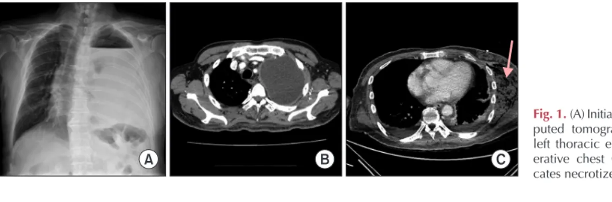
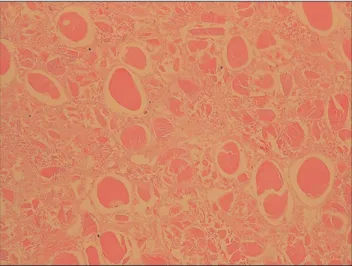
We present a case study of necrotizing fasciitis (NF), a very rare but dangerous complica- tion of chest tube management. A 69-year-old man with shortness of breath underwent thoracostomy for chest tube placement and drainage with antibiotic treatment, followed by a computed tomography scan. He was diagnosed with thoracic empyema. Initially, a non-cardiovascular and thoracic surgeon managed the drainage, but the management was inappropriate. The patient developed NF at the tube site on the chest wall, requiring emergency fasciotomy and extensive surgical debridement. He was discharged without any complications after successful control of NF. A thoracic surgeon can perform both tube thoracostomy and tube management directly to avoid complications, as delayed drainage might result in severe complications.

Keywords: Empyema, Chest tubes, Necrotizing fasciitis, Case report

Copyright

© 2021, The Korean Society for Thoracic and Cardiovascular Surgery

This is an Open Access article distributed under the terms of the Creative Commons Attribution Non-Commercial License (http://creativecommons.org/licenses/

by-nc/4.0) which permits unrestricted non-commercial use, distribution, and reproduction in any medium, provided the original work is properly cited.

Case report

A 69-year-old man was brought to the emergency room due to shortness of breath. The patient had diabetes and a history of cerebral infarction 10 years ago. The sequelae of cerebral infarction included slight motor weakness of the left leg that did not interfere with activities of daily life. He had received conservative treatment for symptoms such as myalgia, fever, and chills at a nearby hospital 2 weeks prior.

He was transferred to Dankook University Hospital for shortness of breath that had started 1 day before admis- sion. Left-sided hydrothorax was observed on an initial chest X-ray conducted in the emergency room. Conse-

quently, thoracostomy was performed for chest tube place- ment. The drainage was also very turbid. Subsequent chest computed tomography (CT) revealed left thoracic empy- ema (Fig. 1). Laboratory tests revealed an initial C-reactive protein level of 30.76 mg/dL, a white blood cell count of 15.71×10

3

/μL, and a negative tuberculosis test result. A pleural fluid culture study, the results of which came back on the sixth hospital day, showed the growth of Strepto- coccus constellatus and an anaerobe. The patient was ad- mitted with the chest tube, and ceftriaxone and clindamy- cin were used as empirical antibiotics. When the total amount of drainage reached 1 L on the day of admission, tube clamping was performed considering re-expansion

Fig. 1. (A) Initial chest X-ray. (B) Com- puted tomography (CT) revealing left thoracic empyema. (C) Preop- erative chest CT; the arrow indi- cates necrotized muscle.

A B C

https://doi.org/10.5090/jcs.20.125

pISSN: 2765-1606 eISSN: 2765-1614

J Chest Surg. 2021;54(5):404-407

(2)

405

Sangwook Chun, et al. Necrotizing Fasciitis due to Chest Tube Complication

http://www.jchestsurg.org

JCS

edema. The next day, natural draining resumed. From that day onward, redness, warmth, swelling, and tenderness were observed in the left flank. On the following day, symptoms worsened, and another CT scan was performed.

On chest CT, signs of infection, including gas formation, were found in the soft tissue and the muscle layer of the left chest wall (Fig. 1). At this point, an internal medicine doctor contacted cardiovascular and thoracic surgery de- partment, and we performed surgery immediately, after making a diagnosis of necrotizing fasciitis (NF). Surgery was performed under general anesthesia, with the patient in the right lateral position. Wide sterilization and draping were performed from the axilla to the buttock. An approx- imately 60-cm horizontal incision was made along the left flank from the scapula tip to the anterior superior iliac spine. There was a large amount of pus, and the anatomy of the muscle was indistinguishable due to severe infective necrosis. Necrotic tissues were removed, including those at the subcutaneous tissue, latissimus dorsi, teres muscle, ser- ratus anterior muscle, and gluteus muscle, using a scalpel and electrocautery (Fig. 2). The surgery was completed with an open-dressing using betadine wet gauze. NF was confirmed by pathology (Fig. 3). The antibiotics were switched to meropenem and vancomycin as empirical therapy. Acinetobacter baumannii complex growth was noted in cultures of samples collected during surgery;

therefore, we changed vancomycin to tigecycline based on antibiotic sensitivity. Wound irrigation and open dressing were performed daily under consultation with the ortho- pedic surgery department. On postoperative day 6, we ap- plied negative-pressure wound therapy. Extubation was performed on postoperative day 12, and the patient was transferred to the general ward. On postoperative day 35, delayed wound closure was performed. C-reactive protein levels decreased gradually after surgery and reached 0.31

mg/dL at discharge. The patient was discharged from the hospital on day 98 and currently continues to be monitored via follow-up through outpatient visits.

The study was approved by the Institutional Review Board of Dankook University Hospital (IRB approval no., 2020-09-018). The requirement for informed consent was waived.

Discussion

NF refers to extensive necrosis of the superficial fascia, resulting in widespread damage to the surrounding tissues and extreme systemic toxicity regardless of the specific bacterial etiology. NF was first described by Hippocrates in approximately 500 BC. Despite advances in understanding this disease and improvements in medical treatment, NF-related mortality remains high (at 25%–30%) and has

Fig. 2. Wound pictures. (A) Imme- diately postoperative. (B) Shoelace- sutured wound. (C) Before delayed wound repair.

A B C

Fig. 3. Microscopic image of removed tissue reveals necrotic tis-

sues without nuclei (H&E, ×200).

(3)

406

https://doi.org/10.5090/jcs.20.125

http://www.jchestsurg.org

JCS

not improved significantly [1]. NF is caused not only by β -hemolytic streptococci, but also by a number of different organisms [2]. Therefore, in recent years, NF has been re- garded a clinical entity, not a specific bacterial infection.

The incidence of NF is nearly 1,000 cases per year in the United States. NF incidence has increased from 1980 to 2000, but the exact reason remains unclear. Although it is extremely rare, NF is a life-threatening condition requiring early aggressive intervention in the form of salvage.

The initial injuries are minor in 80% of NF patients. Re- cently, NF has been reported to occur predominantly at the extremities and on the trunk. Infection involves the subcu- taneous tissue, but may extend downward to the fascia or muscle. The depth of the primary infection site is import- ant and related to mortality [1]. The risk factors for poly- microbial NF include obesity, chronic renal failure, alco- holism, trauma, surgical incision, and indwelling catheters.

The reason for NF in our case seems have been indwelling catheter-tube thoracostomy.

The signs and symptoms vary, including subcutaneous and fascial necrosis, cellulitis, edema, disorientation or ap- athy, and skin discoloration, with only 10%–40% of pa- tients presenting these classic signs. Local erythema and swelling, along with pain, are the most common signs of NF [1].

Radiographic modalities are sometimes needed to con- firm the diagnosis of NF. CT is the most sensitive diagnos- tic modality because it can show inflammatory changes such as fascial edema, thickening, abscesses, and gas for- mation. In our case, CT showed necrotized muscle with gas formation. Although radiologic modalities are ad- vanced and conventionally used, surgery remains the gold standard modality for the diagnosis of NF.

The basic treatment for NF is surgical debridement.

Some studies have reported that the most important deter- minant of mortality is the timing and adequacy of initial debridement. Wong et al. [3] reported a 9-fold increase in mortality if the procedure was delayed for over 24 hours from the time of hospital admission. Some studies also showed remarkable increases in mortality with delays and inadequate operative interventions.

Antibiotics also play an important role in improving sys- temic sepsis and bacterial spread. Traditionally, an empiri- cal regimen including penicillin and clindamycin was rec- ommended to target Gram-positive and anaerobic organisms.

However, the recommended initial antibiotic regimen has recently changed because of the emergence of resistant mi- croorganisms [1].

The successful treatment of NF requires coordination

between surgical management and medical care. No fac- tors other than rapid widespread operative debridement and appropriate antibiotics have an effect on mortality.

Although uncommon, complications associated with tube thoracostomy do occur and include subcutaneous emphysema, chest wall hematoma, hemothorax, lung lac- eration, chylothorax, and injury to mediastinal structures, the diaphragm, or intra-abdominal viscera [4]. NF of the chest wall was first reported by Pingleton and Jeter [5] in 1983, and its incidence is extremely rare worldwide.

Adequate drainage of early parapneumonic effusions is usually achieved with simple tube thoracostomy. However, the success rate deteriorates to 35%–39% in cases of true empyema [4]. The suction drainage technique is safe, quick, and relatively painless. It can also clear large amounts of pus, resulting in rapid clinical improvement [6]. As men- tioned above, if pleural infection is confirmed, then imme- diate and complete drainage is necessary.

It would be best for a thoracic surgeon to perform tube thoracostomy and tube management directly, but there are cases where other departments have to provide assistance due to the shortage of thoracic surgeons. In such cases, im- proper drainage of empyema and complications resulting from this are rare, but sufficient care is required. Likewise, it is necessary to be wary of re-expansion edema, a compli- cation that may occur due to rapid drainage of effusion;

however, in cases of infective effusion, it is necessary to be aware that drainage may be delayed, resulting in serious complications, and to always closely monitor the proper drainage of the chest tube. As there may be a lack of un- derstanding of this condition in other departments, thor- ough education and immediate inter-department consulta- tions are required.

Conflict of interest

No potential conflict of interest relevant to this article was reported.

ORCID

Sangwook Chun: https://orcid.org/0000-0003-1561-442X Gyeongho Lee: https://orcid.org/0000-0001-5928-3939 Kyoung Min Ryu: https://orcid.org/0000-0001-8461-6010

References

1. Sarani B, Strong M, Pascual J, Schwab CW. Necrotizing fasciitis:

current concepts and review of the literature. J Am Coll Surg

(4)

407

Sangwook Chun, et al. Necrotizing Fasciitis due to Chest Tube Complication

http://www.jchestsurg.org

JCS

2009;208:279-88.

2. Rea WJ, Wyrick WJ Jr. Necrotizing fasciitis. Ann Surg 1970;172:957- 64.

3. Wong CH, Chang HC, Pasupathy S, Khin LW, Tan JL, Low CO.

Necrotizing fasciitis: clinical presentation, microbiology, and deter- minants of mortality. J Bone Joint Surg Am 2003;85:1454-60.

4. de Hoyos A, Sundaresan S. Thoracic empyema. Surg Clin North Am 2002;82:643-71.

5. Pingleton SK, Jeter J. Necrotizing fasciitis as a complication of tube thoracostomy. Chest 1983;83:925-6.

6. Cummin AR, Wright NL, Joseph AE. Suction drainage: a new ap-

proach to the treatment of empyema. Thorax 1991;46:259-60.

수치

Fig. 1. (A) Initial chest X-ray. (B) Com- Com-puted tomography (CT) revealing  left thoracic empyema
Fig. 3. Microscopic image of removed tissue reveals necrotic tis- tis-sues without nuclei (H&E, ×200).

참조

관련 문서

A study on the safety analysis of steam generator tube plugging level upward in Hanbit Unit #5,6..

As a result, the development of Internet media allowed netizens to participate in Internet journalism, which is limited through media convergence, but can

Using this leap motion, a surgeon can perform the virtual shoulder arthroscopic surgery skillfully and reduce mistakes in surgery.. The simulation using leap motion shows

As a result, this study suggests some desirable teaching methods to gain knowledge of traditional Korean music in a familiar way, as well as a simple and effective teaching

Second, as a result of bootstrapping results the to digital communication competencies mediate relationship between high-performance human resource management

At the end of the study, a reevaluation of each study case was performed with the same questionnaire. The result shows there has been a meaningful result in the group

These optimum design requirements include: i) a minimum number of tube turns for a PHP operable in top heat mode, ii) the use of R-123 as working fluid with 50 % charging

Laminar flow through a tube (pipe) of constant diameter → Poiseuelle flow.. for incompressible