An inflammatory myofibroblastic tumor is a benign inflammatory tumefaction of unknown etiology. It is relatively rare but could occur at various locations throughout the body (1). Inflammatory myofibroblastic tumors are generally considered a benign mass and in- clude inflammatory cells, histiocytes, and fibroblasts (1).
Nevertheless, aggressive features that may mimic a ma- lignant tumor have been described. We present a malig- nancy mimicking inflammatory myofibroblastic tumor in the left upper lobe which extended to the chest wall and spinal canal with bony destruction.
Case Report
A 46-year-old man presented with pain on the left
shoulder and upper back for three months. He had numbness on both lower legs and had difficulty walking and had no previous medical issues.
A chest radiograph showed an ill-defined increased opacity of about 6cm in diameter located in the left up- per lung zone above the aortic arch (Fig. 1A). The con- trast enhanced computed tomography (CT) scans showed strongly enhanced soft tissue mass in the apico- posterior segment of the left upper lobe, which extend- ed to the chest wall and left 3rd, and 4th rib destruction (Fig. 1B). The mass also extended into the spinal canal with thecal sac compression. T1-weighted images of magnetic resonance (MR) images demonstrated a het- erogeneous mass with a signal intensity slightly greater than that of skeletal muscle (Fig. 1C). It was slightly hy- perintense on T2-weighted images (Fig. 1D), and moder- ately enhanced on gadolinium-enhanced images (Fig.
1E). The signal intensity of the bone marrow of T3 and T4 vertebral bodies was diffusely decreased on T1- and T2-weighted images.
Laboratory examinations revealed increased free kap- pa light chain (35.70 mg/L) and increased serum free
J Korean Soc Radiol 2010;63:145-148
─ 145 ─
Aggressive Pulmonary Inflammatory Myofibroblastic Tumor with Chest Wall Invasion: A Case Report1
Sung Hee Park, M.D., Soon Gu Cho, M.D., Hyeon Gyu Yi, M.D.
2, Suk Jin Choi, M.D.
3, Kyung Hee Lee, M.D.
1
Department of Radiology, Inha University, School of Medicine
2
Department of Internal Medicine, Inha University, School of Medicine
3
Department of Pathology, Inha University, School of Medicine Received January 27, 2010 ; Accepted April 20, 2010
Address reprint requests to : Kyung Hee Lee, M.D., Department of Radiology, Inha University Hospital, 3-ga, Shinheung-dong, Jung-gu, Incheon 400-711, Korea.
Tel. 82-32-890-2769 Fax. 82-32-890-2743 E-mail: [email protected]
Inflammatory myofibroblastic tumor is a benign lesion in pathology. However it may be associated with the invasion of adjacent structures mimicking malignancy. We de- scribe a case of aggressive pulmonary inflammatory myofibroblastic tumor with chest wall invasion and spine destruction.
Index words : Lung Neoplasms
Tomography, X-Ray Computed Thorax
Magnetic Resonance Imaging
Granuloma, Plasma Cell
Lung Diseases
Sung Hee Park, et al : Aggressive Pulmonary Inflammatory Myofibroblastic Tumor with Chest Wall Invasion
─ 146 ─
A B
C D
E
Fig. 1. Pulmonary inflammatory myofibroblastic tumor with bone destruction and spinal canal invasion in a 45-year-old man.
A. Chest radiograph shows ill-defined increased opacity (arrow) in the left upper lobe above the aortic arch.
B. Contrast-enhanced axial CT scan reveals a well-enhanced soft tissue mass (arrow) in the apicoposterior segment of the left up- per lobe which extended to the chest wall with left 3 rd, and 4th rib destruction. The mass also invaded into the spinal canal with cord compression.
C-E. Axial MR image shows a heterogeneous mass (arrow) with
signal intensity slightly greater than that of the skeletal muscle
on a T1-weighted image, slightly hyperintense on a T2-weighted
image and moderately enhanced on a gadolinium-enhanced T1-
weighted image.
lambda light chain (35.20 mg/L). The serum kappa to lambda ratio was within normal limits (normal refer- ence range, 0.26-1.65). The serum protein electrophore- sis showed increased gamma-globulin fraction with a polyclonal feature. Thus, we could rule out a plasmacy- toma, a malignant plasma cell tumor.
A percutaneous needle biopsy was performed.
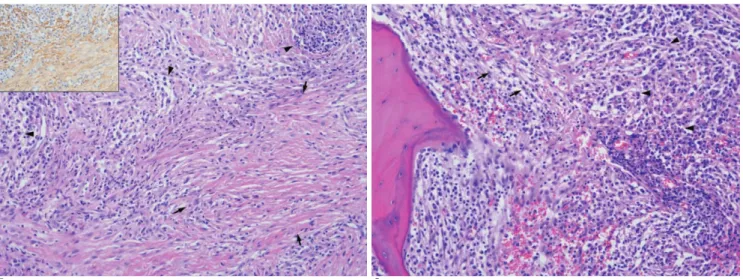
Microscopic examination revealed a marked infiltration of plasma cells. Subsequently, a surgical biopsy with de- bulking operation was done. The mass could not be completely removed because of its invasion into the spinal canal and vertebral body. A histologic examina- tion demonstrated that the lesion was composed of in- flamed fibrous connective tissue showing compact fasci- cular spindle cell proliferation (Fig. 1F) and trabecular bone with massive plasmacytic infiltration (Fig. 1G).
Immunophenotypically, the myofibroblasts were nega- tive for ALK1 (anaplastic large cell lymphoma kinase 1) and positive for desmin and smooth muscle actin. The plasma cells were polyclonal for the immunoglobulin light chain.
Radiotherapy was administered (45 Gy), with no cor- ticosteroid therapy and the patient had no significant side effects except for mild back pain.
Discussion
An inflammatory myofibroblastic tumor is a relatively rare benign inflammatory process that can occur in vari-
ous organs of the body, lung, orbit, nasal sinuses, liver, spleen, pancreas, bowel, kidney, urinary bladder, testis, heart, lymphatic system, skin and so on (1). However, the lung is the most frequently involved organ (2). The pathogenesis of the inflammatory myofibroblastic tu- mor is uncertain. Histopathologic examination reveals infiltration of polygonal lymphocytes, plasma cells, and macrophages without the evidence of atypia, and is composed of various inflammatory cells. Therefore, an inflammatory myofibroblastic tumor is thought to be an inflammatory process rather than a neoplasm (3).
The patients who have a pulmonary inflammatory myofibroblastic tumor frequently complain of nonspe- cific symptoms such as cough, fever, dyspnea, cyanosis, hemoptysis, and chest pain (3, 4).
The vast majority of pulmonary inflammatory myofi- broblastic tumors occur as solitary, well-defined masses.
They typically occur in an intraparenchymal location, but may occur in the trachea and the bronchi (5).
Nevertheless, extrapulmonary extensions suggestive of malignancy have been described in some previous re- ports (2, 3) and have been found to invade the medi- astinum and diaphragm (2), as well as the thoracic verte- bra (3). The case we report here showed an ill-defined lung mass with infiltration to the adjacent structures in- cluding vertebrae and ribs with imaging findings indi- cating severe destruction. However, no laboratory evi- dence of plasmacytoma was found, and immunoperoxi- dase reactions for light chains evidently demonstrated
J Korean Soc Radiol 2010;63:145-148
─ 147 ─
F G
Fig. 1. F. Microscopically, there is fascicular proliferation of plump spindle cells featuring myofibroblast (arrows) and inflammatory infiltrates mainly composed of plasma cells (arrow heads) (H & E, ×200). Inset shows myofibroblast with positive staining for smooth muscle actin.
G. The marrow space of bone show extensive infiltration of mature plasma cells (arrow heads) along with loose myofibroblastic
(arrows) proliferation.
the polygonal nature of the plasma cells.
On chest radiograph, pulmonary inflammatory myofi- broblastic tumor typically appears as a solitary, periph- eral, sharply circumscribed, lobulated mass (6). CT fea- tures including contrast enhancement and the patterns of calcification have been reported as variable and non- specific (5). However, they most commonly appeared with heterogeneous attenuation and enhancement on CT scans. On T1-weighted images, these tumors have intermediate signal intensity, and have high signal inten- sity on T2-weighted images (6). In our case, the mass had an ill-defined border and showed homogeneous en- hancement on CT scans. On MR image, the mass was hypointense on T1-weighted images, iso- to slight hyper- intense on T2-weighted images and markedly enhanced after gadolinium was administered.
The radiologic differential diagnosis for an inflamma- tory myofibroblastic tumor occurring as a solitary pul- monary nodule includes primary or secondary neo- plasm, hamartoma, chondroma, hemangioma, granulo- ma, and pulmonary sequestration (6). However, the dif- ferential diagnosis may be difficult in a case with aggres- sive features. In our case, the initial diagnosis was a ma- lignant tumor, which originated from lung or chest wall.
Complete surgical resection is considered the most ef- fective treatment of an inflammatory myofibroblastic tumor (7). Steroid therapy or radiation therapy could be effective in surgically unresectable cases (4). Our case
was impossible to make the complete resection because of invasion to the adjacent structures. Our case study underwent radiation therapy after an excisional biopsy.
In conclusion, an inflammatory myofibroblastic tu- mor is a benign lesion of unknown etiology. However, it may demonstrate locally aggressive behavior mimicking a malignant tumor. We reported an extremely rare case of an inflammatory myofibroblastic tumor that invaded into the chest wall and spinal canal.
References
1. Boutarbouch M, Arkha Y, Rifi L, Derraz S, El Ouahabi A, El Khamlichi A. Intradural cervical inflammatory pseudotumor mim- icking epidural hematoma in a pregnant woman: case report and review of the literature. Surg Neurol 2008;69:302-305
2. Corneli G, Alifano M, Forti Parri S, Lacava N, Boaron M. Invasive inflammatory pseudo-tumor involving the lung and the medi- astinum. Thorac Cardiovasc Surg 2001;49:124-126
3. Hong HY, Castelli MJ, Walloch JL. Pulmonary plasma cell granu- loma inflammatory pseudotumor with invasion of thoracic verte- bra. Mt Sinai J Med 1990;57:117-121
4. Kim JH, Cho JH, Park MS, Chung JH, Lee JG, Kim YS, et al.
Pulmonary inflammatory pseudotumor-a report of 28 cases.
Korean J Intern Med 2002;17:252-258
5. Bahadori M, Liebow AA. Plasma cell granulomas of the lung.
Cancer 1973;31:191-208
6. Narla LD, Newman B, Spottswood SS, Narla S, Kolli R.
Inflammatory pseudotumor. Radiographics 2003;23:719-729 7. Scott L, Blair G, Taylor G, Dimmick J, Fraser G. Inflammatory
pseudotumors in children. J Pediatr Surg 1988;23:755-758 Sung Hee Park, et al : Aggressive Pulmonary Inflammatory Myofibroblastic Tumor with Chest Wall Invasion
─ 148 ─
대한영상의학회지 2010;63:145-148
흉벽침범을 동반한 폐의 염증성근섬유모세포종:
증례 보고11
인하대학병원 영상의학과
2
인하대학병원 내과
3