Parachordoma is an extremely rare soft tissue tumor with unique immuno-histochemical features, and it can arise from the arm, thigh, leg, chest wall, and buttock (1-6). The histogenesis of parachordoma is still contro- versial. These tumors display a slow growing nature, but they also show locally destructive growth. After surgery, local recurrence has been reported in some cas- es (1, 2).
We report the imaging features of histologically con- firmed parachordoma arising from the chest wall.
Case Report
A 32-year-old man visited our hospital and presented with right upper back pain, but he was without any res- piratory symptoms. The physical examination showed neither a palpable mass nor skin changes. Chest plain
radiograph revealed a smooth margined, pleural-based mass of the right hemithorax (Fig. 1A), and a CT scan was performed for further evaluation of the mass.
About a 5 cm-sized, pleural-based, non-calcified soft tis- sue mass was noted in the right lateral chest wall. The mass was smoothly marginated and it had caused ero- sion of the adjacent 5 th and 6th ribs with focal destruc- tion and hypertrophy (Fig. 1B). Contrast-enhanced CT scan revealed a homogeneous non-enhancing soft tissue mass of the right lateral chest wall. A small portion of the mass had penetrated chest wall (Fig. 1C). There was no significant lymph node enlargement noted.
Technetium-99 m methyl di-phosphonate (Tc-99m MDP) whole body bone scan showed an area of faint in- creased radioactivity at the right 6th rib region. After considering all imaging features, we thought there was the possibility of a malignant peripheral nerve sheath tu- mor or parosteal malignant bone tumor such as os- teosarcoma or metastasis.
Percutaneous core biopsy was performed with 19.5- gauge coaxial biopsy needle (AUTOVACⓇ, Bard- Angiomed, karlsruhe, Germany) under fluoroscopic guidance. The pathological analysis showed tiny clus- ters of cells with vacuolated cytoplasm in the back- ground of chondroid matrix; there was positive staining for keratin, vimentin, and S-100 protein, which is sug- gestive of parachordoma (Fig 1D, 1E). En-bloc resection
J Korean Radiol Soc 2004;51:295-298
─ 295 ─
Parachordoma of the Chest Wall: Case Report1
Seung Hyun Cho, M.D., Nak Kwan Sung, M.D., Kyung-Jae Jung, M.D., Young Hwan Lee, M.D., Young Chan Park, M.D., Ho Kyun Kim, M.D.,
So yoon Park, M.D.2, Ki Sung Park, M.D.3, Sung Min Ko, M.D.4
We report radiologic findings in a case of chest wall parachordoma in a 32-year-old male with right upper back pain. The plain radiograph and CT scan of the chest re- vealed a soft tissue mass in the right lateral chest wall with rib erosion. En-bloc surgi- cal resection with chest wall reconstruction was performed.
Index words :Soft tissue, neoplasms Neoplasms, CT Parachordoma
11Department of Diagnostic Radiology, School of Medicine, Catholic University of Daegu
2Department of Diagnostic Pathology, School of Medicine, Catholic University of Daegu
3Department of Thoracic Surgery, School of Medicine, Catholic University of Daegu
4Department of Diagnostic Radiology, School of Medicine, Keimyoung University
Received April 12, 2004 ; Accepted August 4, 2004
Address reprint requests to : Kyung-Jae Jung, M.D., Department of Diagnostic Radiology, School of Medicine, Catholic University of Daegu, 3056-6, Daemyung 4-Dong, Nam-gu. Daegu 705-718, Korea.
Tel. 82-53-650-4329 Fax. 82-53-650-4339 E-mail: [email protected]
Seung Hyun Cho, et al: Parachordoma of the Chest Wall
─ 296 ─
A B
C
E
D
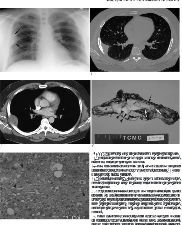
Fig. 1. A 32-year-old man with parachordoma of the chest wall.
A. Chest plain radiograph shows a smoothly marginated, pleur- al-based mass in the right hemithorax.
B. The mass causes contiguous 5 th, 6 th rib erosion with focal destruction and hypertrophy on the non-enhanced CT scan with a bone window setting.
C. Contrast-enhanced CT scan shows a homogeneous, non-en- hancing soft tissue mass with focal wall penetration in the right lateral chest.
D. The resected surface of the tumor was lobulated, firm solid and firm, myxo-cartilaginous in consistency, and pale gray in color. It was un-encapsulated and seated beneath the perios- teum of an involved rib. Note the mass (black arrows) deeply in- vading the rib (open arrows). The periosteum (arrowheads) is el- evated.
E. Photomicrograph of surgical specimen shows the tumor cells embedded in a chondroid-like matrix with myxoid degenera- tion. Most of the tumors are made up of cords and small indis- tinct lobules of polygonal eosinophilic cells with clean, vacuolat- ed cytoplasm (H & E, ×100).
with chest wall reconstruction was successfully per- formed. The mass was originating from the rib, and the periosteum of the rib was elevated by the mass. The pathologic review of the resected right chest wall mass confirmed the previous histologic diagnosis.
Discussion
Parachordoma was first reported as ‘chordoma pe- riphericum’ by Laskowski in 1955 (8) and it was re- described as ‘parachordoma’ by Dabska in 1977 (1). To our knowledge, it has been fewer than 40 reported cases since the first description. Parachordoma grows very slowly, but it has a tendency of local destruction. After surgery, local tumor recurrence has been reported in some cases (1, 2).
The origin of these tumors is still uncertain; the origin has been suggested as ectopic nests of notochord (3), schwann or other neuron-related cells (4), specialized synovial cells (1), and totipotential mesenchymal cells (5). Parachordoma and chordoma have some similar characters of local aggressiveness and the light micro- scopic features. Chordoma generally involves the axial skeleton, from the spheno-occipital area to the sacro- coccygeal area, and this tumor is thought to originate from notochordal remnants. However, parachordoma can be located at any site.
On gross pathologic examination, the tumor showed as a lobulated, un-encapsulated, grayish-white solid mass with focal fibrous bands and a smooth glistening appearance (7). On light microscopic examination, para- chordoma is similar to chordoma and extra-skeletal myxoid chondrosarcoma. Immuno-histochemical stud- ies are thought to be helpful for the differential diagno- sis. Parachordoma and chordoma are positive for cytok- eratin, and this is unlike the extraskeletal myxoid chon- drosarcoma. However, parachordoma is especially neg- ative for cytokeratin 1/10, unlike the chordoma (6, 9).
The simple radiographic findings of most parachordo- mas are well-demarcated soft tissue tumors with or without contiguous bone erosion. Our case also showed a smoothly marginated, pleural-based mass with focally adjacent rib erosion. The CT findings of chest wall para- chordoma have been reported as a homogeneous soft tissue density tumor without signs of rib erosion or pul- monary connection by Gimferrer et al (10). Contrast-en- hanced CT scan of our case also revealed a homoge- neous non-enhancing soft tissue mass of the right lateral chest wall, but the CT scan also showed adjacent rib
erosion. The contrast-enhanced CT scans of most para- chordomas arising from sites other than chest wall re- veal non-enhancing, homogeneous soft tissue density (2, 7). In our opinion, the light microscopic findings of abundant myxoid, chondroid and hyaline background probably explains the lack of enhancement of parachor- doma. Because of local aggressiveness, this soft tissue tumor can involve the adjacent bone. The Technetium- 99 m methyl di-phosphonate(Tc-99 m MDP) whole body bone scan showed a faint, increased radioactivity at the right 6th rib area that was the site of bone involve- ment in our case. Small areas of calcification in the tu- mor were reported by Karabela-Bouropoulou et al (7).
Neither internal hemorrhage nor necrosis has been re- ported in these tumors. MRI series have been per- formed for imaging characterization in two cases by Koh et al (5) and Ishida et al (2); the scans revealed soft tissue masses showing low signal intensity on the T1-weighted images, high signal intensity on the T2-weighted images, and heterogeneous contrast enhancement.
The differential diagnosis of a soft tissue mass in the chest wall includes lymphoma, neurogenic tumor, plas- macytoma, primary bone tumor, soft tissue sarcoma, hemangioma, hemangiopericytoma, desmoid tumor, metastasis, and etc. All these tumors including para- chordoma have similar imaging features. In our opinion, parachordoma displays nonspecific imaging findings. In our case, the imaging features of parachordoma, in brief, were good demarcation, little enhancement, no calcification, local aggressiveness, and homogeneous soft tissue density without any necrosis or hemorrhage.
In summary, we report here on a case of parachordo- ma of the chest wall, and this is an exceedingly rare soft tissue tumor. In most cases, differentiation between parachordoma and other chest wall tumors is very diffi- cult. Parachordoma has nonspecific imaging features and it must be included in the differential diagnosis of chest wall tumors.
References
1. Dabska M. Parachordoma: a new clinicopathologic entity. Cancer 1977;40:1586-1592
2. Ishida T, Oda H, Oka T, Imamura T, Machinami R. Parachordoma:
an ultrastructural and immunohistochemical study. Virchows Arch A Pathol Anat Histopathol 1993;422:239-245
3. Shin H, Mackay B, Ichinose H, Ayala AG, Romsdashl MM.
Parachordoma. Ultrastruct Pathol 1994;18:249-256
4. Hirokawa M, Manabe T, Sugihara K. Parachordoma of the but- tock: an immunohistochemical case study and review. Jpn J Clin Oncol 1994;24:336-339
J Korean Radiol Soc 2004;51:295-298
─ 297 ─
5. Koh JS, Chung JH, Lee SY, Cho KJ. Parachordoma of the tibia: re- port of a rare case. Pathol Res Pract 2000;196:269-273
6. Flope AL, Agoff SN, Willis J, Weiss SW. Parachordoma is im- munohistochemically and cytogenetically distinct from axial chor- doma and extraskeletal myxoid chondrosarcoma. Am J Surg Pathol 1999;23: 1059-1067
7. Karabela-Bouropoulou V, Skourtas C, Liapi-Avgeri G, Mahaira H.
Parachordoma: a case report of a vary rare soft tissue tumor.
Pathol Res Pract 1996;192:972-978
8. Laskowski J, Zarys O. Pathology of tumors. In Kolodziejska H.
Warsaw, Poland: PZWL, 1955:91-99
9. Fisher C. Parachordoma exists-but what is it?. Adv Anat Pathol 2000;7:141-148
10. Gimferrer JM, Baldo X, Montero CA, Ramirez J. Chest wall para- chordoma. Eur J Cardiothorac Surg 1999;16:573-575
Seung Hyun Cho, et al: Parachordoma of the Chest Wall
─ 298 ─
대한영상의학회지 2004;51:295-298
흉벽 유척삭종: 증례 보고1
1대구가톨릭대학교 의과대학 진단방사선과학교실
2대구가톨릭대학교 의과대학 해부병리학교실
3대구가톨릭대학교 의과대학 흉부외과학교실
4계명대학교 의과대학 진단방사선과학교실
조승현・성낙관・정경재・이영환・박영찬・김호균・박소윤2・박기성3・고성민4
저자들은 우상배통을 호소하는 32세 남자환자의 흉벽에서 생긴 유척삭종의 방사선학적 소견을 보고한다. 단순흉부 촬영과 CT촬영에서 늑골의 미란을 동반한 우외측 흉벽의 연부조직의 종괴를 보였고, 외과적 절제술과 흉곽재건술을 시행하였다.