Invasive lobular carcinoma accounts for 5-15% of all breast cancers (1). Moreover, it differs from invasive ductal carcinoma, the most common histologic subtype of breast cancer, not only with respect to histologic and mammographic characteristics, but also in the pattern of metastatic spread (2).
While the spread to the lymph nodes, lung and liver is common in both ductal and lobular carcinomas, inva-sive lobular carcinoma has been found to metastasize frequently to the gastrointestinal tract, peritoneum and retroperitoneum, and the gynecological organs (3-5). To the best of our knowledge, no previous cases of distant subcutaneous metastatic nodules from invasive lobular carcinoma, except for chest wall recurrence, has been reported. We report a case of tumor infiltration into the
anterior abdominal wall presenting as a palpable mass 3 years after the treatment of an invasive lobular carcino-ma of the breast.
Case Report
A 64-year-old woman presented at our hospital with palpable nodules at the lower and mid abdominal wall that had appeared a month earlier. Three years prior, she underwent a right partial mastectomy and sentinel lymph node (LN) biopsy due to breast carcinoma. Surgical histopathology revealed a 2.2×1.3 cm invasive lobular carcinoma of histologic grade III. No metastasis was found in the axillary lymph node sampling from the sentinel LN biopsy. There was no evidence of distant metastases at the time of surgery. Consequently, the breast cancer was consistent with stage IIb. Following the surgical procedure, she received adjuvant chemotherapy, radiotherapy at a total of 5940 cGy dose, and hormone therapy (aromatase inhibitor). There was neither evidence of recurrence nor metastatic lesions during the follow-up examinations (six monthly ultra-sound, mammography, and annual abdominal-pelvic
J Korean Soc Radiol 2011;64:611-614
─ 611 ─
Abdominal Wall Metastasis from an Invasive Lobular
Carcinoma of the Breast:
A Case Report
1Hana Kim, M.D., Eun Ju Son, M.D., Song Mi Noh, M.D.2, Woo-hee Jung, M.D.2, Ji Hyun Youk, M.D., Jin Chung, M.D.
1Department of Radiology, Gangnam Severance Hospital, Yonsei
University College of Medicine, Korea
2Department of Diagnostic Pathology, Gangnam Severance Hospital,
Yonsei University College of Medicine, Korea Received January 6, 2011 ; Accepted April 15, 2011
Address reprint requests to : Eun Ju Son, M.D., Department of Radiology, Gangnam Severance Hospital, Yonsei University College of Medicine, 146-92, Dogok-dong, Gangnam-gu, Seoul 135-720, Korea.
Tel. 82-2-2019-3510 Fax. 82-2-3462-5472 E-mail: [email protected]
Breast cancer is one of the most common malignancies in women. Breast cancer fre-quently metastasizes to the bones, lungs, and liver. However, the recurrence of distant soft-tissue metastasis except to the chest wall is extremely rare. Here, we describe our experience with a patient in whom invasive lobular carcinoma of the breast with metastasis to the abdominal wall presented as subcutaneous nodules without local re-currence.
Index words :Breast, Neoplasm Carcinoma, Lobular Neoplasm Metastasis Abdominal Wall
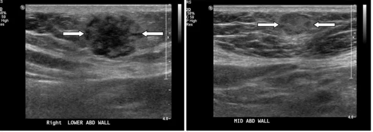
and chest CT) over the three years period after surgery. Upon admission, the patient presented with hard pal-pable nodules in the abdomen. However, mammo-graphic findings were noted in Category 2, and the nod-ules were found to be benign. Sonographic examination (Philips-Advanced Technology Laboratories, Bothell, WA, USA) of the abdominal wall showed a lobulated heterogeneous echoic mass measuring about 2.0×1.4 cm in the subcutaneous layer of the right lower abdomi-nal wall, and a 1.1×0.8 cm sized hyperechoic nodule in the mid abdominal wall (Fig. 1). These lesions were con-sidered as suspicious abnormalities which suggest the presence of metastasis from breast cancer or another
malignant lesion (sarcoma). PET/CT scan was per-formed using F-18 FDG (PET camera; Allegro, Philips, Cleveland, Ohio, CT; Somatom Plus Power, Siemens, Erlangen, Germany). They showed two instances of in-creased FDG uptakes along the abdominal wall, which corresponded to the subcutaneous nodules on CT scan (Fig. 2). US-guided core-needle biopsy for the nodule in the mid abdominal wall was performed and the patholo-gy revealed the metastatic lesion from breast cancer. A histopathologic examination revealed an invasive lobu-lar carcinoma with a prominent signet ring cell feature (Fig. 3) and the tumor was positive for estrogen but not for progesterone receptors. The result of
immunohisto-Hana Kim, et al: Abdominal Wall Metastasis from an Invasive Lobular Carcinoma of the Breast
─ 612 ─
A B
Fig. 1. A 64-year-old woman with a right partial mastectomy performed 3 year ago.
A. US image demonstrates a lobulated heterogeneous echoic mass measuring about 2.0×1.4 cm in the subcutaneous layer of the right lower abdominal wall (white arrows).
B. Another 1.1 cm large hyperechoic nodule at the mid abdominal wall is seen (white arrows).
A B
Fig. 2. PET-CT scan of the patient.
A. Increased FDG uptake along the abdominal wall which corresponds to the 2.0 cm sized nodule on CT scan and noted on US im-age (white arrow)
B. Another FDG uptake lesion is noted at the mid abdominal wall corresponding to the 1.1 cm sized round nodule on CT scan (white arrow).
chemical staining was negative for E-caderine, but the tumor expressions of HER2 and p53 were positive. The patient received additional chemotherapy.
Discussion
Some reports suggest that the metastatic spread of in-vasive lobular carcinoma shows a particular pattern (1, 6). Compared with the behavior of invasive ductal carci-noma, the behavior of an invasive lobular carcinoma is quite different (4, 5). Although invasive lobular carcino-ma carcino-may metastasize as frequently as invasive ductal car-cinoma to the lung, pleura, liver, and lymph nodes, it al-so has been found to frequently metastasize to an atypi-cal site such as the gastrointestinal tract, peritoneum and retroperitoneum, and gynecological organs (1, 2, 4, 5). Some studies found that the rates of metastasis (inva-sive lobular carcinoma vs. inva(inva-sive ductal carcinoma) to the gastrointestinal system (4.5% vs. 0.2%), gynecologic organs (4.5% vs. 0.8%), peritoneum-retroperitoneum (3.1% vs. 0.6%), adrenal glands (0.6% vs. 0%), bone-marrow (21.2% vs. 14.4%), and lung-pleura (2.5% vs. 10.2%) were documented (4).
Some literature reviews found metastatic lesions of in-vasive lobular carcinoma presented in the chest wall (2).
There was also a report on a case of tumor infiltration into the fasciae of the anterior muscular compartment of the thigh, which presented as an edema in the lower ex-tremity 10 years after the treatment of an invasive lobu-lar carcinoma of the breast (7). Thus, distant metastasis to the soft-tissue metastasis from invasive lobular carci-noma was extremely rare. To our best knowledge, there were no previously reported cases of distant subcuta-neous metastasis from invasive lobular carcinoma.
The molecular mechanisms of the different metastatic patterns between invasive ductal carcinoma and inva-sive lobular carcinoma have not been fully outlined. Nevertheless, it has been reported that the loss of the E-cadherin function, a cell-cell adhesion molecule fre-quently altered in invasive lobular carcinoma (8), could be related with local and metastatic tumor progression (9). A study reported that in a series of metastatic inva-sive lobular carcinomas, the complete loss of E-cadherin expression in the primary was documented in 88% of the cases (10). Thus, it is assumed that the down-regula-tion of E-cadherin might affect or be involved in tumor growth and dissemination condition (9). Lobular carci-noma is a distinct subtype of breast carcicarci-noma that dif-fers from infiltration into ductal carcinoma with a histo-logic appearance. Clinicians should notice when facing the metastatic relapse of invasive lobular carcinoma, that multiple metastatic sites are likely to be observed in at least 25% of cases. Radiologists often play an integral role in examining patients with lobular carcinoma to evaluate metastasis and to assess treatment response.
In conclusion, we described a case of multiple subcu-taneous metastatic nodules in the abdominal wall from an invasive lobular carcinoma of the breast. Invasive lobular carcinoma of the breast can metastasize to the abdominal wall, presenting as a soft tissue mass. The ra-diologist should consider metastasis from invasive lobu-lar carcinoma of the breast when subcutaneous nodules are present.
References
1. Sastre-Garau X, Jouve M, Asselain B, Vincent-Salomon A, Beuzeboc P, Dorval T, et al. Infiltrating lobular carcinoma of the breast: clinicopathologic analysis of 975 cases with reference to da-ta on conservative therapy and meda-tasda-tatic patterns. Cancer 1996;77:113-120
2. Winston CB, Hadar O, Teitcher JB, Caravelli JF, Sklarin NT, Panicek DM, et al. Metastatic lobular carcinoma of the breast: pat-terns of spread in the chest, abdomen, and pelvis on CT. AJR Am J Roentgenol 2000;175:795-800
3. Harris M, Howell A, Chrissohou M, Swindell RI, Hudson M, J Korean Soc Radiol 2011;64:611-614
─ 613 ─
Fig. 3. Photomicrograph of the core biopsy specimen (×100). A photomicrograph of a histologic specimen from abdominal wall mass shows isolated cells and small cords of cells an “Indian file” pattern (arrow) that is characteristic of lobular car-cinoma and diffusely infiltrating skeletal muscle fibers (arrow-head) (H and E, 100).
Sellwood RA. A comparison of the metastatic pattern of infiltrating lobular carcinoma and infiltrating duct carcinoma of the breast. Br
J Cancer 1984;50:23-30
4. Borst MJ, Ingold JA. Metastatic patterns of invasive lobular versus invasive ductal carcinoma of the breast. Surgery 1993;114:637-641 5. Lamovec J, Bracko M. Metastatic pattern of infiltrating lobular
car-cinoma of the breast: an autopsy study. J Surg Oncol 1991;48:28-33 6. Kidney DD, Cohen AJ, Butler J. Abdominal metastases of
infiltrat-ing lobular breast carcinoma: CT and fluoroscopic imaginfiltrat-ing find-ings. Abdom Imaging 1997;22:156-159
7. El Khoury M, Cherel P, Becette V, De Maulmont C, Costes V, Talma V, et al. Unusual soft-tissue metastasis of an invasive
lobu-lar carcinoma mimicking fasciitis. AJR Am J Roentgenol 2004;182: 745-747
8. Rasbridge SA, Gillett CE, Sampson SA, Walsh FS, Millis RR. Epithelial (E-) and placental (P-) cadherin cell adhesion molecule expression in breast carcinoma. J Pathol 1993;169:245-250 9. Van Aken E, De Wever O, Correia da Rocha AS, Mareel M.
Defective E-cadherin/catenin complexes in human cancer.
Virchows Arch 2001;439:725-751
10. Ferlicot S, Vincent-Salomon A, Medioni J, Genin P, Rosty C, Sigal-Zafrani B, et al. Wide metastatic spreading in infiltrating lobular carcinoma of the breast. Eur J Cancer 2004;40:336-341
Hana Kim, et al: Abdominal Wall Metastasis from an Invasive Lobular Carcinoma of the Breast
─ 614 ─ 대한영상의학회지 2011;64:611-614