• 검색 결과가 없습니다.

Minute Pulmonary Meningothelial-Like Nodules Simulating Hematogenous Lung Metastasis: A Case Report

N/A
N/A
Protected

Academic year: 2021

Share "Minute Pulmonary Meningothelial-Like Nodules Simulating Hematogenous Lung Metastasis: A Case Report"

Copied!
4
0
0

로드 중.... (전체 텍스트 보기)

전체 글

(1)

Minute Pulmonary Meningothelial-Like Nodules Simulating Hematogenous Lung Metastasis: A Case Report

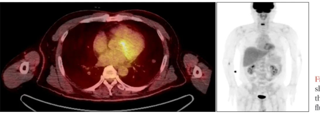
A 52-year-old man was referred to our clinic for an 11.3 mm nodule in the left lower lobe that was discovered on a chest computed tomography (CT) scan. Eleven small nodules were subsequently found in both lungs. Initially, we performed a transthoracic needle aspiration using CT scan guidance. The pathologic report showed a few clusters of atypical cells that were suspicious for malignancy. The positron emission tomography images revealed multiple lung nodules scattered throughout both lungs. The largest nodule (11.3 mm) in the left lower lobe did not have any discernible fludeoxyglucose uptake. For pathologic confirmation, we consulted a thoracic surgeon to perform the video-assisted thoracoscopic surgery. The final diagnosis was minute pulmonary meningothelial-like nodules (MPMNs). MPMNs are benign in nature, and only a few cases require treatment. However, when clinicians are suspicious of potential malignancy, a pathological correlation is essential, even if the final diagnosis is MPMNs.

Keywords: Lung Neoplasms; Neoplasm Metastasis; Immunohistochemistry

many subsequent reports. Initially, MPMNs were thought to resemble chemodectomas. However, ultrastructural, and immunohistochemical findings suggest their similarity to me- ningiomas

2

. Some studies reported that MPMNs were likely reactive in nature

3

, and associated with pulmonary thrombo- embolism, respiratory bronchiolitis-associated interstitial lung disease, severe cardiac disease, and pulmonary adenocarci-

noma

2,4-6

. The incidence of MPMNs varies from 0.3% to 9.5% at

autopsy or surgical specimens

2,3,5,7

. There are just few MPMNs studies in Korea

8,9

. We report a case of MPMNs mimicking the appearance of hematogenous lung metastasis on a chest computed tomography (CT) scan.

Case Report

A 52-year-old man was referred to our clinic for an 11.3 mm nodule in the left lower lobe discovered on a chest CT scan. We found eleven small nodules of various sizes in both lungs, but there was no definite lymph node enlargement in the mediastinum or either hilum (Figure 1). The patient had complained of a persistent cough and mild dyspnea for sev- Copyright © 2013

The Korean Academy of Tuberculosis and Respiratory Diseases.

All rights reserved.

Sang Kook Lee, M.D.

1

, Gi Jeong Kim, M.D.

2

, Young Jae Kim, M.D.

1

, Ah Young Leem, M.D.

1

, Eu Dong Hwang, M.D.

1

, Se Kyu Kim, M.D., Ph.D.

1

, Joon Chang, M.D., Ph.D.

1

, Young Ae Kang, M.D., Ph.D.

1

and Song Yee Kim, M.D.

1

Departments of

1

Internal Medicine and

2

Pathology, Yonsei University College of Medicine, Seoul, Korea

Introduction

Minute pulmonary meningothelial-like nodules (MPMNs) are small, benign, and often incidentally discovered in sur- gical or autopsy specimens of the lung. Since Korn et al.

1

first described these small lesions in 1960, there have been

CASE REPORT

http://dx.doi.org/10.4046/trd.2013.75.2.67

ISSN: 1738-3536(Print)/2005-6184(Online) • Tuberc Respir Dis 2013;75:67-70

67

Address for correspondence: Song Yee Kim, M.D.

Division of Pulmonology, Department of Internal Medicine, Yonsei University College of Medicine, 50 Yonsei-ro, Seodaemun-gu, Seoul 120- 752, Korea

Phone: 82-2-2228-1940, Fax: 82-2-393-6884 E-mail: [email protected]

Received: Feb. 10, 2013 Revised: May 6, 2013 Accepted: May 23, 2013

cc

It is identical to the Creative Commons Attribution Non-Commercial

License (http://creativecommons.org/licenses/by-nc/3.0/).

(2)

SK Lee et al.

68 Tuberc Respir Dis 2013;75:67-70 www.e-trd.org

eral months. He had a history of secondary erythrocytosis, and was a current smoker with a history of 30 pack-years. He had no family history of cancer. His secondary erythrocytosis was mild, and he did not take any medication for it. His hemo- globin level was 18.2 g/dL, and the rest of the laboratory data revealed no abnormal results.

Initially, we decided to perform a biopsy, and consulted with the chest radiologist to perform a transthoracic needle aspira- tion using CT scan guidance. The pathologic report showed a few clusters of atypical cells that were suspicious for malig- nancy. However, cells acquired during needle aspiration were scant, so we could not make a definite diagnosis. For further evaluation, we carried out a positron emission tomography (PET) scan, esophagogastricduodenoscopy (EGD), and colo- noscopy. There was no evidence of malignancy on the EGD or colonoscopy. The PET images showed multiple lung nodules scattered in both lungs. The largest one was 11.3 mm in the left lower lobe and did not have any discernible fludeoxyglu- cose uptake (Figure 2). We could not rule out the possibility of hematogenous lung metastasis from an unknown primary lesion.

For pathologic confirmation, video-assisted thoracoscopic surgery (VATS) was performed while excising a lung paren- chyma sample of 3.5×1×0.6 cm. Analysis revealed a relatively well-circumscribed gray-white solid mass, measuring 1.1 cm in diameter. Microscopically, the surgical lung biopsy

specimen showed an interstitial proliferation of cytologi- cally bland oval to blunted spindle cells. The cells enveloped the small blood vessels, and seemed to form whorls with Figure 1. Chest computed tomography showing an 11.3 mm well-defined nodule in the left lower lobe without any gross signs of calcification, and multiple small nodules of various sizes in both lungs.

Figure 2. Positron emission tomography showing the largest nodule (11.3 mm) in the left lower lobe without any discernible fluorodeoxyglucose uptake.

Figure 3. Surgical lung biopsy specimen showing the typical histo- logical features of minute pulmonary meningothelial-like nodules.

An interstitial proliferation of cytologically bland oval to blunted

spindle cells is shown. The cells enveloped the small blood vessels,

and seemed to form whorls with dispersed nuclear chromatin (H&E

stain, ×200).

(3)

Pulmonary meningothelial-like nodules

http://dx.doi.org/10.4046/trd.2013.75.2.67 69

www.e-trd.org

dispersed nuclear chromatin (Figure 3). Immunohistochemi- cal analysis was performed, and the sample was positive for epithelial membrane antigen (EMA), vimentin, CD56, and progesterone. Cytokeratin, thyroid transcription factor-1, and CD34 were negative. Finally, the diagnosis of MPMNs was established, and there was no evidence of malignancy. On the second postoperative day, the patient was discharged without any complications. Currently, he is being followed-up with at our hospital and is doing well.

Discussion

The origin of multiple pulmonary nodules may be benign or malignant, but multiple pulmonary nodules that are larger than 1 cm in diameter are most likely due to metastatic dis- ease

10

. In this case, the patient presented with a 1.1 cm lung nodule in the left lower lobe on a chest CT, and we were ini- tially suspicious of hematogenous lung metastasis. After VATS, the final pathologic report showed MPMNs. MPMNs are lung nodules often incidentally found in pulmonary parenchymal specimens.

When Korn et al.

1

described MPMNs for the first time in 1960, they were thought to be benign proliferations of chemo- receptor cell precursors. Since then, there have been refutative studies showing that MPMNs have no endocrine granules, and are similar to meningothelial cells

2,4

. Therefore, the name of MPMNs was generally accepted in place of so-called pul- monary chemodectomas

4

. MPMNs are commonly asymp- tomatic, and their etiology is still unknown. According to one study, MPMNs are most frequently seen in the sixth decade, predominantly in women, and involve the upper lung lobes more often than lower lobes

4

. A study reported that MPMNs are found three times more often in the right lung than in the left

2

. The diameters of MPMNs range from 100 µm to 3 mm.

The prevalence of MPMNs is variable in each study, ranging from about 0.3–9.5% at autopsy or surgical resection

2,3,5,7

. In surgically resected specimens, the incidence increases up to 7–13.8%

3,6

. One study reported a 48% incidence rate in exten- sively-sampled lobectomies

11

. These differences in incidence rate might be caused by the study groups’ variable main dis- eases.

Several studies suggested that MPMNs might be associated with chronic lung diseases such as pulmonary thromboem- bolism, respiratory bronchiolitis-associated interstitial lung disease, desquamative interstitial pneumonia, and atypical ad- enomatous hyperplasia

2,5

. Association with congestive heart failure was also suggested

2

. Chronic pulmonary diseases may stimulate the lung, and induce the formation of MPMNs, such as stretching or stiffening of the alveolar septa, or hypoxia, ischemia, parenchymal destruction, or some combination of these factors. The incidence rate of MPMNs has been reported to decrease in acute lung injury

11

. When we consider the

results of previous studies, MPMNs are more likely to come from a reactive origin than a neoplastic origin. Also, there are no reports of cases in pediatric lungs, so we may exclude a congenital origin

11

. In one study about loss of heterozygosity, researchers found that isolated MPMNs lacked mutational damage, consistent with a reactive origin. However, multiple MPMNs show greater genetic instability, and more frequently have loss of heterozygosity. This suggests that MPMNs might represent the transition between a reactive and neoplastic proliferation

7

.

Although curative surgical excision is sometimes needed, MPMNs usually run a benign course, and conservative treat- ment is favorable. However, we should not overlook MPMNs, because some studies have described an association between MPMNs and lung cancer. Niho et al.

3

reported that MPMNs were found in 10% patients with lung adenocarcinoma. In Mizutani al.’s study

6

, MPMNs were found more often in pa- tients with malignant pulmonary tumors (especially lung adenocarcinoma) than in those with benign disease (7.3% vs.

2.5%, p=0.44) Although there is no guideline for follow-up in patients with MPMNs, we will follow the National Compre- hensive Cancer Network guideline for lung cancer screening

12

in this case. We will perform the low dose chest CT annually considering the risk of heavy smoker and history of MPMNs.

Immunohistochemical studies showed that MPMNs stained positive for EMA, vimentin, and progesterone and negative for cytokeratin, actin, CD34, S-100 protein, chromogranin, synaptophisyn, and neuron-specific enolase

3,4,13

. Our immu- nohistochemical staining results corresponded with those of previous studies. Niho et al.

3

reported that half of MPMNs showed immunoreactivity for the progesterone receptor and suggests a role for sex-steroid hormones in the control of their growth. MPMNs in this case exhibited the immunoreactivity for the progesterone, therefore, close follow-up is needed for the growth of MPMNs.

In the present case, the sizes of MPMNs were larger than described in other studies. The pathological and immuno- histochemical examinations provided a definite diagnosis of MPMNs. MPMNs may often be an incidental finding, and most cases do not require special treatment. However, when there is no confident clinical diagnosis, such as in our case, a pathological correlation is essential, despite the eventual diag- nosis of MPMNs.

In summary, we report a case of MPMNs simulating the appearance of hematogenous lung metastasis on a chest CT scan. Minute pulmonary meningothelial-like nodules should be considered in the differential diagnosis of small lung nod- ules.

References

1. Korn D, Bensch K, Liebow AA, Castleman B. Multiple minute

(4)

SK Lee et al.

70 Tuberc Respir Dis 2013;75:67-70 www.e-trd.org

pulmonary tumors resembling chemodectomas. Am J Pathol 1960;37:641-72.

2. Churg AM, Warnock ML. So-called “minute pulmonary che- modectoma”: a tumor not related to paragangliomas. Cancer 1976;37:1759-69.

3. Niho S, Yokose T, Nishiwaki Y, Mukai K. Immunohistochemi- cal and clonal analysis of minute pulmonary meningothelial- like nodules. Hum Pathol 1999;30:425-9.

4. Gaffey MJ, Mills SE, Askin FB. Minute pulmonary menin- gothelial-like nodules: a clinicopathologic study of so- called minute pulmonary chemodectoma. Am J Surg Pathol 1988;12:167-75.

5. Spain DM. Intrapulmonary chemodectomas in subjects with organizing pulmonary thromboemboli. Am Rev Respir Dis 1967;96:1158-64.

6. Mizutani E, Tsuta K, Maeshima AM, Asamura H, Matsuno Y.

Minute pulmonary meningothelial-like nodules: clinicopath- ologic analysis of 121 patients. Hum Pathol 2009;40:678-82.

7. Ionescu DN, Sasatomi E, Aldeeb D, Omalu BI, Finkelstein SD, Swalsky PA, et al. Pulmonary meningothelial-like nodules: a genotypic comparison with meningiomas. Am J Surg Pathol 2004;28:207-14.

8. An J, Park H, Han J, Kim TS, Choi YS, Choi MS, et al. Diffuse

pulmonary meningotheliomatosis: a case report. Korean J Pathol 2011;45(Suppl 1):S32-5.

9. Kfoury H, Arafah MA, Arafah MM, Alnassar S, Hajjar W.

Mimicry of minute pulmonary meningothelial-like nodules to metastatic deposits in a patient with infiltrating lobular carcinoma: a case report and review of the literature. Korean J Pathol 2012;46:87-91.

10. Ginsberg MS, Griff SK, Go BD, Yoo HH, Schwartz LH, Pan- icek DM. Pulmonary nodules resected at video-assisted thoracoscopic surgery: etiology in 426 patients. Radiology 1999;213:277-82.

11. Mukhopadhyay S, El-Zammar OA, Katzenstein AL. Pul- monary meningothelial-like nodules: new insights into a common but poorly understood entity. Am J Surg Pathol 2009;33:487-95.

12. National Comprehensive Cancer Network. NCCN clinical practice guidelines in oncology. Lung cancer screening. Ver- sion 1.2012 [Internet]. Fort Washington: National Compre- hensive Cancer Network; 2012 [cited 2013 Jun 1]. Available from: http://www.nccn.org.

13. Suster S, Moran CA. Diffuse pulmonary meningotheliomato-

sis. Am J Surg Pathol 2007;31:624-31.

수치

Figure 3. Surgical lung biopsy specimen showing the typical histo- histo-logical features of minute pulmonary meningothelial-like nodules

참조

관련 문서

3) In aspect of occupational attitude, almost half of the students chose their occupation for remuneration in the matter of a woman's holding an occupation

systemic circulation, in the right ventricle and oxygenated blood from the lungs, or pulmonary circulation, in the left ventricle, as in birds and mammals.. Two vessels,

* in large objects with small surface area A to volume V ratio (A/V) the physical and chemical properties are primarily defined by the bulk (inside). * in small objects with

• Small lot sizes in both the production process and deliveries from suppliers field a number of benefits that enable JIT systems to operate effectively.. • With small

Commerce of New York and one of the well-known importers handling various Travel Bags and we are writing you with a keen desire to open an account with you.. As you will see

In sows, ovulatory size (8 mm to 11 mm) follicles develop only during the follicular phase after luteal regression.. Prior to luteal regression, the number of small antral

a prospective analysis of 50 cases. Preoperative staging of non-small-cell lung cancer with positron-emission tomography. Staging of non- small-cell lung cancer

1 John Owen, Justification by Faith Alone, in The Works of John Owen, ed. John Bolt, trans. Scott Clark, "Do This and Live: Christ's Active Obedience as the