Usefulness of an Implantable Loop Recorder in Patients with Syncope of an Unknown Cause
Gu Hyun Kang,
1Ju Hyeon Oh,
1Woo Jung Chun,
1Yong Hwan Park,
1Bong Gun Song,
1June Soo Kim,
2Young Keun On,
2Seung Jung Park,
2and June Huh
31Department of Medicine, Samsung Changwon Hospital, Sungkyunkwan University School of Medicine, Changwon;
Departments of 2Medicine and 3Pediatrics, Cardiac and Vascular Center, Samsung Medical Center, Sungkyunkwan University School of Medicine, Seoul, Korea.
Received: April 6, 2012 Revised: August 18, 2012 Accepted: August 20, 2012
Corresponding author: Dr. June Soo Kim, Division of Cardiology, Department of Medicine, Cardiac and Vascular Center, Samsung Medical Center, Sungkyunkwan University School of Medicine,
50 Irwon-dong, Gangnam-gu, Seoul 135-710, Korea.
Tel: 82-2-3410-3414, Fax: 82-2-3410-3417 E-mail: [email protected]
∙ The authors have no financial conflicts of interest.
© Copyright:
Yonsei University College of Medicine 2013 This is an Open Access article distributed under the terms of the Creative Commons Attribution Non- Commercial License (http://creativecommons.org/
licenses/by-nc/3.0) which permits unrestricted non- commercial use, distribution, and reproduction in any medium, provided the original work is properly cited.
Purpose: The mechanisms underlying syncope remain unknown in about 20% of patients with recurrent syncope. The implantable loop recorder (ILR) has been shown to be a useful diagnostic tool in patients with unexplained syncope even af- ter negative initial evaluations. Nevertheless, ILR has rarely been used in clinical practice. Materials and Methods: This study included 18 consecutive patients who had an ILR implanted at our center because of recurrent unexplained syncope after extensive diagnostic tests between February 2006 and June 2011. Results: Di- agnosis was confirmed in 10 (55.6%) of the 18 enrolled patients (13 males, 61±15 years). The confirmed diagnoses included sick sinus syndrome (n=6, 60%), ad- vanced atrioventricular block (n=2, 20%) and ventricular tachyarrhythmia (n=2, 20%). The mean follow-up durations of the total study subjects and the diagnosed patients were 11.3±10.6 months and 5.6±9.2 months, respectively. Of the 10 diag- nosed patients, 8 (80%) were diagnosed within 6 months of loop recorder implanta- tion. Conclusion: ILR may be a valuable and effective diagnostic tool for patients with unexplained syncope.
Key Words: Implantable loop recorder, syncope
INTRODUCTION
Syncope is a frequent symptom in the general population. However, it is often dif- ficult to diagnose the cause of syncope because diverse diseases and factors are in- volved in the occurrence of syncope.1 Although several diagnostic tests can be per- formed, the cause of syncope is unclear in 5-25.5% of patients with syncope.2,3 If the cause of syncope is not correctly diagnosed, patients are at risk of physical in- jury from recurrent syncope. In some patients with cardiac or arrhythmic causes, delay in correct diagnosis and treatment may lead to sudden cardiac death.2,4 Re- cently, use of an implantable loop recorder (ILR) and a standardized diagnostic pathway have led to breakthroughs in reducing the number of unknown causes of syncope.1,2,4,5 The ILR has become the diagnostic tool of choice in unexplained syncope even after negative initial evaluations. However, domestic adoption and
than 3 seconds. A follow-up visit was set up after symptom- atic events or every 3 months in asymptomatic subjects in order to retrieve the times and dates of episodes of brady- cardia or tachycardia as well as the corresponding electro- cardiographic tracings from the memory of the ILR.
Statistical analysis
When the data had normal distributions, they were present- ed as mean±standard deviation (SD). For abnormal distri- butions, medians (inter-quartile range) were used. SPSS for Windows (version 17.0, SPSS, Chicago, IL, USA) was used for all statistical analyses.
RESULTS
Clinical characteristics
Eighteen consecutive patients were identified from Febru- ary 15, 2006 to June 20, 2011. Thirteen patients (72.2%) were male. The mean age (±SD) of the patients was 61.2 (±15.0) years. Several underlying diseases were noted in more than one-fourth of the patients, among which hyper- tension was the most common (n=8, 44.4%). Coronary heart disease, diabetes mellitus, hyperlipidemia and cardio- myopathy were noted in 6 (33%), 5 (27.8%), 5 (27.8%) and 5 (27.8%) patients, respectively. The mean left ventricular ejection fraction (±SD) was 59.6% (±8.1). The mean esti- mated interval (±SD) between syncope or presyncope events was 164 (±219.5) days. The median number (inter- quartile ranges) of syncope or presyncope events was 5 (3- 16). The mean estimated duration of syncope (±SD) was 145.5 (±182.2) seconds. The mean follow-up duration (±SD) of for all study subjects was 11.3 (±10.6) months. In addi- tion, it took a mean of 5.6 (±9.2) months to diagnose pa- tients (n=10, 55.6%) (Table 1).
All but 3 patients (patients 15, 16 and 17) received EPSs before implantation of ILR. EPSs were performed after anti- arrhythmic medications were stopped. Intervals were mea- sured at first conduction; thereafter, antegrade and retrograde refractory periods were determined. Finally, for tachycardia induction, the high right atrium, right ventricular outflow tract and right ventricular apex were stimulated using pro- grammed stimuli. The results were not diagnostic in 15 pa- tients. CAG was performed on 11 patients. Ten (55.6%) of the 18 patients was successfully diagnosed using the ILR.
Despite a short interval (3 days) between symptoms, patient 6 received ILR implantation because diagnosis was not pos- implementation of the ILR has been slow in Korea. Thus,
we analyzed our patients who underwent ILR implantation.
MATERIALS AND METHODS
Study population
According to guidelines,1 the ILR is indicated for early evaluation of 1) patients with recurrent syncope of uncer- tain origin absent of high risk criteria and a high likelihood of recurrence within battery longevity of the device, as well as 2) high-risk patients in whom a comprehensive evalua- tion did not demonstrate the cause of syncope or lead to a specific treatment. Our indication mirrored guidelines of European Society of Cardiology (ESC).
Between February 2006 and June 2011, 18 patients had an ILR implanted at our center due to an unknown cause of syncope even after undergoing several diagnostic tests. As an initial evaluation of syncope, careful medical history tak- ing, physical examination, electrocardiography (ECG), Holt- er monitoring, head-up-tilt test (HUTT) and treadmill test were performed. Invasive studies such as coronary angiog- raphy (CAG) and electrophysiologic studies (EPSs) were performed prior to ILR implantation in some patients.
Event recording and follow-up after ILR implantation The ILR (Reveal DX, Medtronic Inc., Minneapolis, MN, USA) is a rectangular device measuring 62×19×8 mm and weighing 15 grams. To implant the device, a 2.0-cm inci- sion was made in the left chest area. Blunt dissection was then employed to create a 6-cm subcutaneous pocket. The device can be implanted under local anesthesia in 15-20 minutes.
The ILR stores representative rhythm strips when the heart rate exceeds preset limits (automatic activation) or when a patient presses a button on the accompanying actua- tor (patient activation). These records can be transmitted to a physician for review during subsequent office visits.
In all patients, the device was programmed with the fol- lowing settings: 40 minutes of total electrocardiographic re- cording consisting of three patient-activation recordings of a 10 min duration (8 minutes before and 2 minutes after symptom onset) and five automatic activation recordings of a 2 min duration. The following parameters were set for au- tomatic activation: heart rate <40 beats per minute (four consecutive measurements); heart rate >160 beats per min- ute (16 consecutive measurements) and asystole lasting more
After 4.4 months using the ILR, syncope with seizure-like movements occurred while sleeping at night. The patient lost consciousness transiently due to ventricular tachycardia (VT) and ventricular fibrillation (VF) lasting for 90 sec- onds. The arrhythmia started with monomorphic VT, which was then changed into polymorphic VT, VF and monomor- phic VT.6 He did not have any genetic disorder. Patient 16 also had a short interval (9 days) between symptoms. How- ever, diagnosis could not be confirmed over several previ- ous hospital visits despite a total 72 episodes of syncope.
Holter monitoring and event recording at our hospital also could not confirm a diagnosis. Therefore, ILR was per- formed without EPSs and sick sinus syndrome (SSS) was diagnosed 8 days after ILR implantation (Table 2).
Documented arrhythmia and management
SSS (n=6, 60%) was the most common diagnosis among the 10 diagnosed patients. Two patients (20%) were diagnosed with VT and VF. Advanced atrioventricular block (AVB) was also diagnosed in 2 patients (20%) (Fig. 1). Nine patients (90%) were diagnosed successfully within 6.5 months. Only 1 patient (10%) required 30.8 months to arrive at a diagnosis (Fig. 2).
In SSS patients (n=6), diagnosis was confirmed more fre- quently through the patient-activation mode (n=4) than sible using Holter monitoring and EPSs. Furthermore, the
patient’s pattern of symptoms was accompanied by shock.
Table 1. Clinical Characteristics of Study Subjects
Variable Total
No. of enrolled patients 18 (100)
Gender
Male 13 (72.2)
Age at enrollment (yrs) 61.2±15.0
Underlying disease
Diabetes mellitus 5 (27.8)
Hypertension 8 (44.4)
Hyperlipidemia 5 (27.8)
Smoking 4 (22.2)
Cardiomyopathy 5 (27.8)
Coronary heart disease 6 (33.3) History of cerebrovascular disease 2 (11.1)
LV ejection fraction (%) 59.6 (±8.1)
Interval of events (day) 164 (±219.5) Number of syncope and presyncope 5 (3-16) Duration of syncope (seconds) 145.5±182.2 F/U duration of total study subjects (months) 11.3±10.6 F/U duration of diagnosed patients 5.6±9.2 F/U duration of undiagnosed patients 18.4±7.7 No. of diagnosed patients 10 (55.6) F/U, follow-up; LV, left ventricle.
Data are presented as n (%) or interquartile ranges.
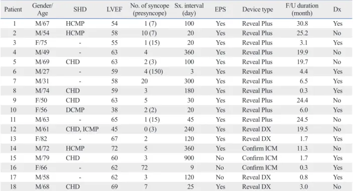
Table 2. Individual Data of Clinical Characteristics Patient Gender/
Age SHD LVEF No. of syncope (presyncope) Sx. interval
(day) EPS Device type F/U duration
(month) Dx
1 M/67 HCMP 54 1 (7) 100 Yes Reveal Plus 30.8 Yes
2 M/54 HCMP 58 10 (7) 20 Yes Reveal Plus 25.2 No
3 F/75 - 55 1 (15) 20 Yes Reveal Plus 3.1 Yes
4 M/49 - 63 4 360 Yes Reveal Plus 19.9 No
5 M/69 CHD 63 2 (3) 100 Yes Reveal Plus 19.7 No
6 M/27 - 59 4 (150) 3 Yes Reveal Plus 4.4 Yes
7 M/31 - 58 20 300 Yes Reveal Plus 6.5 Yes
8 M/74 CHD 59 3 180 Yes Reveal Plus 0.3 Yes
9 F/50 CHD 63 5 30 Yes Reveal Plus 24.4 No
10 F/56 DCMP 38 2 (2) 20 Yes Reveal Plus 6.0 Yes
11 M/63 - 65 1 (15) 45 Yes Reveal Plus 24.5 No
12 M/61 CHD, ICMP 45 0 (3) 240 Yes Reveal DX 19.5 No
13 F/82 - 67 2 120 Yes Reveal DX 1.7 Yes
14 M/72 HCMP 72 5 360 Yes Confirm ICM 11.3 No
15 M/79 CHD 60 3 900 No Confirm ICM 1.7 Yes
16 F/66 - 62 72 9 No Confirm ICM 0.3 Yes
17 M/58 - 62 3 120 No Reveal DX 0.8 Yes
18 M/68 CHD 69 7 25 Yes Reveal DX 3.0 No
SHD, structural heart disease; LVEF, left ventricular ejection fraction; Sx, symptom; M, man; F, female; HCMP, hypertrophic cardiomyopathy; DCMP, dilated cardiomyopathy; ICMP, ischemic cardiomyopathy; CHD, coronary heart disease; F/U, follow-up; Dx, diagnosis; ICM, implantable cardiac monitor; EPS, elec- trophysiologic study.
toring. However, no significant tachyarrhythmia was in- duced with programmed electrical stimulation. Therefore, we could not perform radiofrequency catheter ablation on the patient. Because of concerns about unnecessary defi- brillation shock after recovery from the non-sustained VT which had already induced syncope, pharmacological ther- apy with sotalol was performed instead of ICD implanta- tion. Fortunately, syncope has not recurred (Table 3).
DISCUSSION
The etiologies of syncope remain unknown in 21% to 49%
of all patients even after comprehensive conventional work- through the automatic activation mode (n=2). In cases of
ventricular tachyarrhythmia, the patient activation and auto- matic activation modes were used in both cases. Both ad- vanced AVB patients (n=2) were diagnosed through the au- tomatic activation mode.
Although patient 1 was diagnosed with SSS, an implant- able cardiac defibrillator (ICD) was implanted instead of a permanent pacemaker because of accompanied hypertro- phic cardiomyopathy. In patient 17, non-sustained VT, which induced syncope, was diagnosed using the ILR through the patient activation mode without EPSs. Interestingly, synco- pe occurred during short runs of non-sustained VT. Right ventricular out tract VT was suspected based on the non- sustained VT, which was recorded by 2-channel ECG moni-
Fig. 1. The proportion of finally diagnosed arrhythmias by ILR. SSS, sick si- nus syndrome; AVB, atrio-ventricular block; VT, ventricular tachycardia; VF, ventricular fibrillation; ILR, implantable loop recorder.
Fig. 2. Cumulative diagnostic yields of arrhythmic causes of syncope after ILR implantation. ILR, implantable loop recorder.
Months after ILR implantation 0.00
10.00 20.00 30.00 40.00 50.00 60.00
Cumulative diagnostic yields %
1 6 12 18 24 30
SSS60%
VT/VF 20%
AVB20%
Table 3. Documented Arrhythmia and Management
Patient Gender/Age SHD FHx of SCD Diagnosis Activation mode F/U duration (month) Treatment
1 M/67 HCM - SSS, NSVT Patient 30.8 ICD
3 F/75 - - SSS Automatic 3.1 PPM
6 M/27 - Father VF Automatic 4.4 ICD
7 M/31 - - 2nd degree AVB
(Mobitz type 2) Automatic 6.5 PPM
recommend
8 M/74 CHD - SSS Patient 0.3 PPM
10 F/56 DCMP Nephew SSS Patient 6.0 PPM
13 F/82 - - 3rd degree AVB Automatic 1.7 PPM
15 M/79 CHD - SSS Automatic 1.7 PPM
16 F/66 - - SSS Patient 0.3 PPM
17 M/58 - - VT Patient 0.8 MTx (sotalol
100 mg bid) SHD, structural heart disease; FHx, familial history; SCD, sudden cardiac death; F/U, follow-up; M, man; F, female; HCM, hypertrophic cardiomyopathy;
CHD, coronary heart disease; DCMP, dilated cardiomyopathy; SSS, sick sinus syndrome; NSVT, non sustained ventricular tachycardia; VF, ventricular fibril- lation; VT, ventricular tachycardia; AVB, atrioventricular block; ICD, implantable cardiac defibrillator; PPM, permanent pacemaker; MTx, medical treatment;
bid, twice per day.
uation methods in establishing a diagnosis for recurrent un- explained syncope. These results may influence the use of ILR in Korea. However, indications for ILR implantation should be considered in terms of the frequency of syncopal episodes. For example, patient 16 had a short interval be- tween symptoms (9 days) in this study. In addition, the pa- tient was diagnosed just 8 days after ILR implantation. An event recorder was not available for this patient because her syncope occurred suddenly without prodromal symptoms.
Even so, repeated Holter monitoring or prolonged ECG monitoring during hospitalization might also be a useful al- ternative. Giada, et al.14 limited target patients to those who had symptoms less frequently than once a month as part of the inclusion criteria. Further studies are needed to establish guidelines regarding symptom frequency in patients under- going ILR implantation.
Newly developed ILR devices can detect cardiac arrest while its external receiver can alert bystanders to start re- suscitation and to automatically call emergency medical services.15 However, wireless data transmission from the ILR is impossible in Korea because of radio frequency se- lection problems. It is clear that wireless data transmission offers the possibility of alerting care providers when cardi- ac arrests occur, decreasing response times and improving survival. Therefore, wireless data transmission should be instituted as soon as possible along with the use of the ILR.
In conclusion, the ILR may be a valuable and effective tool for determining arrhythmic causes of unexplained syn- cope. Furthermore, this selected Korean patient population behaved very much like patients from other parts of the world. Thus, it is believed that ILRs are equally helpful in evaluating Korean and Asian patients in clinical practice.
Study limitations of the present study
The small sample size and the potential selection bias of the population regarding patient economic status due to non- coverage by insurance in Korea are major limitations of our study. In addition, this study was conducted with a cross- sectional design. Further large-scale prospective studies with a larger sample size are needed to confirm our results.
REFERENCES
1. Task Force for the Diagnosis and Management of Syncope; Euro- pean Society of Cardiology (ESC); European Heart Rhythm As- sociation (EHRA); Heart Failure Association (HFA); Heart Rhythm Society (HRS), Moya A, et al. Guidelines for the diagno-
ups.7,8 Among conventional tests, HUTT (19% to 61%) and postural blood pressure measurement (15% to 33%) show relatively high diagnostic yields.7,8 Our results showed that it was possible to diagnose more than half (55.6%) of the patients with unexplained syncope using the ILR. In previ- ous reports, Boersma, et al.,9 Vitale, et al.10 and Lombardi, et al.5 reported of diagnosis rates of 28%, 32% and 50% using the ILR, respectively, in patients with recurrent unexplained syncope. Recently, in the PICTURE registry, a prospective, multicenter, observational study, ILR-guided diagnosis was established in 78% of unexplained syncope patients.4
Among the arrhythmic causes of the syncope diagnosed using the ILR, SSS was the most common (60%) in the present study, which was similar to the results of previous studies that reported the incidence of SSS to be between 54.5% and 58.3%.5,9 Advanced AVB was reported as the sec- ond most common cause, the occurrence of which ranged from 8.3% to 27.3%.5,9 However, in patients with bundle branch block and negative EPSs, the most commonly re- ported cause was prolonged asystolic pauses, mainly due to sudden-onset paroxysmal AVB.11
Furukawa, et al.12 reported that a quarter of patients who had unexplained syncope needed more than 18 months of follow-up using the ILR to establish a diagnosis. However, most patients (90%) were diagnosed within 18 months in our study. Furthermore, it took less than 6 months to arrive at a diagnosis in 8 of the 10 patients. There were significant differences between the results of previous reports and ours.
These differences were probably due to a high recurrence rate among the selected patients. However, the actuarial curves showed similar patterns of steep rises during the first 6 months (Fig. 2). Furthermore, these patterns were promi- nent in cases of asystolic-type arrhythmias, such as SSS and significant AVB. On the other hand, none of the asys- tolic-type episodes showed a continuously increasing linear curve during the follow-up period up to 48 months.12 With greater advancements in technology, newly-developed de- vices offer a longer longevity of more than 3 years, allow- ing for more convenient and prolonged observation.
In the present study, 3 patients (patients 15, 16 and 17) underwent ILR implantation without EPS. They were all diagnosed successfully within 2 months. The updated ver- sion of the ESC guidelines highlights the use of ILR and recommends its early use in diagnostic workups.1
Krahn, et al.13 and Giada, et al.14 showed, in their random- ized controlled trials, that a strategy of primary monitoring using an ILR is more cost-effective than conventional eval-
9. Boersma L, Mont L, Sionis A, García E, Brugada J. Value of the implantable loop recorder for the management of patients with unexplained syncope. Europace 2004;6:70-6.
10. Vitale E, Ungar A, Maggi R, Francese M, Lunati M, Colaceci R, et al. Discrepancy between clinical practice and standardized indi- cations for an implantable loop recorder in patients with unex- plained syncope. Europace 2010;12:1475-9.
11. Brignole M, Menozzi C, Moya A, Garcia-Civera R, Mont L, Al- varez M, et al. Mechanism of syncope in patients with bundle branch block and negative electrophysiological test. Circulation 2001;104:2045-50.
12. Furukawa T, Maggi R, Bertolone C, Fontana D, Brignole M. Ad- ditional diagnostic value of very prolonged observation by im- plantable loop recorder in patients with unexplained syncope. J Cardiovasc Electrophysiol 2012;23:67-71.
13. Krahn AD, Klein GJ, Yee R, Hoch JS, Skanes AC. Cost implica- tions of testing strategy in patients with syncope: randomized as- sessment of syncope trial. J Am Coll Cardiol 2003;42:495-501.
14. Giada F, Gulizia M, Francese M, Croci F, Santangelo L, Santo- mauro M, et al. Recurrent unexplained palpitations (RUP) study comparison of implantable loop recorder versus conventional di- agnostic strategy. J Am Coll Cardiol 2007;49:1951-6.
15. Arzbaecher R, Hampton DR, Burke MC, Garrett MC. Subcutane- ous electrocardiogram monitors and their field of view. J Electro- cardiol 2010;43:601-5.
sis and management of syncope (version 2009). Eur Heart J 2009;30:2631-71.
2. Brignole M, Ungar A, Bartoletti A, Ponassi I, Lagi A, Mussi C, et al. Standardized-care pathway vs. usual management of syncope patients presenting as emergencies at general hospitals. Europace 2006;8:644-50.
3. Suzuki T, Matsunaga N, Kohsaka S. Diagnostic patterns in the evaluation of patients hospitalized with syncope. Pacing Clin Electrophysiol 2006;29:1240-4.
4. Edvardsson N, Frykman V, van Mechelen R, Mitro P, Mohii-Os- karsson A, Pasquié JL, et al. Use of an implantable loop recorder to increase the diagnostic yield in unexplained syncope: results from the PICTURE registry. Europace 2011;13:262-9.
5. Lombardi F, Calosso E, Mascioli G, Marangoni E, Donato A, Rossi S, et al. Utility of implantable loop recorder (Reveal Plus) in the diagnosis of unexplained syncope. Europace 2005;7:19-24.
6. Shin DH, Kim JS, Park JW, Yim HR, Kim JH, Lee SM, et al. The use of an implantable loop recorder in patients with syncope of unknown origin. Korean Circ J 2008;38:205-11.
7. Kang GH, Oh JH, Kim JS, On YK, Song HG, Jo IJ, et al. Diag- nostic patterns in the evaluation of patients presenting with synco- pe at the emergency or outpatient department. Yonsei Med J 2012;
53:517-23.
8. Pires LA, Ganji JR, Jarandila R, Steele R. Diagnostic patterns and temporal trends in the evaluation of adult patients hospitalized with syncope. Arch Intern Med 2001;161:1889-95.