DOI: 10.4196/kjpp.2009.13.6.409
409
ABBREVIATIONS: CCK, cholecystokinin; GC, guanylate cyclase;
MLCK, myosin light-chain kinase.
Received August 31, 2009, Revised October 29, 2009, Accepted November 13, 2009
Corresponding to: Hyeyoung Kim, Department of Food and Nutrition, Yonsei University College of Human Ecology, 262, Seongsan-no, Seodaemun-gu, Seoul 120-749, Korea. (Tel) 82-2-2123-3125, (Fax) 82-2-364-5781, (E-mail) [email protected]
Altered Gene Expression in Cerulein-Stimulated Pancreatic Acinar Cells: Pathologic Mechanism of Acute Pancreatitis
Ji Hoon Yu1, Joo Weon Lim2, and Hyeyoung Kim2
1Department of Pharmacology, Yonsei University College of Medicine, Seoul 120-752, 2Department of Food and Nutrition, Research Institute of Food & Nutritional Sciences, Brain Korea 21 Project, Yonsei University College of Human Ecology, Seoul 120-749, Korea
Acute pancreatitis is a multifactorial disease associated with the premature activation of digestive enzymes. The genes expressed in pancreatic acinar cells determine the severity of the disease. The present study determined the differentially expressed genes in pancreatic acinar cells treated with cerulein as an in vitro model of acute pancreatitis. Pancreatic acinar AR42J cells were stimulated with 10−8 M cerulein for 4 h, and genes with altered expression were identified using a cDNA microarray for 4,000 rat genes and validated by real-time PCR. These genes showed a 2.5-fold or higher increase with cerulein: lithostatin, guanylate cyclase, myosin light chain kinase 2, cathepsin C, progestin-induced protein, and pancreatic trypsin 2. Stathin 1 and ribosomal protein S13 showed a 2.5-fold or higher decreases in expression. Real-time PCR analysis showed time-dependent alterations of these genes. Using commercially available antibodies specific for guanylate cyclase, myosin light chain kinase 2, and cathepsin C, a time-dependent increase in these proteins were observed by Western blotting. Thus, disturbances in proliferation, differentiation, cytoskeleton arrangement, enzyme activity, and secretion may be underlying mechanisms of acute pancreatitis.
Key Words: Cerulein, Pancreatitis, Acinar cells, DNA microarray
INTRODUCTION
Acute pancreatitis is a multifactorial disease associated with the release of digestive enzymes to the pancreatic in- terstitium and the systemic circulation, as well as with in- creased cytokine production and release (Schoenberg et al., 1990). Cerulein pancreatitis is one of the best-characterized animal models of experimental pancreatitis and exhibits bi- ochemical, morphological, and pathophysiological similarities to various aspects of human pancreatitis (Willemer et al., 1992). Doses of CCK or cerulein, a cholecystokinin (CCK) analog, beyond those that cause the maximum pancreatic secretion of amylase and lipase (Jensen et al., 1989; Sato et al., 1989) result in pancreatitis. The disease is charac- terized by dysregulation of the production and secretion of digestive enzymes, particularly the inhibition of pancreatic secretion and an elevation in serum levels, as well as cyto- plasmic vacuolization, the death of acinar cells, edema for- mation, and infiltration of inflammatory cells into the pan- creas (Schoenberg et al., 1990; Lerch and Adler, 1995). The key events appear to be a premature, intra-pancreatic acti- vation of digestive enzyme granules, but the earliest events that trigger acute pancreatitis are unclear.
Previously we showed that intravenous infusion of cer-
ulein induces hyperamylasemia, inflammation, edema for- mation, and high production of lipid peroxide, an index of oxidative cell damage, in rat pancreas (Choi et al., 1985).
Cytokine expression and secretory responses using CCK were determined in freshly isolated pancreatic acinar cells.
Maximum stimulation of digestive enzymes and cytokines were achieved with 10−9 M CCK (Kim et al., 1996) and 10−8 M CCK (Yu et al., 2002; Yu et al., 2005; Ju et al., 2006;
Yu et al., 2006), respectively.
Stress or injury in acinar cells induces the activation of a signaling mechanisms and intracellular activation of di- gestive enzymes. These early events are translated into long-term responses by the expression of specific genes;
these genes determine the ultimate severity of pancreatitis.
We previously reported that NF-κB, AP-1, and mitogen- activated protein kinase are activated early and induce the expression of cytokines in cerulean-stimulated pancreatic acinar cells (Lee et al., 2003; Ju et al., 2006). We previously reported that cerulein (10−8 M) induces the activation of Ras, NF-κB, AP-1, mitogen-activated protein kinase (p38, ERK, JNK), and JAK2/STAT3 to induce expression of cyto- kines (IL-6, IL-8, IL-1β, TGF-β) and vascular endothelial growth factor-D (VEGF-D) in pancreatic acinar AR42J cells (Yu et al., 2002; Lee et al., 2003; Yu et al., 2005; Ju et al., 2006; Yu et al., 2006; Lee et al., 2007; Yu et al., 2008). In addition, neutrophils activated pancreatic acinar cells to in- duce cytokine expression via NF-κB activation (Kim et al., 1999). Gene chip analysis using 8,000 genes for rat pancre-
Table 1. Altered genes by cerulein
No. Gene Primer sequencesa Foldb
Up-regulated genes Cy5/Cy3
1 Regeneration protein, lithostatin (F) ACACCTTGTATCTGTGCTCAATGTAG 7.86 (Pancreatic stone protein) (R) CAAACTAAAGCTGTTTGCTGTCTGGTA
2 Guanylate cyclase 2C (F) GTGACATTGTCGGTTTCACG 6.13 (R) CAAGGCCATCTTGGAAATGT
3 Myosin light chain kinase 2 (F) CTGACAAGACGGACATGTGG 5.71
(R) AAGTCTTTGGCCTCGTCTGA
4 Cathepsin C (F) TCAGACCCCAATCCTGAGTC 3.76
(R) AACGGAGGCAGTTTTCCTTT
5 Progestin-induced protein (F) CTGGCAAAAACACAGAAGCA 3.35
(R) AGCATCGGCATCTGAACTCT
6 Pancreatic trypsin 2 (F) GGAGGATACACCTGCCAAGA 2.83 (R) TCCTATCGAAGTTGGGATGC
Down-regulated genes Cy3/Cy5 1 Stathin 1 (F) AAGGATCTTTCCCTGGAGGA 2.66
(R) TTCTCCTCTGCCATTTTGCT
2 Ribosomal protein S13 (F) ACCGGCTGGCTCGATACTA 2.50
(R) GCTTGTGTACGCAACAGCAT
aGene sequences used as forward (F) and reverse (R) primers for real-time PCR, bfold is the ratio of Cy5/Cy3for up-regulated genes and Cy3/Cy5 for down-regulated genes.
atic acinar cells isolated from in vivo pancreatitis animal models using cerulein and taurocholate administration showed fifteen differentially expressed genes, including the pro-inflammatory mediators, MCP-1, IL-6, and gro-α as well as the transcription factor, EGR-1 (Ji et al., 2003).
Cerulein (Grady et al., 1996) and taurocholate (Kim et al., 2002) activate stress kinases, including Jun kinase.
Here we determined the gene expression changes after cerulein treatment of pancreatic acinar cells to understand of the pathophysiology of acute pancreatitis. Pancreatic aci- nar AR42J cells were stimulated with 10−8 M cerulein for 4 h. Alterations in gene expression were identified using a cDNA microarray for 4,000 rat genes and validated by real-time RT-PCR. Western blot analysis was performed to confirm changes in protein expression.
METHODS Cell culture
Rat pancreatic acinar AR42J cells (pancreatoma, ATCC CRL 1492) were obtained from the American Type Culture Collection (Manassas, Virginia, USA) and cultured in Dulbecco’s modified Eagle’s medium (Sigma, St. Louis, Missouri, USA) supplemented with 10% fetal bovine serum (GIBCO-BRL, Grand Island, New York, USA) and anti- biotics (100 U/ml penicillin and 100 μg/ml streptomycin) under 44 mM sodium bicarbonate and 10% CO2 environ- ment as recommended (Freshney et al., 1994).
Experimental protocol
Acinar cells were plated at 2×10⁶/ml in a 100-mm culture plate (Falcon 3,047, Becton Dickinson Labware, Lincoln Park, New Jersey, USA) and allowed to attach for 12 h.
The cells were treated with cerulein (10−8 M) and cultured for 4 h. The dose and duration of cerulein treatment in-
duced activation of NF-κB and Janus kinase (Jak)/signal transducer and activator of transcription (Stat), in- flammatory cytokine expression, and hypersecretion (Kim et al., 1996; Yu et al., 2002; Yu et al., 2005; Ju et al., 2006;
Yu et al., 2006).
Probe preparation and cDNA microarray
Total RNA was prepared from cells stimulated with or without cerulein for 4 h by guanidine thiocyanate extraction method (Chomczynski and Sacchi, 1987). Cy3-dUTP or Cy5-dUTP (Amersham Pharmacia Biotec UK Ltd, Bucking- hamshire, UK) was incorporated when 50 μg total RNA was reverse transcribed into cDNA and primed with oligo (dT) primer. A cDNA probe from cells cultured without cer- ulein was incorporated with Cy3 while that from the cells with cerulein was incorporated with Cy5. Cy3- or Cy5-labeled cDNA probe was purified with Chroma-spin 100 columns (Clontech Laboratories, Inc., Palo Alto, Cali- fornia, USA) following the manufacturer’s instructions. A rat gene chip (4,000 genes and 2 housekeeping genes; Geno Check, Ansan, Kyunggi-do, Korea. http://www.genocheck.com) cDNA microarray was prehybridized at room temperature for 2 h in prehybridization buffer (6× SSC, 0.2% SDS, 5×
Denhardt’s solution and 1 mg/ml salmon sperm DNA).
Different fluorescent-labeled cDNA probes were mixed and applied on the microarray following incubation at 62oC for 16 h under humidified conditions. The fluorescent images of the hybridized microarray were scanned with a fluo- rescent laser confocal slide scanner (GMS 418 array, Wallac Laboratories, Atlanta, Georgia, USA). Images and quanti- tative gene expression levels were analyzed by ImaGeneTM II (BioDiscovery, Inc., Marina de Rey, California, USA).
Real-time PCR analysis
Real-time PCR analysis was performed with a SYBRⓇ Green Realtime PCR master mix kit (Toyobo, Osaka, Japan)
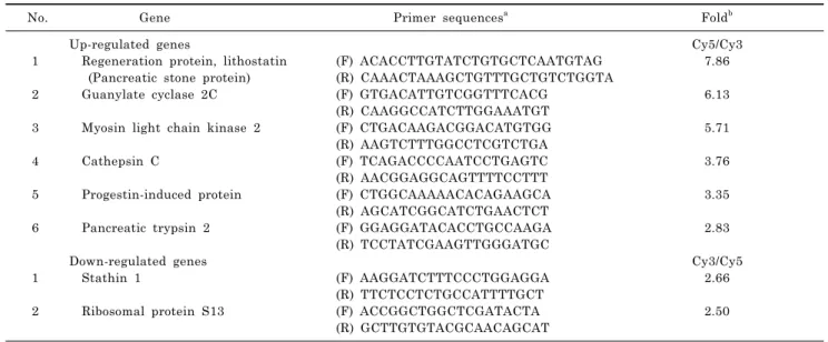
Fig. 1. A representative scatter plot of cDNA microarray analysis and modified Venn diagram according to gene function. (A) AR42J cells stimulated with cerulein (labeled with Cy5) or without cerulein (labeled with Cy3) were labeled and hybridized to the cDNA microarray. Cy5/Cy3 ratios indicate relative expression levels. (B) Venn diagram of genes shows functional overlap. Ceru- lein changed genes related to cell proliferation and differentiation, carcinogenesis, enzyme activity and secretion and cytoskeleton arrangement.
using a Roche Light cycler (Roche Molecular Biochemicals, Mannheim, Germany). Two micrograms of total RNA were reverse transcribed using the M-MLV reverse transcription system (Promega, Madison, Wisconsin, USA) in 20 μl in a thermocycler (Applied Biosystems GeneAmp PCR System 9700, Foster City, USA). Then 1/10 volume of each RT re- action was amplified with SYBR Green master mix (Toyobo, Osaka, Japan) containing 10 μM of customized primers and GAPDH (Table 1); the reactions were measured in a Light Cycler real-time PCR detection system (Roche Molecular Biochemicals). PCR was conducted using the fol- lowing cycling conditions: pre-incubation and denaturation at 95oC for 10 min, followed by amplification with 40 cycles of: denaturation at 95oC for 30 s with a thermal ramp rate of 20oC/s; annealing at 60oC for 5 s with a thermal ramp rate of 20oC/s; amplification at 72oC for 30 s with a thermal ramp rate of 20oC/s. The mRNA levels of target genes were normalized to GAPDH. The primers used in real-time PCR were listed in Table 1. The primers for GAPDH were for- ward, ACCACAGTCCATGCCATCAC and reverse, TCCAC- CACCCTGTTGCTGTA, giving a 460 bp PCR product.
Western blot analysis for guanylate cyclase, myosin light chain kinase 2, and cathepsin C
Cells were treated with cerulein (10−8 M) and cultured for 6 h. The cells were harvested and lysed in Tris-HCl buffer (pH 7.4) containing 0.5% Triton X-100 and a protease in- hibitor cocktail (Boehringer-Mannheim, Indianapolis, Indi- ana, USA) for the determinations of guanylate cyclase, my- osin light chain kinase 2, and cathepsin C. The protein con- centration of each sample was determined by Bradford as- say (Bio-Rad laboratories, Hercules, CA, USA). Protein (50 μg) was separated on 8∼10% SDS-polyacrylamide gel electro- phoresis under reducing conditions, and transferred onto nitrocellulose membranes (Amersham Inc., Arlington Heights, IL) by electroblotting. The transfer of protein and equality of loading in all lanes was verified using reversible staining with Ponceau S. The membranes were blocked using 5%
nonfat dry milk in TBS-T (Tris-buffered saline and 0.15%
Tween 20) for 3 h at room temperature. The proteins were detected with antibodies for guanylate cyclase (1:1,000;
sc-34428), myosin light chain kinase (1:1,000; sc-12450), cathepsin C (1:1,000; sc-74590) and actin (1:1,000; sc-1615) (all from Santa Cruz Biotechnology, Santa Cruz, CA) di- luted in TBS-T containing 5% dry milk, and incubated at 4oC overnight. After washing in TBS-T, the immunoreactive proteins were visualized using goat anti-rabbit and donkey anti-mouse secondary antibodies conjugated to horse radish peroxidase, followed by enhanced chemiluminescence (Am- ersham). Actin was used as a loading control.
RESULTS cDNA microarray
To characterize changes in mRNA expression induced by cerulein, rat pancreatic acinar AR42J cells were stimulated with or without cerulein for 4 h, and then total RNA was extracted. cDNA prepared from total RNA were labeled with Cy5 fluorochrome (with cerulein, red) and Cy3 (without cerulein, green) (Fig. 1A) to indicate relative expression levels. A Cy5/Cy3 ratio of 1 indicates identical expression.
Up- and down-regulated genes
Most genes showed only small differences after cerulein stimulation, indicated by Cy5/Cy3 ratios between 2 and 0.5.
We extracted genes with expression levels more than 2.5 fold higher or lower after cerulein (Table 1). Two house- keeping genes, GADPDH and tubulin, were used as in- ternal controls to correct for mRNA abundance. These genes showed similar intensities of signals in hybridized microarray, and the mean of those control genes were used to normalize the target genes. Cerulein elevated the ex- pression of lithostatin, guanylate cyclase, myosin light chain kinase 2, cathepsin C, progestin-induced protein, and pancreatic trypsin 2. Cerulein down-regulated stathin 1 and ribosomal protein S13. These genes have a variety of functions, including cell proliferation and differentiation (lithostatin, progestin-induced protein, stathin 1, guanylate cyclase 2, trypsin 2), carcinogenesis (lithostatin, progestin-
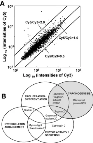
Fig. 2. Time-dependent mRNA ex- pression after cerulein treatment for 8 genes. Relative mRNA expression in AR42J cells treated with cerulein (10−8 M) was assessed by real-time RT- PCR. The internal standard (GAPDH) was coamplified with each gene.
induced protein, ribosomal protein S13, trypsin 2), enzyme activity and secretion (myosin light chain kinase 2, cathe- psin, trypsin 2, guanylate cyclase 2), and cytoskeleton ar- rangement (myosin light chain kinase 2, stathin 1) (Fig. 1B).
Real-time PCR analysis
To confirm these changes in gene expression, cells were stimulated with cerulein for up to 4 h. Real-time PCR anal- ysis showed a time-dependent increase in 6 genes (lithostatin, guanylate cyclase, myosin light chain kinase 2, cathepsin C, progestin-induced protein, and pancreatic trypsin 2) and a time-dependent decrease in 2 genes (stathin 1 and riboso- mal protein S13) (Fig. 2). At 4 h, cerulein increased mRNA
levels of lithostatin, guanylate cyclase, and myosin light chain kinase 2 almost 10-fold, higher than in microarray analysis. Cerulein increased cathepsin C, progestin-induced protein, and pancreatic trypsin 2 about 2.5-fold. Cerulein decreased stathin 1 and ribosomal protein S13 levels about 2.5 fold, similar to changes in the microarray.
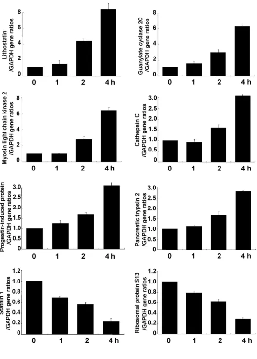
Western blot analysis of guanylate cyclase, myosin light chain kinase 2, and cathepsin C
To confirm changes in protein expression, Western blot analysis was performed using commercially available anti- bodies (Fig. 3). Cells were cultured in the presence of cer- ulein for 6 h, harvested, and lysed. Cerulein increased lev-
Fig. 3. Western blot analysis for guanylate cyclase, myosin light chain kinase 2, and cathepsin C. Cells were cultured with cerulein for 6 h, harvested, lysed, and extracted. Whole cell extracts (50 μg of protein/lane) were loaded, separated by 8∼10% SDS-poly- acrylamide gel electrophoresis, and transferred onto nitrocellulose membranes by electroblotting. The membranes were blocked with 5% nonfat dry milk in TBS-T. The proteins were detected with specific antibodies. After washing in TBS-T, the immunoreactive proteins were visualized using secondary antibodies conjugated to horseradish peroxidase, followed by enhanced chemiluminescence.
Actin was used as a loading control.
els of guanylate cyclase, myosin light chain kinase 2, and cathepsin C, but did not change actin levels.
DISCUSSION
Doses of cerulein beyond those that cause the maximum pancreatic secretion of digestive enzymes results in pan- creatitis (Jensen et al., 1989; Sato et al., 1989). The charac- teristic events of pancreatitis include the dysregulation of digestive enzyme production, cytoplasmic vacuolization, the death of acinar cells, edema formation, and an infiltration of inflammatory cells into the pancreas (Willemer et al., 1992; Lerch and Adler, 1995). The premature activation of digestive enzymes is indicated here as the up-regulation of cathepsin C and trypsin 2 (Willemer et al., 1992; Lerch and Adler, 1995). Cerulein-induced acute pancreatitis shows pro- minent interstitial edema and acinar cell vacuolization in rats (Zhou et al., 1994; Namkung et al., 2004), which was inhibited by a calcium channel blocker (Zhou et al., 1994) and a calpain I inhibitor (Virlos et al., 2004). Therefore, intracellular calcium and calpain activation may be in- volved in the pathogenesis of edema and vacuole formation in cerulein-induced pancreatitis.
The pancreas secretes primarily two types of metabol- ically important proteins: digestive enzymes such as amylase and lipase, and hormones, including insulin and glucagon.
Lithostatin is the only protein secreted from the pancreas that has no known digestive or hormonal activity. Human lithostatin is a 144-residue protein that is identical to the reg protein, expressed in the endocrine compartment of the regenerating pancreas (Watanabe et al., 1990). It contains a trypsin-sensitive cleavage site that is conserved in several species. Tryptic cleavage produces the amino-terminal de- capeptide and a carboxy-terminal peptide of 133 amino acid residues (Graf et al., 2001). The latter has a tendency to precipitate at neutral pH and is the predominant compo- nent of the protein matrix of pancreatic stones (calcium car-
bonate crystals). The physiological role of lithostatin is to stabilize pancreatic secretions that are saturated with cal- cium carbonate, as demonstrated through in vitro assays that show the inhibitory action of lithostatin against nucle- ation and growth of calcium carbonate crystals (Multigner et al., 1983). Thus, lithostatin is secreted into the pancre- atic juice where it inhibits stone formation (Patard et al., 2003). Lithostatin was discovered in regenerating liver or regenerating islets in the pancreas, but not in normal tis- sues (Terazono et al., 1988). Lithostatin expression is low in the normal colon, but up-regulated in Crohn’s diseases and ulcerative colitis (Hartupee et al., 2001) and colorectal tumors (Violette et al., 2003) as a prognostic indicator of tumor survival (Violette et al., 2003). Cerulein up-regulates lithostatin and may support regeneration and proliferation of pancreatic acinar cells, indicating a potential connection between pancreatitis and the development of pancreatic cancer.
Guanylate cyclase (GC) has two forms, soluble and partic- ulate forms, and mediates cGMP production (Wedel and Garbers, 1997). Three isoforms of mammalian membrane GC (GC-A, GC-B, and GC-C) serve as receptors for natru- retic peptides, heat-stable enterotoxin, and guanylin (Drewett and Garbers, 1994). Membrane GC is one polypep- tide chain with high homology in the cytoplasmic domains but differences in extracellular ligand-binding domains (Drewett and Garbers, 1994). Little is known about in- trinsic mechanisms of the regulation of particulate GC.
Dephosphorylation (Potter and Garbers, 1992) and oligome- rization (Lowe, 1992) of GC receptors, as well as association of GC with a regulatory phosphatase (Chinkers, 1994), reg- ulate GC activity. Membrane GC may play a role in the physiology of the exocrine pancreas, particularly in regulat- ing acinar cell growth (Seidler et al., 1989). CCK increases the accumulation of cGMP in pancreatic acinar cells, which activates cytosolic ADP-ribosyl cyclase activity and stim- ulates intracellular Ca2+ stores (Sternfeld et al., 2003).
Therefore, increased GC expression may contribute to cell growth and exocrine function in acinar cells during pancreatitis.
Myosin light-chain kinase (MLCK), first purified from rat pancreas, phosphorylates two light chain subunits of myosin, a doublet with components of 18 and 20 kDa (Bissonnette et al., 1989). The enzyme is completely dependent on Ca2+
and calmodulin. Pancreatic MLCK may regulate myosin phosphorylation and enzyme secretion. Yoshida et al.
(Yoshida et al., 2000) demonstrated that MLCK 4 is an im- portant intracellular mediator during stimulus-secretion coupling of rat pancreatic acinar cells, whereas MLCK 2 has no effect on CCK-induced enzyme secretion. Therefore, MLCK 2 may contribute to cytoskeletal arrangement by mediating myosin phosphorylation and exocrine function by stimulating enzyme secretion in pancreatic acinar cells.
Acute pancreatitis increases intracellular chymotrypsin activity (Piotrowski et al., 2003). Two other enzymes with chymotrypsin-like activity, proteasome and lysosomal cath- epsin A, exist in the pancreas (Piotrowski et al., 2003).
Cathepsin C is a dipeptidyl peptide hydrolase acting on di- peptide esters and amides (Rojas-Espinosa et al., 1975).
Cerulein-induced up-regulation of cathepsin C may hydro- lyze pancreatic dipeptides and induce acinar cell damage during acute pancreatitis.
Progestin induces the differentiation of both endometrial stromal and epithelial cells, acting as the “differentiating”
or “growth limiting” hormone in the endometrium (Bulun
et al., 2006). This progestin effect is mediated by progester- one receptors in stromal cells (Kurita et al., 2000). In con- trast, progestins control mammary gland tumorigenesis af- ter binding to progesterone receptors (Carnevale et al., 2007). The progesterone receptor functions either as a tran- scription factor or as a signaling activator in a breast cancer cell line (Carnevale et al., 2007). Progestin initiates Wnt-be- ta-catenin signaling for proliferation and differentiation in rat uterine stromal cells (Rider et al., 2006). A progesterone antagonist prevented BRCA1-mediated mammary tumori- genesis in mice, suggesting anti-progesterone treatment may be effect for breast cancer prevention in individuals with BRCA1 mutation (Poole et al., 2006). Treatment of progesterone stimulates cell proliferation within the islets of Langerhans in rats (Nieuwenhuizen et al., 1999). There- fore, cerulein-induced increases in progestin may increase cell proliferation and relate pancreatitis and pancreatic cancer.
The pancreas is an important endocrine and exocrine se- cretory organ in mammals. Many digestive enzymes are synthesized in pancreatic acinar cells (Gorelick and Otani, 1999). Under normal conditions, these enzymes remain in- active in isolated zymogen granules inside pancreatic aci- nar cells (Kassell and Kay, 1973) and only become active after entering the small intestine. The activation of a key enzyme in zymogen granules, trypsin, requires proteolytic activation by cleavage of the propeptide, which can be com- pleted in the duodenum through activation by the brush border endoprotease, enteropeptidase (Kassell and Kay, 1973). This initial activation of trypsin can further activate trypsinogen into active trypsin and other zymogens, such as chymotrypsinogen, protelastase, and prophospholipase to their active states (Gorelick and Otani, 1999). During acute pancreatitis, these digestive enzymes are pre- maturely activated before leaving the pancreas and start digesting the pancreas to lead to acute pancreatitis (Steer, 1999). Lithostatin contains a trypsin-sensitive site, and up-regulated trypsin 2 may cleave lithostatin to tryptic cleavage products, including a carboxy-terminal peptide of 133 amino acids. In addition, trypsin is activated in pancre- atic cancer cells (Chen et al., 2009) to stimulate growth and adhesiveness in an autocrine manner (Giancotti and Mainiero, 1994). The stage and type of carcinoma is related to the level of trypsin associated with cell invasion and ex- tracellular matrix degradation (Koivunen et al., 1991; Walz and Fenton, 1994). Therefore, up-regulation of trypsin 2 in pancreatic acinar cells may contribute to the development of pancreatic cancer.
Stathmin, a major microtubule-destabilizing protein, is down-regulated by cerulein. In general, stathmin interacts directly with soluble tubulin to form a complex that seques- ters free tubulin and impedes the polymerization of micro- tubules (Belmont and Mitchison, 1996). The depolymerizing activity of stathmin is turned off upon its phosphorylation during the onset of mitosis, leading to formation of the mi- totic spindle. Conversely, reactivation of stathmin by de- phosphorylation is necessary before the cells exit mitosis and enter a new interphase (Rubin and Atweh, 2004). In addition to its role in mitosis and cell cycle progression, stathmin is also involved in diverse cell functions, such as cell proliferation and differentiation (Larsson et al., 1995).
Stathmin is expressed in actively proliferating cells (Iancu et al., 2001), including liver regeneration after partial hep- atectomy (Koppel et al., 1993) and hepatic ischemia-re- perfusion injury (Barone et al., 2005), whereas its expre-
ssion is dramatically decreased upon the induction of differ- entiation and cessation of proliferation of leukemia cells (Melhem et al., 1991), and in the later stages of mega- karyocyte maturation (Rubin et al., 2003). Stathmin is abundantly expressed in fetal liver, but dramatically de- creased in adult liver (Bièche et al., 2003). Cerulein may induce differentiation and cessation of proliferation by de- creasing stathin expression, but cerulein also increased lithostatin and progestin, two genes that increase cell pro- liferation, indicating an imbalance between cell prolifera- tion and differentiation.
Ribosomal protein S13 is found in the head region of the small subunit, where it interacts with the central protub- erance of the large ribosomal subunit and with the P site-bound tRNA through its extended C terminus (Cukras and Green, 2005; Noller et al., 2005). The bridging inter- actions between the large and small subunits are dynamic and are critical in the molecular motions of the translation cycle. S13 provides a direct link between the tRNA-binding site and the movements in the head of the small subunit seen during translocation, thereby providing signal trans- duction (Cukras and Green, 2005). The expression level of ribosomal protein S13 was lower in NK/T cell lymphoma than in normal lymphocytes, indicating that it plays a role in the development of the NK/T cell lymphoma (Yang et al., 2006). Cerulein decreases S13 expression, indicating disturbances in translation or signal transduction may be involved in the pathogenesis and/or development of pancreatitis.
In our previous studies, cerulein induced the expression of cytokines (IL-6, IL-8, IL-1β, TGF-β) and VEGF-D by the activation of NF-κB, AP-1, Mitogen-activated protein kin- ases, and Jak2/Stat3 in pancreatic acinar AR42J cells (Yu et al., 2002; Lee et al., 2003; Yu et al., 2005; Ju et al., 2006;
Yu et al., 2006; Lee et al., 2007; Yu et al., 2008). Here, cer- ulein up-regulated 6 genes (lithostatin, guanylate cyclase, myosin light chain kinase 2, cathepsin C, progestin-induced protein, pancreatic trypsin 2) and down-regulated 2 genes (stathin 1, ribosomal protein S13) that are related to pro- liferation, differentiation, carcinogenesis, cytoskeletal ar- rangement, enzyme activity, and secretion. These changes may accompany inflammatory events. Since lithostatin, progestin-induced protein, trypsin, and ribosomal protein S13 are involved in carcinogenesis, the relationship be- tween pancreatitis and the development of pancreatic can- cer requires further study. Additional in vivo studies should also be performed for comparison to human pathophysiology.
ACKNOWLEDGEMENTS
This study was supported by the Basic Science Research Program through the National Research Foundation of Korea (NRF) funded by the Ministry of Education, Science, and Technology (R11-2007-040-01002-0) (to H Kim). H Kim is grateful to Brain Korea 21 Project, College of Human Ecology, Yonsei University.
REFERENCES
Barone S, Okaya T, Rudich S, Petrovic S, Tenrani K, Wang Z, Zahedi K, Casero RA, Lentsch AB, Soleimani M. Distinct and sequential upregulation of genes regulating cell growth and cell cycle progression during hepatic ischemia-reperfusion injury.
Am J Physiol Cell Physiol 289: C826−C835, 2005.
Belmont LD, Mitchison TJ. Identification of a protein that interacts with tubulin dimers and increases the catastrophe rate of microtubules. Cell 84: 623−631, 1996.
Biéche I, Maucuer A, Laurendeau I, Lachkar S, Spano AJ, Frankfurter A, Lévy P, Manceau V, Sobel A, Vidaud M, Curmi PA. Expression of stathmin family genes in human tissues:
non-neural-restricted expression for SCLIP. Genomics 81: 400−
410, 2003.
Bissonnette M, Kuhn D, de Lanerolle P. Purification and charac- terization of myosin light-chain kinase from the rat pancreas.
Biochem J 258: 739−747, 1989.
Bulun SE, Cheng YH, Yin P, Imir G, Utsunomiya H, Attar E, Innes J, Julie Kim J. Progesterone resistance in endometriosis: Link to failure to metabolize estradiol. Mol Cell Endocrinol 248: 94−
103, 2006.
Carnevale RP, Proietti CJ, Salatino M, Urtreger A, Peluffo G, Edwards DP, Boonyaratanakornkit V, Charreau EH, Bal de Kier Joffe E, Schillaci R, Elizalde PV. Progestin effects on breast cancer cell proliferation, proteases activation, and in vivo development of metastatic phenotype all depend on progesterone receptor capacity to activate cytoplasmic signaling pathways.
Mol Endocrinol 21: 1335−1358, 2007.
Chen N, Zou J, Wang S, Ye Y, Huang Y, Gadda G, Yang JJ. Desig- ning protease sensors for real-time imaging of trypsin activation in pancreatic acinar cells. Biochemistry 48: 3519−3526, 2009.
Chinkers M. Targeting of a distinctive protein-serine phosphatase to the protein kinase-like domain of the atrial natriuretic peptide receptor. Proc Natl Acad Sci USA 91: 11075−11079, 1994.
Choi JY, Kim KH. Effects of small molecular antioxidants on cerulein-induced acute pancreatitis in rat. Korean J Physiol Pharmacol 2: 629−635, 1998.
Chomczynski P, Sacchi N. Single-step method of RNA isolation by acid guanidinium thiocyanate-phenol-chloroform extraction. Anal Biochem 62: 156−159, 1987.
Cukras AR, Green R. Multiple effects of S13 in modulating the strength of intersubunit interactions in the ribosome during translation. J Mol Biol 349: 47−59, 2005.
Drewett JG, Garbers DL. The family of guanylyl cyclase receptors and their ligands. Endocr Rev 15: 135−162, 1994.
Freshney RI. Culture of Animal Cells; A Manual for Basic Tech- nique. 3th ed. John Wiley and Sons Inc, New York, p 71−103, 1994.
Giancotti FG, Mainiero F. Integrin-mediated adhesion and signaling in tumorigenesis. Biochim Biopys Acta 1198: 47−64, 1994.
Gorelick FS, Otani T. Mechanisms of intracellular zymogen activation. Baillieres Best Pract Res Clin Gastroenterol 13: 227−
240, 1999.
Grady T, Dabroski A, Williams JA, Logsdon CD. Stress-activated protein kinase activation is the earliest direct correlate to the induction of secretagogue-induced pancreatitis in rats. Biochem Biophys Res Commun 227: 1−7, 1996.
Graf R, Schiesser M, Scheele G.A, Marquardt K, Frick TW, Mumann RW, Bimmler D. A family of 16-kDa pancreatic secretory stress proteins form highly organized fibrillar structures upon tryptic activation. J Biol Chem 276: 21028−21038, 2001.
Hartupee JC, Zhang H, Bonaldo MF, Soares MB, Dieckgraefe BK.
Isolation and characterization of a cDNA encoding a novel member of the human regenerating protein family: Reg IV.
Biochim Biophys Acta 1518: 287−293, 2001.
Iancu C, Mistry SJ, Arkin S, Wallenstein S, Atweh GF. Effects of stathmin inhibition on the mitotic spindle. J Cell Sci 114: 909−
916, 2001.
Jensen RT, Wank SA, Rowley WH, Sato S, Gardner JD. Interaction of CCK with pancreatic acinar cells. Trends Pharmacol Sci 10:
418−423, 1989.
Ji B, Chen X-Q, Misek DE, Kuick R, Hanash S, Ernst S, Najarian R, Logsdin CD. Pancreatic gene expression during the initiation of acute pancreatitis: identification of EGR-1 as a key regulator.
Physiol Genomics 14: 59−72, 2003.
Ju KD, Yu JH, Kim H, Kim KH. Role of mitogen-activated protein kinases, NF-κB, and AP-1 on cerulein-induced IL-8 expression
in pancreatic acinar cells. Ann N Y Acad Sci 1090: 368−374, 2006.
Kassell B, Kay J. Zymogens of proteolytic enzymes. Science 180:
1022−1027, 1973.
Kim H, Kim KH. Secretory response of cultured acinar cells of rat pancreas to cholecystokinin. Yonsei Medical J 37: 405−411, 1996.
Kim JY, Kim KH, Lee JA, Namkung W, Sun AQ, Anathanarayanan M, Suchy FJ, Shin DM, Muallem S, Lee MG. Transporter- mediated bile acid uptake causes Ca2+-dependent cell death in rat pancreatic acinar cells. Gastroenterology 122: 1941−1953, 2002.
Kim H, Seo JY, Cho SH, Kim KH. Lipid peroxidation, NF-κB activation and cytokine production in neutrophil-stimulated pancreatic acinar cells. Kor J Physiol Pharmacol 3: 521−528, 1999.
Koivunen E, Saksela O, Itkonen O, Osman S, Huhtala ML, Stenman UH. Human colon carcinoma, fibrosarcoma and leukemia cell lines produce tumor-associated trypsinogen. Int J Cancer 47: 592
−596, 1991.
Koppel J, Loyer P, Maucuer A, Rehak P, Manceau V, Guguen- Guillouzo C, Sobel A. Induction of stathmin expression during liver regeneration. FEBS Lett 331: 65−70, 1993.
Kurita T, Beitel LK, Cooke PS, Lydon G.R, Cunha JP. Paracrine regulation of epithelial progesterone receptor and lactoferrin by progesterone in the mouse uterus. Biol Reprod 62: 831−838, 2000.
Larsson N, Melander H, Marklund U, Osterman O, Gullberg M.
G2/M transition requires multisite phosphorylation of onco- protein 18 by two distinct protein kinase systems. J Biol Chem 270: 14175−14183, 1995.
Lee JW, Kim KH, Kim H. Role of vascular endothelial growth factor-D (VEGF-D) on IL-6 expression in cerulein- stimulated pancreatic acinar cells. Ann NY Acad Sci 1095: 129−133, 2007.
Lee JW, Seo J, Kim H, Chung JB, Kim KH. Signal transduction of cerulein-induced cytokine expression and apoptosis in pancreatic acinar cells. Ann NY Acad Sci 1010: 104−108, 2003.
Lerch MM, Adler G. Experimental animal models of acute pancre- atitis. Int J Pancreatol 15: 159−170, 1994.
Lowe DG. Human natriuretic peptide receptor-A guanylyl cyclase is self-associated prior to hormone binding. Biochemistry 31:
10421−10425, 1992.
Melhem RF, Strahler JR, Hailat N, Zhu XX, Hanash SM. Involve- ment of OP18 in cell proliferation. Biochem Biophys Res Commun 179: 1649−1655, 1991.
Multigner L, De Caro A, Lombardo D, Campese D, Saries H.
Pancreatic stone protein, a phosphoprotein which inhibits calcium carbonate precipitation from human pancreatic juice.
Biochem Biophys Res Commun 110: 69−74, 1983.
Namkung W, Han W, Luo X, Muallem S, Cho KH, Kim KH, Lee MG. Protease-activated receptor 2 exerts local protection and mediates some systemic complications in acute pancreatitis.
Gastroenterology 126: 1844−1859, 2004.
Nieuwenhuizen AG, Schuiling GA, Liem SM, Moes H, Koiter TR, Uilenbroek JT. Progesterone stimulates pancreatic cell prolife- ration in vivo. Eur J Endocrinol 140: 256−263, 1999.
Noller HF, Hoang L, Fredrick K. The 30S ribosomal P site: a function of 16S rRNA. FEBS Lett 579: 855−858, 2005.
Patard L, Lallemand JY, Stoven V. An insight into the role of human pancreatic lithostathine. JOP 4: 92−103, 2003.
Piotrowski Z, Mysliwiec P, Gryko M, Ostrowska H, Baltaziak M.
Chymotrypsin-like activity in rat tissues in experimental acute pancreatitis. Rocz Akad Med Bialymst 48: 61−65, 2003.
Poole AJ, Li Y, Kim Y, Lin SC, Lee WH, Lee EY. Prevention of Brca1-mediated mammary tumorigenesis in mice by a proge- sterone antagonist. Science 314: 1467−1470, 2006.
Potter LR, Garbers DL. Dephosphorylation of the guanylyl cyclase-A receptor causes desensitization. J Biol Chem 267: 14531−14534, 1992.
Rider V, Isuzugawa K, Twarog M, Jones S, Cameron B, Imakawa K, Fang J. Progesterone initiates Wnt-beta-catenin signaling but estradiol is required for nuclear activation and synchronous
proliferation of rat uterine stromal cells. J Endocrinol 191: 537−
548, 2006.
Rojas-Espinosa O, Arce-Paredez P, Dannenberg AM, Kamaenetz RL. Macrophage esterase: identification, purification and pro- perties of a chymotrypsin-like esterase from lung that hydrolyses and transfers nonpolar amino acid esters. Biochim Biophys Acta 22: 161−179, 1975.
Rubin CI, Atweh GF. The role of stathmin in the regulation of the cell cycle. J Cell Biochem 93: 242−250, 2004.
Rubin CI, French DL, Atweh GF. Stathmin expression and megakaryocyte differentiation: a potential role in polyploidy. Exp Hematol 31: 389−397, 2003.
Sato S, Stark HA, Martinez J, Beaven M A, Jensen R T, Gardner JD. Receptor occupation, calcium mobilization, and amylase release in pancreatic acini: effect of CCK-JMV-180. Am J Physiol 257 (Gastrointest Liver Physiol 20): G202−G209, 1989.
Schoenberg MH, Bruchler M, Gaspar M, Stinner A, Younes M, Melzner I, Bültmann B, Beger HG. Oxygen free radicals in acute pancreatitis of the rat. Gut 31: 1138−1143, 1990.
Seidler NW, Jona I, Vegh M, Martonos A. Cyclopiazonic acid is a specific inhibitor of the Ca2+-ATPase of sarcoplasmic reticulum.
J Biol Chem 264: 17816−17823, 1989.
Steer ML. Early events in acute pancreatitis. Baillieres Best Pract Res Clin Gastroenterol 13: 213−225, 1999.
Sternfeld L, Krause E, Guse AH, Schulz I. Hormonal control of ADP-ribosyl cyclase activity in pancreatic acinar cells from rats.
J Biol Chem 278: 33629−33636, 2003.
Terazono K, Yamamoto H, Takasawa S, Shiga S, Yonemura Y, Tochino Y. A novel gene activated in regenerating islets. J Biol Chem 263: 2111−2114, 1988.
Violette S, Festor E, Pandreu-Vasile I, Mitchell V, Adida C, Lesu- ffleur T. Reg IV, a new member of the regenerating gene family, is overexpressed in colorectal carcinomas. Int J Cancer 103: 185
−193, 2003.
Virlos I, Mazzon E, Serraino I, Genovese T, Di Paola R, Thie- merman C, Siriwardena A, Cuzzocrea S. Calpain I inhibitor ameliorates the indices of disease severity in a murine model of cerulein-induced acute pancreatitis. Intensive Care Med 30:
1645−1651, 2004.
Walz DA, Fenton JW. The role of thrombin in tumor cell metastasis.
Invasion Metastasis 14: 303−308, 1994.
Watanabe T, Yonekura H, Terazono K, Yamamoto H, Okamoto H.
Complete nucleotide sequence of human reg gene and its expression in normal and tumoral tissues. The reg protein, pancreatic stone protein, and pancreatic thread protein are one and the same product of the gene. J Biol Chem 265: 7432−7439, 1990.
Wedel BJ, Garbers DL. New insights on the functions of the guanylyl cyclase receptors. FEBS Lett 410: 29−33, 1997.
Willemer S, Elsasser HP, Adler G: Hormone-induced pancreatitis.
Eur Surg Res 24: 29−49, 1992.
Yang F, Liu WP, He MX, Tang QL, Zhao S, Zhang WY, Xia QJ, Li GD. Real-time fluorescence quantitative PCR in detecting ribosome protein S13 (RPS13) gene expression in NK/T cell lymphoma. Sichuan Da Xue Xue Bao Yi Xue Ban 37: 464−466, 2006.
Yoshida H, Nozu F, Lankischo TO, Mitamura K, Owyang C, Tsunoda Y. A possible role for Ca(2+)/calmodulin-dependent protein kinase IV during pancreatic acinar stimulus-secretion coupling. Biochim Biophys Acta 1497: 155−167, 2000.
Yu JH, Lim JW, Kim H, Kim KH. NADPH oxidase mediates interukin-6 expression in cerulein-stimulated pancreatic acinar cells. Int J Biochem Cell Biol 37: 1458−1469, 2005.
Yu JH, Lim JW, Namkung W, Kim H, Kim KH. Suppression of cerulein-induced cytokine expression by antioxidants in pancreatic acinar cells. Lab Inv 82: 1359−1368, 2002.
Yu JH, Kim KH, Kim H. SOCS 3 and PPAR-gamma ligands inhibit the expression of IL-6 and TGF-bata by regulating JAK2/STAT3 signaling in pancreas. Int J Biochem Cell Biol 40: 677−688, 2008.
Yu JH, Kim KH, Kim H. Suppression of IL-1beta expression by the Jak 2 inhibitor AG490 in cerulein-stimulated pancreatic acinar cells. Biochem Pharmacol 72: 1555−1562, 2006.
Zhou W, Shen F, Miller JE, Han Q, Olson MS. Evidence of altered cellular calcium in the pathogenetic mechanism of acute pan- creatitis in rats. J Surg Res 60: 147−155, 1996.