CASEREPORT INTRODUCTION OpticNerveInjurySecondarytoEndoscopicSinusSurgery:anAnalysisofThreeCases
5
0
0
전체 글
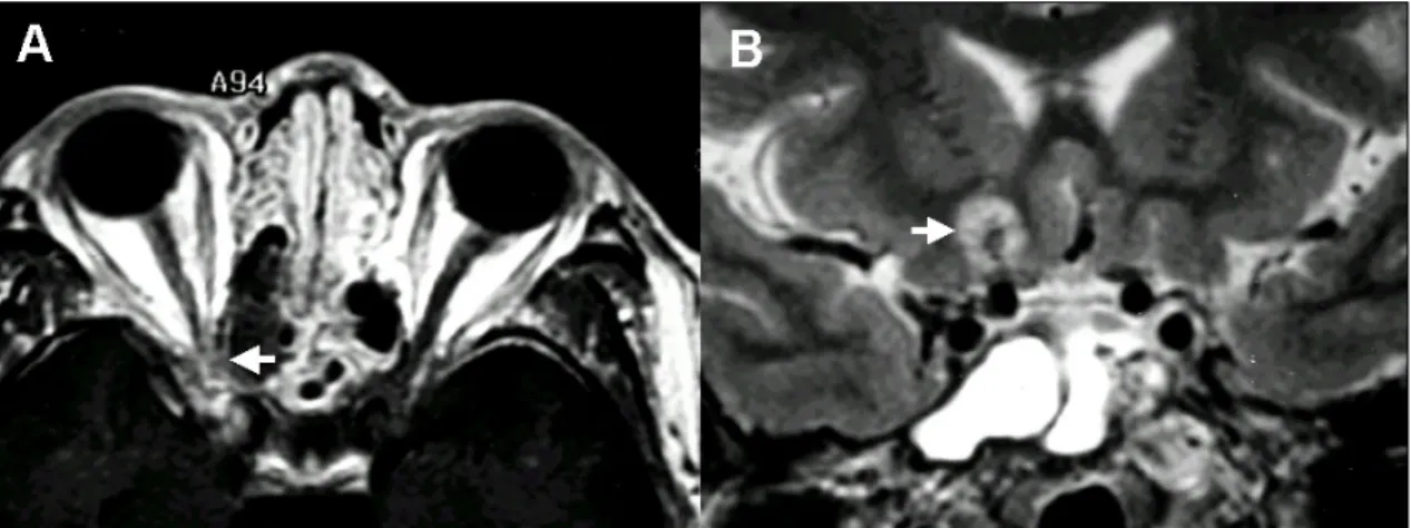
(2) Optic Nerve Injury after ESS. left endoscopic sinus surgery that had been performed 13 years previously at our hospital and two other later endoscopic sinus surgeries at a private hospital. During the hospital stay in our department, he complained of a postnasal drip, visual acuity deficit and visual field deficit. A physical examination showed no evidence of exophthalmos, subconjunctival hemorrhage or ocular motility abnormalities. His visual examination showed a right eye vision of 0.05 and a left eye vision of 0.5. Visual field tests showed deficits in all areas except in the inferior-lateral portion (Fig. 1), and intact light reflexes. On the second day of admission of our hospital, orbital CT was performed, and revealed no observable damage in the lamina papyracea or the extraocular muscles. However, an orbital MRI showed a defect of the superolateral wall of the right sphenoid sinus with an exposed optic nerve (Fig. 2). Because a fundoscopic examination revealed neuronal atrophy, 250 mg of Solumedrol was given intravenously for four days. Ten days after the completion of this symptomatic treatment, a visual field examination revealed a slight visual field improvement (Fig. 1), but no change in visual acuity. The patient was discharged on the 20th hospital day, and did not experienced any change in his symptoms over the 6-month postsurgical period. Case 2. Total blindness The case involved a 42 year-old male who underwent endoscopic sinus surgery under general anesthesia at an another medical center one week. prior to being admitted to our department, complaining of a loss of left visual acuity. Immediately after surgery, no deficits in ocular motility were observed, but the left eye had no light perception. An examination of the left eye also showed proptosis, subconjunctival hemorrhage and ecchymosis, hence emergency lateral canthotomy and orbital massage were performed at that medical center. In addition, acetazolamide IV and dexamethasone IV were given but his vision did not recover. The orbital CT from the medical center did not show any damage of the left lamina papyracea, but revealed a hematoma around the left medial rectus muscle and a possible partial defect in the left optic nerve. The patient was referred to our hospital 7 days after surgery, and. Fig. 1. Visual field defect (dark area) was noted in the whole area, except in the small inferomedial portion of the field, on the day of surgery (A). The visual field defect (dark area) improved but an extensive visual field defect remained, except in the inferomedial portion of the field 8 days after sinus surgery (B) in Case 1.. Fig. 2. Axial MRI showing a defect of the superolateral wall of the right sphenoid sinus with an exposed optic nerve (A, white arrow). Coronal MRI showing high signal intensity above the internal carotid artery (B, white arrow), possibly due to an intraparenchymal hemorrhage in Case 1. Yonsei Med J Vol. 46, No. 2, 2005.
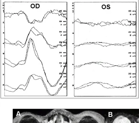
(3) Jin Young Kim, et al.. Fig. 3. Visual evoked potential test findings 8 days after sinus surgery. A flat wave for the left eye indicated complete loss of visual activity in Case 2.. Fig. 4. The loss of the fat line around the medial rectus muscle and enhanced signal intensity around the medial rectus and posterior to the optic nerve (white arrow) in T1 and T2-weighted MRIs in Case 2.. on physical examination, persisting subconjunctival hemorrhage and a mid-dilated pupil were noted. Fundoscopic examination did not reveal any abnormalities. On the second day of admission (8 days after surgery), a visual evoked potential (VEP) test revealed an absence of left optic nerve activity (P100: not detected, flat wave) (Fig. 3). Orbital MRI showed resolution of the hematoma surrounding the left medial rectus muscle but also showed loss of the medial rectus muscle fat line. Contrast enhancement around the left medial rectus muscle and behind the left optic nerve was also observed (Fig. 4). The patient was Yonsei Med J Vol. 46, No. 2, 2005. treated with prednisolone orally for 10 days, and then discharged. Orbital MRI was performed 2 months after surgery. It revealed decreased contrast enhancement around the medial rectus muscle but a persisting fat line loss and a continued contrast enhancement behind the left optic nerve. Visual acuity remained zero. Case 3. Total blindness The case involved is a 53 year-old female who experienced a loss of right visual acuity after endoscopic sinus surgery under local anesthesia at a local clinic one day prior to being admitted to.
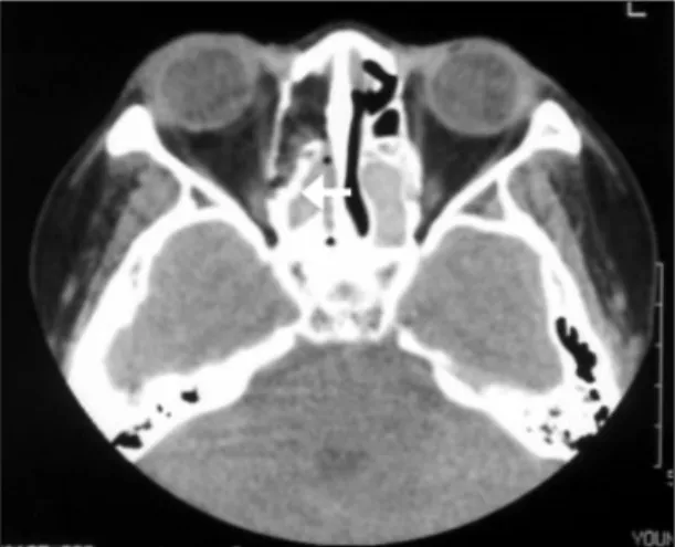
(4) Optic Nerve Injury after ESS. Fig. 5. Axial CT showing a small right medial wall fracture and a possible partial defect in the right optic nerve. A thick bony septa was seen in the ethmoid area (white arrow) in Case 3.. Fig. 6. Orbital MRI showing a mild enhancement of the posterior half of the right optic nerve sheath (white arrow) and a dark signal focus of medial rectus muscle near the right orbit apex in Case 3.. our department. Her past surgical history included both endoscopic sinus surgery, which had been performed 15 years previously at another local ENT clinic. Immediately after recent right endoscopic sinus surgery, the right eye had no light perception. The orbital CT from the local radiology center showed a small right medial wall fracture. (Fig. 5) A thick bony septa observed in the right ethmoid area. (Fig. 5) On the first day of admission in our hospital (2 days after surgery), steroid therapy was started for traumatic optic neuropathy. Steroid pulse therapy was. treated for 4 days, and then prednisolone orally for 10 days. On the second day of admission, a visual evoked potential (VEP) test revealed the absence of right optic nerve activity (P100: not detected, flat wave). After steroid treatment for 2 weeks, light perception was absent. Orbital MRI was performed on the second day of admission and showed a mild enhancement of the posterior half of the right optic nerve sheath, and a dark signal focus of medial rectus muscle near the right orbit apex (Fig. 6). Two months after ESS, her visual acuity remained zero. Optic nerve injury that occurs during ESS usually happens when the operation is performed under general anesthesia. Under local anesthesia, optic nerve injury can be prevented because the patient complains of pain the moment that the lamina papyracea is broken. Optic nerve injury under general anesthesia may occur under the following circumstances; 1) When the surgeon relies on his own experience rather than precise anatomical knowledge; 2) When preoperative CT has not been fully reviewed, or 3) When the operation is conducted without frequently checking the position and direction of the endoscope. Blindness is a rare complication, and can result from; direct damage to the optic nerve by surgical instruments, loss of blood supply to the optic nerve, or indirect damage to the optic nerve due to optic nerve compression by a retrobulbar hematoma. However, in many cases, it is not easy to determine whether blindness has been caused by direct or indirect damage. The reason why direct damage to the optic nerve occurs especially around the posterior ethmoid sinus, may be explained by the proximity of the lamina papyracea and the optic nerve, and the fact that it is difficult for the surgeon to be aware of the break through the lamina papyracea because of the thin fat layer at this site. Morever, aerated posterior ethmoid air cells alongside the optic canal (Onodi cells) occur in a few people and these may place the optic nerve at greater risk of injury during ethmoidectomy.2-4 The mechanisms of blindness caused by indirect injury are a loss of blood supply to the retina or optic nerve compression. Retinal blood supply is sustained by branches of the internal ophthalmic artery, namely the posterior ciliary artery and Yonsei Med J Vol. 46, No. 2, 2005.
(5) Jin Young Kim, et al.. central retinal artery.5 Since the central retinal artery and medial posterior ciliary artery run along the inferolateral and medial portions of the optic nerve respectively, they can be easily injured during ESS.6-8 Total blindness, that is irreversible damage, may result from central retinal artery injury due to a loss of blood to the retina; however, ischemic optic neuropathy is more likely to occur when the medial posterior ciliary artery is injured, because it is not a major feeding vessel.6 Retrobulbar hematoma due to optic nerve compression can be easily diagnosed by proptosis and a stony hard orbit. Measures undertaken to release the pressure, such as orbital massage, lateral canthotomy, and steroid administration can prevent blindness to some degree.9,10 In this study, all three cases presented with optic nerve damage at the posterior ethmoid sinus, and cases 2 and 3 were revision cases. Case 2 was under general anesthesia and case 1 and 3 were under local anesthesia. In cases 1, and 3, direct damage to the optic nerve was presumed since the patient suddenly felt a sharp pain during the operation. In case 3, the surgeon mistook the thick ethmoid bony septum as the middle turbinate during operation. Retrobulbar hematoma was accompanied in case 2 only. In that case, early orbital apex decompression or optic nerve decompression would be helpful because lateral canthotomy and steroids did not bring the vision back. Since no improvement of blindness was observed despite steroid administration in all 3 cases and after the management of retrobulbar hematoma in case 2, we suggest that in the majority of cases blindness may be irreversible and that the prevention of optic nerve damage by a thorough preoperative review of the patient is most important. Especially in revision cases, thickening of the. Yonsei Med J Vol. 46, No. 2, 2005. ethmoid septa and displacement of the ethmoid septa can cause the incorrect recognition of structures in the operative field and therefore careful attention is needed. Also, in case of a lateralized middle turbinate, severe septal mucosal hypertrophy, and high septal deviation, care must be taken because the endoscope can easily head laterally. It is important to frequently check the location and direction of the endoscope during surgery, to avoid optic nerve injury. In addition, surgeons must have precise knowledge of the detailed anatomy through cadaver dissections, the ability to interpret the PNS CT scan and experienced procedural surgical skills.. REFERENCES 1. Kennedy DW, Senior BA. Endoscopic sinus surgery: a review. Otolaryngol Clin North Am 1997;30:313-30. 2. Rene C, Rose GE, Lenthall R, et al. Major orbital complications of endoscopic sinus surgery. Br J Ophthalmol 2001;85:598-603. 3. Driben JS, Bolger WE, Robles HA, Cable B, Zinreich J. The Reliability of Computerized Tomographic Detection of the Onodi (sphenoethmoid) Cell. Am J Rhinol 1998;12:105-11. 4. Yeoh KH, Tan KK. The optic nerve in the posterior ethmoid in Asians. Acta Otolaryngol (Stockh) 1994;114: 329-36. 5. Newell F. Ophthalmology. St. Louis. The CV Mosby Company; 1969. p.26-7. 6. Rootman J, Stewart B, Goldberg RA. Orbital surgery. Philadelphia: Lippincott; 1995. p.79-146. 7. Hayreh SS, Dass R. The ophthalmic artery. II: Ontraorbital course. Br J Ophthalmol 1962;46:165-85. 8. Hayreh SS. The ophthalmic artery. III: Branches. Br. J. Ophthalmol 1962;46:212-47. 9. Stankiewiez JA. Blindness and Intranansal Endoscopic Ethmoidectomy: Prevention and management. Otolaryngol Head Neck Surg 1989;101:320-9. 10. Buus DR, Tse DT, Farris BK. Ophthalmic complications of sinus surgery. Ophthalmology 1990;97:612-9..
(6)
수치
관련 문서
The infraoptic anterior cerebral artery (large arrow, C and D) arising contralateral ICA at the level of ophthalmic artery origin (open arrow, C) passes under the right optic
Orbital imaging scans showed fusiform enlargement of the inferior and medial rectus muscles with significant contrast enhancement in the right eye6. A calcified nodule embedded in
T1- weighted orbital coronal MRI showing noticeably reduced mass in the right inferior rectus muscle (A, arrow) and normal left lower eyelid area (B, arrowhead)..
(A) Fundus picture of right eye showing subhyaloid hemorrhage, (B) contrast enhanced computed tomography scan orbit sugges- tive of right orbital proptosis with infective foci
The arrow indicates bulging enhancing mass in right orbit apex, extending to cavernous sinus with fatty marrow signal loss and enhancement of right lesser
Orbital magnetic resonance imaging reveals asymmetric enlargement of right medial rectus muscle and superior rectus/levator palpebrae superioris muscle complex
While meningioma located in optic nerve sheath, parasel- lar region, and medial sphenoidal wing causes visual loss by its direct compressive effect, large intracranial
