J Korean Soc Radiol 2017;77(3):143-147 https://doi.org/10.3348/jksr.2017.77.3.143
INTRODUCTION
Orbital metastasis from solid tumor is a rare entity usually found in patients with multiple organ metastases. The common primary tumor sites are breast, prostate gland, lung, etc. Typical imaging features of orbital metastasis include bone destruction with contiguous mass, extraocular muscle mass, and diffuse in- traconal mass formation (1, 2).
Small cell lung cancer (SCLC) is a well-known malignancy, having an early spread and multi-organ involvement; however,
there are only a few reported cases of orbital metastases present as diffuse and large tumor destroying the bony orbit (3-5).
Herein, we describe the image findings of a solitary extraocular muscle metastasis of SCLC, presenting as a discrete extraocular muscle mass with mild homogeneous enhancement.
CASE REPORT
A 74-year-old man presented with right ocular pain radiating to the right temporal area, of 1-month duration. He developed
Solitary Orbital Metastasis Presenting as Extraocular Muscle Mass in Lung Cancer Patient: A Case Report
외안근 비대의 형태로 나타난 폐암 환자의 고립성 안와 전이: 증례 보고
Hae Won Kim, MD
1, Jee Young Kim, MD
1*, Soo Ah Im, MD
2, Joo Wan Park, MD
3, Chan Kwon Park, MD
4, Yosep Chong, MD
5Departments of 1Radiology, 3Ophthalmology, 5Hospital Pathology, Yeouido St. Mary’s Hospital, College of Medicine, The Catholic University of Korea, Seoul, Korea
2Department of Radiology, Seoul St. Mary Hospital, College of Medicine, The Catholic University of Korea, Seoul, Korea
4Division of Pulmonary and Critical Care Medicine, Department of Internal Medicine, Yeouido St. Mary’s Hospital, College of Medicine, The Catholic University of Korea, Seoul, Korea
A 74-year-old man presented with right ocular pain and diplopia. Contrast enhanced orbital computed tomography showed a bulging mass with homogenous density and mild homogeneous enhancement, at the right medial rectus muscle belly. Mag- netic resonance imaging of the orbit showed a bulging mass along the medial rectus muscle, with superiorly displaced medial rectus muscle. The mass showed homoge- nous hyperintensity to extraocular muscle on T2-weighted images, and mild rim en- hancement on contrast enhance T1-weighted images. Differential diagnosis of images included primary lesion of the extraocular muscle such as lymphoma, inflammatory pseudotumor and myositis. Chest X-ray incidentally revealed a left hilar mass. Histo- pathologic diagnosis of the hilar mass was confirmed as small cell lung cancer. A total body 18F-fluorodeoxy glucose positron emission tomography scan revealed ab- normal uptake only in the left hilar region and the right orbit. Histopathologic diag- nosis of the extraocular muscle mass was metastasis of the small cell lung cancer. Al- though there are few reports of small cell lung cancer with orbital metastasis, solitary extraocular muscle metastasis without other systemic metastasis is quite rare. Here, we report the case with an initial presentation of a solitary EOM metastasis, in a pa- tient with small cell lung cancer.
Index terms Extraocular Muscle Small Cell Lung Cancer Metastasis
Computed Tomography Magnetic Resonance Imaging
Received June 17, 2016 Revised September 27, 2016 Accepted April 11, 2017
*Corresponding author: Jee Young Kim, MD Department of Radiology, Yeouido St. Mary’s Hospital, College of Medicine, The Catholic University of Korea, 10 63-ro, Yeongdeungpo-gu, Seoul 07345, Korea.
Tel. 82-2-3779-2017 Fax. 82-2-783-5288 E-mail: [email protected]
This is an Open Access article distributed under the terms of the Creative Commons Attribution Non-Commercial License (http://creativecommons.org/licenses/by-nc/4.0) which permits unrestricted non-commercial use, distri- bution, and reproduction in any medium, provided the original work is properly cited.
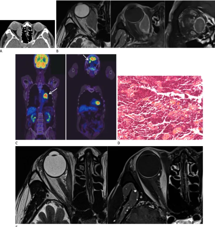
Fig. 1. A 74-year-old man with right ocular pain.
A. Contrast-enhanced axial CT image of the orbit shows a bulging mass within the right medial rectus muscle belly, with mild homogenous en- hancement. There is no adjacent fat infiltration and bone erosion.
B. T2-weighted axial image shows a bulging mass of the right medial rectus muscle belly, with homogenous signal intensity. There are no adja- cent fat infiltrations. Gadolinium based contrast enhanced T1-weighted axial and coronal images show mild rim enhancement of the bulging mass with superiorly displaced medial rectus. There is no adjacent fat infiltration.
C. Initial 18F-FDG PET scan shows increased FDG uptake in the right orbit and left hilar region (arrows).
D. Photomicrograph of biopsy specimen from the right medial rectus muscle. Hematoxylin-eosin staining (× 20) shows densely packed small cells with scant cytoplasm. Histologic diagnosis is metastatic small cell lung cancer.
E. Follow up T2-weighted axial and contrast enhanced T1-weighted axial images after 6 weeks of chemotherapy, show decrease in size of the bulg- ing mass of the right medial rectus muscle.
FDG = fluorodeoxy glucose, PET = positron emission tomography E
C D
A B
diplopia and aggravation of orbital pain 10 days prior to inves- tigation. Motility examination of the right eye revealed im- paired medial gaze. Ophthalmologic examination indicated normal visual acuity in both eyes, with normal anterior and posterior segments. He had no significant medical history. Lab- oratory tests showed the white blood cell count and C-reactive protein levels were within the normal limit. Initial clinical diag- nosis included inflammatory pseudotumor and cavernous si- nus syndrome, since the patient presented with right ocular pain.
Contrast enhanced orbital computed tomography (CT) ob- tained 35 seconds after contrast injection, revealed a bulging mass at the right medial rectus muscle belly with mild homoge- neous enhancement. Adjacent orbital fat infiltration or bony destruction was absent (Fig. 1A). Magnetic resonance imaging (MRI) of the orbit showed a bulging mass along the medial rec- tus muscle with superiorly displaced medial rectus muscle. The mass showed homogenous hyperintensity to extraocular muscle on T2-weighted images, and mild rim enhancement on contrast enhanced T1-weighted images. Perilesional fat infiltration was absent (Fig. 1B). Based on the image features, differential diag- nosis included primary tumor of the extraocular muscle, such as lymphoma or inflammatory pseudotumor or myositis.
Plain chest radiograph for routine check-up incidentally re- vealed a left hilar mass. Consecutive CT of the chest showed con- glomerated lymphadenopathy in the left hilum. A total body
18F-fluorodeoxy glucose positron emission tomography scan revealed abnormal uptake in only the left hilar region and the right orbit (Fig. 1C). These findings raised a possibility that the orbital mass could be a metastasis from lung cancer.
The patient underwent transbronchial needle aspiration for the left hilar mass, and the histopathologic diagnosis confirmed SCLC. Successive incisional biopsy of the right medial rectus muscle confirmed metastatic carcinoma from SCLC (Fig. 1D).
Patient was finally diagnosed as SCLC with solitary orbital me- tastasis. The patient started chemotherapy with platinum-based regimen. After 6 cycles of chemotherapy, the orbital symptom improved, and the extraocular mass showed marked decrease in size (Fig. 1E).
DISCUSSION
Metastasis to the soft tissues of the orbit is relatively uncom-
mon (2). Hematogenous metastases to the orbit are most com- monly the result of breast carcinoma in women, and carcinoma of the lung, kidney, or prostate in men. Imaging appearances of orbital metastasis are various, and may occur in any part of the orbital compartments. Many metastases target the bony wall of the orbit, affecting the orbital tissues as the metastasis expands (6). Shields et al. (2) reported that 41 of 64 orbital metastatic tu- mors showed a diffuse configuration, and the remaining 23 cases were round to ovoid and well-circumscribed, depending on the tumor origin. Multiplicity of lesions suggests a diagnosis of metastatic disease (6).
Extraocular muscle metastases are rare, and discrete extraoc- ular muscle metastases are even rarer, constituting only 9% of orbital metastases (7). The majority of patients with metastases of the extraocular muscle not only have a prior diagnosis of pri- mary malignancy at presentation, but also the metastasis occurs late in the course of the systemic malignancy. The most fre- quently affected extraocular muscle is the medial rectus, fol- lowed by the lateral rectus, the superior rectus, and the inferior rectus (7). There are few case reports of orbital metastases from SCLC (3-5), and they usually present as diffuse soft tissue mass involving the extraocular muscle with wall destruction of the bony orbit. However, our case presented with a solitary mass within the extraocular muscle. There are some reports of me- tastasis confined to the extraocular muscle from other prima- ries, such as carcinoid tumor or prostate cancer, involving single or multiple extraocular muscles (8). These lesions are well-de- fined, fusiform, or round masses within the extraocular muscle belly, with heterogeneous and minimal enhancement on the MR images. Our case revealed similar image features.
Differential diagnosis of extraocular muscle lesions includes lymphoma, inflammatory pseudotumor, or myositis. Lympho- ma appears more commonly as diffuse ill-defined orbital disease rather than a well-circumscribed, round or oblong mass (9).
However, radiological and clinical signs of both lymphoma and extraocular muscle metastasis are non-specific, and include pain- less muscle enlargement or masses with heterogeneous contrast enhancement (10). Inflammatory myositis often extends ante- riorly to involve tendon insertion, and may show a ragged, fluffy border of the involved muscle with infiltration and obliteration of the contiguous fat, particularly in the peripheral surgical space between the periosteum of the orbital wall and the mus-
cle cone (6).
In our case, the patient had no previous history of malignancy.
Initial chest radiograph played an important role in the final di- agnosis. Char et al. (1) reported that 11 of 31 (25%) patients had no known primary malignancy at the time of diagnosis of or- bital metastasis. Therefore, metastasis should be included in the differential diagnosis of patients presenting as a discrete mass of EOM muscle with minimal and heterogeneous enhancement on MR images, even though the patient has no known primary malignancy.
In conclusion, we described here a case of solitary extraocu- lar muscle metastasis from SCLC, presenting as bulging mass of the extraocular muscle. CT and MR showed a discrete mass of the right medial rectus muscle belly, with homogenous signal intensity and mild rim enhancement. Although solitary EOM metastasis is rare, a better understanding of imaging and clini- cal features would be useful in accurate diagnosis of orbital mass.
REFERENCES
1. Char DH, Miller T, Kroll S. Orbital metastases: diagnosis and course. Br J Ophthalmol 1997;81:386-390
2. Shields JA, Shields CL, Brotman HK, Carvalho C, Perez N, Eagle RC Jr. Cancer metastatic to the orbit: the 2000 Robert
M. Curts Lecture. Ophthal Plast Reconstr Surg 2001;17:346- 354
3. Spaide RF, Granger E, Hammer BD, Negron FJ, Paglen PG.
Rapidly expanding exophthalmos: an unusual presentation of small cell lung cancer. Br J Ophthalmol 1989;73:461-462 4. Mena AM, Pardo J. Orbital metastasis as the initial mani- festation of small cell lung cancer. Acta Ophthalmol Scand 2002;80:113-115
5. Henning M, Hu Q, Siegelmann-Danieli N. Orbital metastasis as the presenting symptom of extensive stage small cell lung cancer. Eur J Intern Med 2008;19:65-66
6. Cunnane ME, Sepahadari AR, Gardiner M. Pathology of the eye and orbit. In: Som PM, Curtin HD, eds. Head and neck imaging, 5th ed. St. Louis: Elsevier, 2011:591-756
7. Lacey B, Chang W, Rootman J. Nonthyroid causes of extra- ocular muscle disease. Surv Ophthalmol 1999;44:187-213 8. Gupta A, Chazen JL, Phillips CD. Carcinoid tumor metastases
to the extraocular muscles: MR imaging and CT findings and review of the literature. AJNR Am J Neuroradiol 2011;32:
1208-1211
9. Meltzer DE. Orbital imaging: a pattern-based approach. Ra- diol Clin North Am 2015;53:37-80
10. Surov A, Behrmann C, Holzhausen HJ, Kösling S. Lymphomas and metastases of the extra-ocular musculature. Neurora- diology 2011;53:909-916
외안근 비대의 형태로 나타난 폐암 환자의 고립성 안와 전이: 증례 보고
김혜원
1· 김지영
1* · 임수아
2· 박주완
3· 박찬권
4· 정요셉
574세 남환이 오른쪽 안구 통증과 복시를 주소로 내원하였다. 조영증강 전 전산화단층촬영 영상에서 오른쪽 안구의 내측 곧은 근육 내에 균질한 음영의 종괴가 있었으며, 조영증강 영상에서 종괴는 경도의 균질한 조영증강을 보였다. 자기공명영 상 T2 강조영상에서 종괴는 외안근보다 높은 신호강도를 보였고, 조영증강 후 T1 강조영상에서 종괴는 테두리 모양의 조 영 증강을 보였으며, 외안근은 위쪽으로 밀려 있었다. 감별진단으로 외안근의 원발성 병변인 림프종, 근육염 또는 염증거짓 종양을 고려하였다. 기본검사로 시행한 단순흉부촬영에서 왼쪽 폐문부의 종괴가 발견되어 조직검사를 시행하였으며 소세 포 폐암으로 진단되었다. 양전자방출단층촬영술 영상에서 왼쪽 폐문부와 오른쪽 안와에만 국소 fluorodeoxy glucose 섭취 를 보였다. 오른쪽 안구의 내측 곧은 근육에서 조직검사를 시행하였고 병리진단은 폐암의 외안근 전이로 확인되었다. 소세 포 폐암 환자에서 안와 전이에 대한 보고가 있었으나 외안근에 국한된 종괴로 나타나는 경우는 매우 드물어 증례 보고를 하고자 한다.
가톨릭대학교 의과대학 여의도성모병원 1방사선과학교실, 3안과학교실, 5병원병리학교실,
2가톨릭대학교 의과대학 서울성모병원 방사선과학교실, 4가톨릭대학교 의과대학 여의도성모병원 호흡기내과