Received: November 7, 2017 Revised: November 30, 2017 Accepted: December 6, 2017 JOURNAL OF
TRAUMA AND INJURY
CASE REPORT
J Trauma Inj 2017;30(4):227-230 http://doi.org/10.20408/jti.2017.30.4.227
Correspondence to Jae Hun Kim, M.D.
Department of Trauma Surgery, Pusan Na- tional University Hospital, 179 Gudeok-ro, Seo-gu, Busan 49241 Korea
Tel: +82-51-240-7369 Fax: +82-51-240-7719
E-mail: [email protected]
http://www.jtraumainj.org eISSN 2287-1683
pISSN 1738-8767
Copyright © 2017 The Korean Society of Trauma
This is an Open Access article distributed under the terms of the Creative Commons Attribution Non-Commercial License (http://creativecommons.org/licenses/by-nc/4.0/) which permits unrestricted noncommercial use, distribution, and reproduction in any medium, provided the original work is properly cited.
Rectus Sheath Hematoma Caused by Noncontact Strenuous Exercise
Gil Hwan Kim, M.D., Jae Hun Kim, M.D., Ho Hyun Kim, M.D.
Department of Trauma Surgery, Pusan National University Hospital, Busan, Korea
Rectus sheath hematoma (RSH) is an uncommon but well-documented clinical condi- tion. It is usually caused by direct trauma or anticoagulation, although there are many other causes. However, RSH after noncontact strenuous exercise is very rare. We present a rare case of RSH after playing volleyball without direct trauma that was successfully treated by angiographic embolization.
Keywords: Abdominal wall; Sports; Angiography
INTRODUCTION
Rectus sheath hematoma (RSH) with inferior epigastric artery injury is an uncommon but well-documented clinical condition [1]. It is usually associated with abdominal trauma or anticoagulation. Other causes of RSH include abdominal surgery, old age, asthma, coughing, and subcutaneous drug injection into the abdomen [2]. Some cases of RSH due to direct trauma or other causes have been reported, but very few for RSH caused due to noncontact strenuous exercise. We present a rare case of RSH after play- ing volleyball without direct trauma in a healthy individual.
CASE REPORT
A 41-year-old woman with right lower quadrant abdominal pain presented to the emergency department. She had experienced abdominal pain after playing volleyball a week previously, which soon resolved with rest. But, the pain recurred after spiking the ball while playing volleyball on the day of admission. The pain aggravated by strength- ening her abdomen and subsided with rest. There was no history of direct abdominal trauma. She had no medical history and was not on any medication. Her vital signs
228 http://doi.org/10.20408/jti.2017.30.4.227
Journal of Trauma and Injury Volume 30, Number 4, December 2017
were stable: blood pressure, 110/60 mmHg; pulse rate, 87 beats/min; respiratory rate, 16 breaths/min; and body temperature, 36.2°C. Physical examination revealed ten- derness and a palpable mass in the right lower quadrant of the abdomen. Laboratory data, including hemoglobin
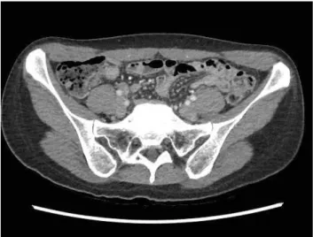
levels, in the normal range. Computed tomography (CT) revealed a 6×4×18 cm rectus sheath hematoma with ex- travasation in the right lower rectus abdominis muscle (Fig. 1). In addition, angiography revealed bleeding of the inferior epigastric artery branch (Fig. 2). Angiographic embolization was performed, and extravasation was not observed 3 days later (Fig. 3). The patient was discharged Fig. 1. Abdominal CT shows rectus sheath hematoma on (A) right lower quadrant with extravasation (arrow) and (B) on right rectus abdominis muscle (arrow). CT: computed tomography.
A B
Fig. 2. Angiography shows extravasation of lower branch of right inferi- or epigastric artery (arrow).
Fig. 3. Extravasation of right lower quadrant was not observed 3 days after angiographic embolization (arrow).
229
http://www.jtraumainj.org Gil Hwan Kim, et al. RSH due to Noncontact Strenuous Exercise
on day 7 without complications. On follow-up CT at 3 months, RSH was completely absorbed (Fig. 4).
DISCUSSION
RSH is an uncommon cause of abdominal pain, with an incidence of approximately 1.8%. It usually occurs in women in their fifth or seventh decades [3]. Anatomically, the posterior sheath does not exist below the arcuate line of Douglas. Because there is no protection from the poste- rior rectus sheath, abdominal pressure is directly applied to the inferior epigastric vessels [2].
Various causes of RSH include abdominal trauma, anti- coagulation therapy, abdominal surgery, old age, asthma, coughing, and drug injection into the abdomen. RSH due to twisting or sudden position change without a direct blow, such as that during yoga, tennis, and volleyball have been rarely reported [2].
RSH due to strenuous exercise has been described in tennis players, mainly due to trunk hyperextension with eccentric contraction of the abdominal musculature fol- lowed by concentric contraction during trunk flexion.
The nondominant rectus abdominis muscle is at a high risk because of its primary involvement in the overhead swinging motion, regardless of the sport [4]. Teske [5]
reported that RSH was more common on the right side (60%) and in the lower abdominal quadrants (80%).
RSH was diagnosed based on patient history and phys- ical examination. It is important to distinguish RSH from other abdominal diseases such as appendicitis or diverticulitis. The patient may have a rigid abdomen and tenderness. A palpable mass or bluish discoloration can be found on the injured abdomen. CT is the radiological modality of choice for diagnosing RSH, revealing any extravasation in case of ongoing bleeding from the epigas- tric vessels [6,7].
Most cases of RSH are usually self-limiting and can be managed conservatively in stable patients [7]. However, blood loss can be massive and even fatal. In cases of RSH with unstable vital signs, extravasation of epigastric ves- sels observed on CT, expanding hematoma, or ruptured peritoneum, angiographic or surgical intervention may be needed. Surgical management can be performed in an emergency patient with hypovolemic shock. In most cas- es, identifying the bleeding focus is difficult in the setting of an expanding hematoma. There is a report on proxi- mal ligation of inferior epigastric artery [8]. At present, angiographic embolization is an accepted first line choice for interventional management of RSH [9]. It is very safe and effective, with an overall success rate of 90% and no procedure-related complications [10].
Taken together, although RSH is rare and can be caused by various causes, we should know that noncontact strenuous exercise is one of the causes. Therefore, when a patient presents with abdominal pain after strenuous exercise, clinicians should consider RSH and avoid misdi- agnosis.
ACKNOWLEDGEMENTS
This work was supported by clinical research grant from Pusan National University Hospital in 2017.
REFERENCES
1. Sha r ma H, Shek hawat NS, Bha nda r i S, Memon B, Memon MA. Rectus sheath haematoma: a rare presenta- tion of non-contact strenuous exercises. Br J Sports Med 2007;41:688-90.
Fig. 4. Rectus sheath hematoma was completely absorbed 3 months later.
230 http://doi.org/10.20408/jti.2017.30.4.227
Journal of Trauma and Injury Volume 30, Number 4, December 2017
2. Choi Y, Lee D. A case of rectus sheath hematoma caused by yoga exercise. Am J Emerg Med 2009;27:899.e1-2.
3. Klingler PJ, Wetscher G, Glaser K, Tschmelitsch J, Schmid T, Hinder RA. The use of ultrasound to differentiate rectus sheath hematoma from other acute abdominal disorders.
Surg Endosc 1999;13:1129-34.
4. Cruz AR, Mautner K. Rectus Abdominis Injury in Elite Volleyball Players. Int J Athl Ther Train 2016;21:18-22.
5. Teske JM. Hematoma of the rectus abdominis muscle: re- port of a case and analysis of 100 cases from the literature.
Am J Surg 1946;71:689-95.
6. Salemis NS, Gourgiotis S, Karalis G. Diagnostic evaluation and management of patients with rectus sheath hematoma.
A retrospective study. Int J Surg 2010;8:290-3.
7. Hatjipetrou A, Anyfantakis D, Kastanakis M. Rectus sheath hematoma: a review of the literature. Int J Surg 2015;13:267- 71.
8. Mantelas M, Katsiki N, Antonitsis P, Kyurdzhieva E, Mikhailidis DP, Hatzitolios A. Rectus sheath hematoma: a simplified emergency surgical approach. Open Cardiovasc Med J 2011;5:4-5.
9. Morrissey B, Harmon M, McEniff N, McMahon G. Inferi- or epigastric artery injury following blunt trauma treated by catheter embolization. J Vasc Med Surg 2014;2:124.
10. Sobkin PR, Bloom AI, Wilson MW, LaBerge JM, Hastings GS, Gordon RL, et al. Massive abdominal wall hemorrhage from injury to the inferior epigastric artery: a retrospective review. J Vasc Interv Radiol 2008;19:327-32.