전대뇌동맥(anterior cerebral artery)의 기시와 관련된 기 형은 매우 드물다. 이러한 기형은 보통 경동맥-전대뇌동맥간 연결(carotid-ACA anastomosis) 혹은 시각신경 하방 경로 를 취하는 전대뇌동맥(infraoptic course of ACA)이라는 용 어로 약 30예가 지금까지 보고되었다(1, 2). 하지만 반대측 내경동맥(internal carotid artery)에서 기시하는 시각신경 하 방 전대뇌동맥(infraoptic ACA)은 지금까지 보고된 증례가 없 으며, 저자들은 자기공명 혈관조영술에서 이러한 기형을 경험 하였기에 문헌 고찰과 함께 보고하고자 한다.
증례 보고
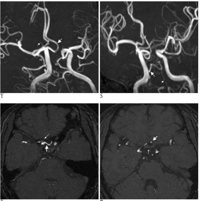
47세 남자 환자가 약 20일 전부터 시작된 둔한 느낌의 두 통을 주소로 내원하였다. 두통은 어느 한 부위로 국한되지 않 고 전반적인 양상이며, 내원 당시 시행한 이학적 검사 및 신 경학적 검사에서도 뚜렷한 이상 소견은 없었다. 환자는 5년 전 위폴립으로 폴립제거술을 시행한 것 외에는 과거력이나 가 족력에서 특이사항은 없었다. 내원 후 시행한 뇌 자기공명영 상과 3-D time of flight 자기공명 혈관조영술에서 왼쪽 전 대뇌동맥이 눈동맥(ophthalmic artery)이 기시하는 부위의 오 른쪽 내경동맥에서 기시하고 바탕영상(sourse image)에서 이 혈관은 오른쪽 시각신경(optic nerve)의 아래를 지나 시각교 차(optic chiasm)의 앞, 양쪽 시각신경의 사이에서 위로 뻗어 전교통동맥(anterior communicating artery)과 만나는 것을 관찰하였다. 또한 오른쪽 내경동맥에서 기시한 지속 삼차동맥 (persistent trigeminal artery)이 뇌기저동맥(basilar artery) 으로 연결되는 것과 왼쪽 내경동맥에서 A1 분절의 무형성증 을 관찰하였다(Figs. 1A-1D).
환자는 두통에 대한 대증요법 치료 후 증상이 호전되어 퇴 원하였다.
고 찰
전대뇌동맥의 A1 분절은 보통 내경동맥이 두 가지로 나뉘 는 부위에서 기시하여 동측의 시각신경이나 시각교차의 위를 넘어 앞쪽, 안쪽 방향으로 뻗어 전교통동맥과 연결된다. 전대 뇌동맥에서 전교통동맥으로 연결되는 부위의 해부학적 변이 는 매우 흔하며, 두 개의 A1 분절사이에 다양한 정도의 비대 칭성이 약 80%의 환자에서 발견된다(3). 그러나 시각신경 하 방 경로를 취하는 전대뇌동맥은 매우 드물어서 Robinson이 1959년 사체 해부에서 처음 발견한 이래 지금까지 약 30예 정도가 보고되었다(2). 시각신경 하방 전대뇌동맥은 보통 눈 동맥(ophthalmic artery)이 기시하는 부위 근처의 동측 내경 동맥에서 기시하여 동측 시각신경의 아래를 지나 안쪽으로 뻗 어 양쪽 시각신경 사이에서 위로 이동하여 전교통동맥과 합 치게 된다. 이는 고식적 혈관조영술과 자기공명 혈관조영술에 서 비슷한 모양으로 보인다. 그러나 자기공명 혈관조영술의 경우 높은 공간해상도의 바탕 영상을 분석하여 전대뇌동맥과 시각신경 혹은 시각교차와의 관계를 잘 파악할 수 있다.
지금까지 보고된 시각신경 하방 전대뇌동맥과는 다르게 본 증례에서는 왼쪽 전대뇌동맥이 오른쪽 내경동맥의 눈동맥이 나오는 부위 근처에서 기시하여 동측의 시각신경의 아래를 지 나 시각교차의 전방, 양쪽 시각신경 사이를 지나 위쪽으로 진 행하여 정상적인 전교통동맥과 연결되어 있었고 원위부의 오 른쪽 내경동맥은 정상적인 위치에서 전대뇌동맥과 중대뇌동 맥으로 나뉘어지며, 왼쪽 내경동맥은 바로 중대뇌동맥과 연결 되는 것을 자기공명 혈관조영술과 바탕영상에서 확인하였고 왼쪽 A1 분절은 관찰되지 않았다(Figs. 1A-1D). 이와 같이 반대측의 내경동맥에서 기시하는 시각신경 하방 전대뇌동맥 은 여러 문헌의 고찰에서 보고된 바 없는 것으로 보인다.
태생학적 배경은 내경동맥의 조기분기(early bifurcation of ICA) (4), 시각교차 전 문합의 확장(enlargement of prechi- asmal anastomosis) (5, 6), 원시 등측 안동맥과 배측 안동 대한영상의학회지 2005;53:165-167
─ 165 ─
반대측 내경동맥에서 기시한 시각신경 하방 전대뇌동맥: 증례 보고1
강 명 진
매우 드문 기형인 반대측 내경동맥의 눈동맥이 나오는 부위에서 기시하는 시각신경 하방 전대뇌동맥을 자기공명 영상과 자기공명 혈관조영술에서 우연히 발견하였기에 이를 보고한 다.
1동아대학교 의과대학 진단방사선과학교실
이 논문은 2005년 5월 31일 접수하여 2005년 6월 28일에 채택되었음.
맥 간 태중 교통의 지속(persistence in uterocommunication between the primitive dorsal and ventral ophthalmic artery) (5, 7), 원시 후각동맥과 원시 상악동맥 분지들 사이의 연결 (anastomosis between branches of primitive olfactory and primitive maxillary arteries) (5, 6) 등으로 이런 혈관 기형 을 설명하고 있다. 특히 시각교차 전 문합에 기여하는 동맥들 로는 안동맥의 가지, 상 뇌하수체 동맥(superior hypophy- seal artery)이 있으며, 이 가설은 본 예에서와 같이 반대측 시각신경 하방 전대뇌동맥이 있고 동측에 정상적인 시각신경
상방의 A1 분절이 같이 존재하는 것을 설명할 수 있다(2).
윌리스환 내의 다른 변이와 마찬가지로 시각신경 하방 전 대뇌동맥의 경우에도 대뇌 동맥류의 빈도가 높다(2, 7). 동맥 류가 발생하는 가장 흔한 부위는 전대뇌동맥-전교통동맥 콤 플렉스(ACA-AcomA complex)이나, 그외 어느 부위에서나 생길 수 있다(1). 또한 경동맥-뇌기저동맥간 문합(carotid- basilar artery anastomosis) (8), 내경동맥의 무형성증(9), A1 분절의 저형성 혹은 무형성(7), 융합된 뇌량주위 동맥 (fused pericallosal artery) (10), 얼기모양 전교통동맥(flex- 강명진: 반대측 내경동맥에서 기시한 시각신경 하방 전대뇌동맥
─ 166 ─
A B
C D
Fig. 1. A-D. Angiographic (A, B) and source images of a 3D TOF MR angiography (C, D). The left anterior cerebral artery (large ar- row, A) is arising from the right ICA at the origin of the ophthalmic artery (small arrow, A). Both posterior communicating arteries (open arrows, A) are directly connected to both posterior cerebral arteries. The right persistent trigeminal artery (large arrow, B) is arising from right internal carotid artery and anastomosing with hypoplastic basilar artery (small arrow, B). Note left A1 segment agenesis.
The infraoptic anterior cerebral artery (large arrow, C and D) arising contralateral ICA at the level of ophthalmic artery origin (open arrow, C) passes under the right optic nerve (small arrow, C), and then ascends anterior to the optic chiasm (small arrow, D) to join the normal positioned anterior communicating artery (large arrow, D).
iform AcomA) (10) 등 다른 혈관 기형과 동반되기도 한다.
본 증례에서는 내경동맥에서 기시한 지속 삼차동맥(persis- tent trigeminal artery)이 뇌기저동맥으로 연결되는 것과 반 대측 내경동맥에서 A1 분절의 무형성증이 동반되어 있었다.
고식적 혈관조영술은 침습적인 검사이며 위험한 합병증을 일으킬 가능성이 항상 존재하여 매우 제한적으로 시행하고 있 으며 자기공명 혈관조영술은 비용이 많이 드는 단점에도 불 구하고 환자에게 고통을 주지 않고 손쉽게 뇌혈관 구조를 파 악할 수 있어서 현재 광범위하게 사용하고 있다. 이로 인해 드문 대뇌혈관 기형을 관찰할 기회는 더욱 증가하였으므로 세 심하게 관찰할 필요가 있다. 특히 전교통동맥의 동맥류가 있 을 때에는 반드시 동반된 전대뇌동맥의 기형이 있는지에 대 해 확인하여 수술 시 접근 방법이나 중재적 시술 시 접근 경 로를 결정하여야 한다.
저자들은 자기공명 혈관조영술에서 우연히 발견된 드문 혈 관 기형인 반대측 내경동맥에서 기시한 시각신경 하방 전대 뇌동맥 1예를 경험하였기에 보고하는 바이다.
참 고 문 헌
1. Curtis A Given II, P Pearse Morris. Recognition and importance of an infraoptic anterior cerebral artery: case report. AJNR Am J
Neuroradiol 2002;23:452-454
2. Spinnato S, Pasqualin A, Chioffi F, Da Pian R. Infraoptic course of the anterior cerebral artery associated with an anterior communi- cating artery aneurysm: anatomic case report and embryological considerations. Neurosurgery 1999;44:1315-1319
3. Maurer J, Maurer E, Perneczky A. Surgically verified variations in the A1 segment of the anterior cerebral artery: report of two cases.
J Neurosurg 1991;75:950-953
4. Rosenorn J, Ahlgren P, Ronde F. Preoptic origin of the anterior cerebral artery. Neuroradiology 1985;27:275-277
5. Nutik S, Dilenge D. Carotid-anterior cerebral artery anastomosis:
case report. J Neurosurg 1976;44:378-382
6. Padget DH. The development of the cranial arteries in the human embryo. Contrib Embryol 1948;32:207-262
7. Odake G. Carotid-anterior cerebral artery anastomosis with aneurysm: case report and review of the literature. Neurosurgery 1988;23:654-658
8. Teal JS, Rumbaugh CL, Segall HD, Bergeron RT. Anomalous branches of the internal carotid artery. Radiology 1973;106:567-573 9. Turnbull I. Agenesis of the internal carotid artery. Neurology
1975;12:588-590
10. McCormick W. A unique anomaly of the intracranial arteries of man. Neurology 1969;19:77-80
대한영상의학회지 2005;53:165-167
─ 167 ─
J Korean Radiol Soc 2005;53:165-167
Address reprint requests to : Myong-Jin Kang, M.D., Department of Diagnostic Radiology, Dong-A University College of Medicine, 1, 3-Ga Dongdaeshin-dong, Seo-gu, 602-715 Pusan, Korea.
Tel. 82-51-240-5354 Fax. 82-51-253-4931 E-mail: [email protected]
Infraoptic Anterior Cerebral Artery Arising from Contralateral Internal Carotid Artery: Case Report1
Myong-Jin Kang, M.D.
1Department of Diagnostic Radiology, College of Medicine Dong-A University
We report an unusual case of an anomalous origin of the anterior cerebral artery from the contralateral inter- nal carotid artery at the level of the origin of the ophthalmic artery, which was discovered incidentally by MRI and MR angiography.
Index words :Cerebral blood vessel, MR Angiography