10.3803/jkes.2009.24.1.33
서 론
1)중격-시신경 형성장애(septo-optic dysplasia, SOD)는 10 만명당 2명 정도의 낮은 유병률을 갖는 드문 선천성 질환으 로 시신경 형성부전, 뇌량이나 투명 중격 등의 중간선 뇌 이 상 및 뇌하수체 형성부전에 의한 뇌하수체기능저하증을 특 징으로 한다[1]. 1956년 de Morsier에 의해 명명된 이후 de Morsier's syndrome으로도 불려진다[2]. 이 질환의 원인으 로 HESX1 돌연변이가 알려져 있으나[5], 가족형 중에서도 일부에서만 발견되며, 대부분의 가족형 및 빈도가 많은 산발 형에서는 아직 원인이 정확히 밝혀져 있지 않다.
대부분의 환자들은 영유아기에 안구 진탕, 시력 저하, 경
접수일자: 2008년 8월 15일 통과일자: 2008년 10월 24일
책임저자: 정윤석, 아주대학교 의과대학 내분비대사내과학교실
* 본 연구는 경기도의 경기도지역협력연구센터사업의 일환으로 수행하였음.
련 등의 증상으로 나타나 추가적인 방사선 검사 등을 통해 진단되나[3], 드물게는 영유아기 이후에 성장 지연이나 성발 달 지연, 요붕증 등의 증상으로 나타나는 경우도 있다[4]. 국 내에서는 주로 안과학회에 보고되다가[5], 최근 요붕증을 동 반한 증례가 내분비학회에 보고된 바 있었다[6].
저자들은 저신장증, 성발달 지연으로 내원한 23세 남자에 서 중격-시신경 형성장애를 진단하고, 원인 감별을 위해 유 전자 검사를 시행하였으며, 뇌하수체호르몬 이상을 진단하 였고, 호르몬 치료를 시행하며 경과 관찰 중인 환자를 경험 하였기에 문헌고찰과 함께 보고하는 바이다.
증 례
환 자: 23세 남자
주 소: 저신장 및 성발달 지연
현병력: 초등학교 3학년 때 저신장으로 인근 병원에서 검 사 결과 성장호르몬 결핍증을 진단받았고 고등학교 1학년
뇌하수체 전엽 호르몬 이상을 동반한 중격-시신경 형성장애 1예
아주대학교 의과대학 내분비대사내과학교실, 신경과학교실1, 안과학교실2, 영상의학교실3
이진우․황의경․김태호․윤형영․정재호․최용원․용석우
1․안재홍
2․김선용
3․김호성
3․정윤석 A Case of Septo-optic Dysplasia Associated with Anterior Pituitary Hormone Abnormalities
Jin-Woo Lee, Eui-Kyung Hwang, Tae-Ho Kim, Hyung-Young Yoon, Jae Ho Jung, Yong Won Choi, Suk-Woo Yong1, Jae-Hong Ahn2, Sun-Yong Kim3, Ho Sung Kim3, Yoon-Sok Chung Departments of Endocrinology and Metabolism, Neurology1, Ophthalmology2, and Radiology3, Ajou University School of Medicine
ABSTRACT
Septo-optic dysplasia (SOD) is a rare congenital malformation syndrome that is manifested by a triad of optic nerve hypoplasia, midline brain abnormalities and hypopituitarism. It is known to be associated with homeobox gene HESX1 mutation in some familial cases.
We experienced a case of SOD in a 23 year-old male who presented with short stature and delayed puberty.
The basal serum levels of testosterone and IGF-1 were low and the prolactin level was high. The combined pituitary stimulation study revealed decreased growth hormone responses. Brain MRI revealed complete agenesis of the corpus callosum, hypoplasia of the anterior pituitary gland and herniation of the third ventricle into the pituitary fossa. On the neurologic and ophthalmologic examinations, there was no definite abnormality except mild optic atrophy on the optical coherence tomography. Genetic analysis using polymerase chain reaction with direct sequencing revealed no HESX1 mutation. (J Korean Endocr Soc 24:33~37, 2009) ꠏꠏꠏꠏꠏꠏꠏꠏꠏꠏꠏꠏꠏꠏꠏꠏꠏꠏꠏꠏꠏꠏꠏꠏꠏꠏꠏꠏꠏꠏꠏꠏꠏꠏꠏꠏꠏꠏꠏꠏꠏꠏꠏꠏꠏꠏꠏꠏꠏꠏꠏꠏꠏꠏꠏꠏꠏꠏꠏꠏꠏꠏꠏꠏꠏꠏꠏꠏꠏꠏꠏꠏꠏꠏꠏꠏꠏꠏꠏꠏꠏꠏꠏꠏꠏꠏꠏꠏꠏꠏꠏꠏꠏꠏꠏꠏꠏꠏꠏ Key Words: Septo-optic dysplasia, hypopituitarism, HESX1 mutation
때까지 성장호르몬을 투여 받았으며, 당시 2차 성징이 나타 나지 않아 시행한 검사에서 남성호르몬 감소 소견 보여 남 성호르몬을 투여 받았으며 1년 전 고프로락틴혈증이 발견되 어 bromocriptine을 복용하며 지내던 중 최근 원인 감별 위 해 시행한 뇌자기공명영상 촬영에서 뇌량 형성부전 및 뇌하 수체 위축 소견이 보여 내분비대사내과에 의뢰되었다.
과거력: 주산기 시절의 특별한 병력은 없었고, 원시 및 난 시, 사시, 치아결손으로 치료받은 과거력이 있었다.
가족력: 뇌질환, 안질환 및 호르몬 이상 질환을 의심할 만 한 가족력은 없었다.
진찰 소견: 내원 당시 생체 활력 징후는 체온 36.6℃, 맥 박 65회/분, 호흡 20회/분, 혈압 126/76 mmHg이었고, 의식 은 명료하였다. 키 167 cm이고 체중 67.3 kg이었다. 호흡음, 심음, 장음은 정상적으로 청진되었고 복부의 팽만, 간 및 비
장 비대 등은 관찰되지 않았다. 사춘기 발달은 Tanner stage 4기에 해당하였다.
혈액검사 소견: 말초혈액검사에서 백혈구 8,300/mm³, 혈 색소 14.1 g/dL, 혈소판 280,000/mm³이었다. 생화학검사에 서 혈액요소질소 11.6 mg/dL, 크레아티닌 1.0 mg/dL, 나트 륨 143 mmol/L, 칼륨 4.8 mmol/L, 염소 105 mmol/L였으 며, 혈액 삼투압 농도 299 mOsm/L, 소변 삼투압 농도 659 mOsm/L로 정상이었다.
호르몬검사 소견: 갑상선자극호르몬 1.97 uIU/mL, 유리 T4 1.16 ng/dL, 부신피질자극호르몬 84 pg/mL로 정상 소견 을 보였고 프로락틴 23.1 ng/mL로 증가된 소견을 보였으며, 테스토스테론 0.30 ng/mL, IGF-1 127 ng/mL (정상치 179~777 ng/mL)로 감소된 소견을 보였고 황체형성호르몬 2.3 mIU/mL, 난포자극호르몬 1.8 mIU/mL로 테스토스테론
A
B
C
D
Fig 1. Brain MRI. A, B. T1-weighted sagittal view showed complete agenesis of corpus callosum and septum pellucidum.
Hypoplasia of anterior pituitary gland and ectopic location of posterior pituitary gland were observed. Herniation of infundibular recess of the third ventricle into pituitary fossa was noted. C. There was no corpus callosum or anterior commisure in T2-weighted axial view. D. Ectopic posterior pituitary gland was well visualized in T1-weighted coronal view.
Table 1. Combined pituitary stimulation test Glucose
(mg/dL)
TSH (μIU/mL)
Prolactin (ng/mL)
GH (ng/mL)
Cortisol (μg/dL )
ACTH (pg/mL)
LH (mIU/mL)
FSH (mIU/mL)
0 min 89 1.97 23.1 0.73 14.2 84 2.3 1.8
30 min 23 12.79 40.8 0.57 12.5 65 5.2 2.8
60 min 96 13.38 43.4 0.98 20.2 85 4.3 2.4
90 min 72 10.80 35.8 0.94 15.3 54 4.6 3.1
120 min 92 9.00 31.8 0.67 13.0 51 3.9 2.7
Interpretation appropriate normal Basal hyperprolactinemia and decreased response
to stimulation
deficiency normal normal deficiency deficiency
농도에 비해 상대적인 감소 소견을 보였다. 복합 뇌하수체 자극 검사에서는 자극에 대한 성장호르몬, 황체형성호르몬 및 난포자극호르몬의 반응이 저하된 소견을 보였다(Table 1).
방사선 소견: 뇌 자기공명영상 촬영에서는 뇌량의 형성부 전 및 투명 중격 결손과 뇌하수체 전엽의 형성저하증과 뇌 하수체 후엽의 이소성 위치가 관찰되었다. 뇌하수체 오목으 로의 제3뇌실의 이탈과 뇌하수체 오목 앞쪽으로 뇌실질의 전반적인 하향 이탈 소견이 관찰되었다(Fig. 1).
안과 소견: 나안시력은 우안 0.8 좌안 1.0이었으며 교정시 력은 양안 1.0이었다. 구면렌즈 대응치는 우안 -0.75D, 좌안 +0.125D이었다. 동공의 대광반사는 정상이었고 안압은 우 안 13 mmHg 좌안 12 mmHg으로 정상이었다. 세극등검사 에서 각막, 전방, 홍채 및 수정체는 정상이었다. 안저검사에 서 시신경은 유두함몰 부위의 경계가 불분명하였으며 유두 주위맥락막위축 (peripapillary chorioretinal atrophy) 소견이
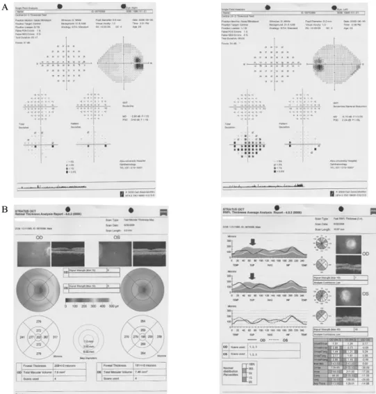
전 방향에 걸쳐 관찰되었다. 황반부를 포함한 중심망막이나 주변부 망막은 정상적인 소견을 보였다. 광간섭단층촬영검 사(optical coherence tomography)에서 두 눈의 시신경 상측 에서 망막신경섬유층의 국소적 결손 소견이 관찰되었다. 시 야검사에서는 양안의 전반적인 감도 저하 소견과 하측 시야 에 국소적인 감도 저하 소견이 산발적으로 발견되었으나 임 상적으로 의미 있는 변화는 아니었다(Fig. 2).
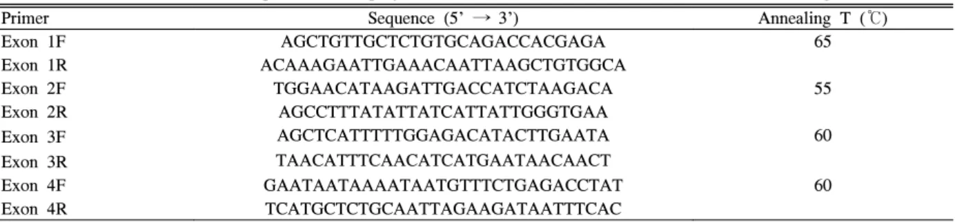
유전자 검사 방법 및 결과: 중합효소연쇄반응을 통하여 유전자 증폭을 하였다. 시발체는 Dattani 등[13]의 논문에 근거하여 바이오니어 회사에 의뢰하여 제작하였다. 주형 DNA 증폭은 Maxime PCR Premix kit (iNtRON, Korea)를 이용하였다. 중합효소연쇄반응은 PTC-200 (MJ Reaserch) 을 사용하였고, 방법은 94℃에서 5분간 변성시킨 후, 94℃에 서 30초간 변성한 후, 각 엑손에 적합한 온도에서 30초간 결 합, 72℃에서 30초간 합성 과정을 30회 시행하였고 최종적 으로 합성 과정을 72℃에서 10분간 시행하였다(Table 2). 중
A
B
Fig. 2. Ophthalmologic examination. A. Visual field analysis showed generalized reduction of sensitivity without significant visual field defect in both eyes. B. Optical coherence tomography showed focal retinal nerve fiber layer defect in superior segments of optic nerve head in both eyes (red arrow) suggesting mild optic nerve hypoplasia.
합효소연쇄반응이 종료된 시료에서 10 μL를 취하여 0.5 μ g/mL의 ethidium bromide가 첨가된 1.8% 아가로즈 겔에 전기 영동하여 확인하였다. 4개의 엑손에 대하여 직접 염기 서열 분석을 마크로젠 회사에 분석 의뢰하여 결과를 얻었다.
중격-시신경 형성장애와 관련하여 기존에 보고된 돌연변이 부위인 Q6H, R160C, S170L, T181A를 포함한 전체 HESX1 유전자 부위에서 해당 돌연변이는 관찰되지 않았다.
임상경과 및 치료: 환자는 뇌량의 형성 장애 및 뇌하수체 기능저하증이 동반된 중격-시신경 형성장애로 진단되었다.
남성호르몬 결핍에 대하여 testosterone undecanoate (Andriol Testocap) 80 mg bid, 고프로락틴혈증에 대하여 bromocriptine mesylate (Parlodel) 1.25 mg (1일 1회) 경구 투여하면서 현재 양호한 상태로 외래 추적 관찰 중이다.
고 찰
1941년 Reeves이 선천성 실명을 보인 생후 7개월 된 영 아에서 시신경 형성부전과 투명중격의 결손이 동반되어 있 던 예를 보고하였고, 1956년 de Morsier[2]이 선천성 시신 경 형성부전 환자의 부검 후 처음으로 중격-시신경 형성장 애로 명명하였으며, 이후 투명 중격의 결손을 보인 36명의 환자 중 9명에서 시신경 형성부전이 동반되어 있었음을 보 고하였다[7]. 1970년에는 Hoyt 등이 중격-시신경 형성장애 가 뇌하수체성 저신장증과 연관되었음을 처음으로 기술하였 고, 같은 해에 Kaplan 등이 중격-시신경 형성장애가 요붕증 및 뇌하수체기능저하증과 관련되며, 이는 간뇌의 병변에 의 한 시상하부 호르몬 및 항이뇨호르몬의 감소에 기인할 것이 라고 기술하였다[8]. 1972년에는 Billson과 Hopkins[9]가 뇌 하수체기능저하 및 요붕증, 선천성 시신경 형성부전을 보인 환자에서 투명 중격은 정상적이었음을 기술하면서 선천성 시신경 형성부전에 동반되는 구조적 이상 및 뇌하수체의 기 능 이상이 다양한 범주로 나타날 수 있음을 보고하였다.
중격-시신경 형성장애는 대부분의 경우 산발형으로 나타 나나[10] 소수에서는 상염색체 열성 유전의 형태를 보이기 도 하며[11], 일부에서는 상염색체 우성으로 나타난다[12].
가족성으로 나타나는 경우 중 일부에서는 3번 염색체에 있
는 호메오박스 유전자인 HESX1 돌연변이가 보고되고 있으 나[13], 본 증례의 경우 해당 돌연변이는 관찰되지 않았다.
그외에도 SOX2, SOX3 유전자의 변이와 관련되었다는 보 고가 있다 [14]. 이 질환의 다른 원인으로 임신 당시 모체의 낮은 연령, 당뇨병의 병력, 임신 중 음주, 코카인, 항전간제 복용 등이 보고되어 왔으나[15~19], 본 증례에서는 해당사 항이 없었다.
임상 양상은 유아기에 시력 저하, 안구 진탕, 사시 등의 안과적 증상으로 가장 먼저 나타나는 경우가 흔하고 시력 장 애의 정도는 거의 정상적인 시력에서부터 실명에 이르기까지 다양하게 나타날 수 있다[3]. 중격-시신경 형성장애에 있어서 시신경 형성부전이 동반되는 빈도는 75%에서 80%로 보고 되었으며 두 눈이 동시에 이환되는 경우가 더 흔하다[2]. 본 증례의 경우 영유아기에 원시, 난시 및 사시 등의 병력이 있 었으나, 안과 검사에서 사시는 발견되지 않았고 경도의 굴절 이상만 발견되었다. 안저검사에서 시신경 형성부전 소견은 확실하지 않았으나, 광간섭단층촬영검사에서 시신경 주위 망막신경섬유층의 경한 위축 소견이 관찰되어 시신경의 국 소적 형성부전이 동반되었음을 시사하였다. 그러나 시력 저 하나 의미 있는 시야결손은 관찰되지 않았다.
뇌하수체기능저하는 신생아기에 저혈당이나 경련, 2주 이 상 지속되는 신생아 황달과 같은 양상으로 나타날 수 있으 며, 이후에는 성장호르몬의 결핍이 가장 많이 동반되기 때문 에 저신장으로 나타나는 경우가 가장 많고, 그 외에 사춘기 지연, 요붕증 등으로 나타나는 경우도 보고되었다[20]. 본 증례에서도 저신장증 및 성발달 지연 등의 증상이 나타났으 나, 치료에 반응을 보였고, 경련이나 요붕증 등 환자 상태의 급격한 변화를 유발할 수 있는 증상들이 없었기 때문에 충 분한 평가가 미루어졌던 것으로 생각된다. 본 증례에서 관찰 되는 고프로락틴혈증의 원인은 제3뇌실이 뇌하수체 오목으 로 이탈되면서 뇌하수체 줄기를 압박하여 발생된 것으로 보 이며 이는 공터키안 증후군과 유사한 기전으로 생각된다.
결론적으로 뇌하수체기능저하증을 보이는 환자에서 영유 아기의 안과적 혹은 신경과적 증상 동반의 과거력이 있을 때 적절한 영상 검사 및 안과 검사를 통해 중격-시신경 형성 장애 등의 선천성 질환을 고려해 보아야 할 것이다.
Table 2. Primer sequences and polymerase chain reaction conditions for the HESX1 gene
Primer Sequence (5' → 3') Annealing T (℃)
Exon 1F AGCTGTTGCTCTGTGCAGACCACGAGA 65
Exon 1R ACAAAGAATTGAAACAATTAAGCTGTGGCA
Exon 2F TGGAACATAAGATTGACCATCTAAGACA 55
Exon 2R AGCCTTTATATTATCATTATTGGGTGAA
Exon 3F AGCTCATTTTTGGAGACATACTTGAATA 60
TAACATTTCAACATCATGAATAACAACT Exon 3R
Exon 4F GAATAATAAAATAATGTTTCTGAGACCTAT 60
Exon 4R TCATGCTCTGCAATTAGAAGATAATTTCAC
요 약
저자들은 저신장증 및 성발달 지연을 보였던 환자에서 중 격-시신경 형성장애를 진단하고, 원인 감별을 위한 유전자 검사에서 HESX1 돌연변이는 관찰되지 않은 상태로, 호르몬 치료를 시행하며 경과 관찰 중인 증례를 경험하였기에 문헌 고찰과 함께 보고하는 바이다.
참 고 문 헌
1. Dattani MT, Robinson IC: The molecular basis for developmental disorders of the pituitary gland in man.
Clin Genet 57:337-346, 2000
2. De Morsier G: Studies on malformation of cranio -encephalic sutures. III. Agenesis of the septum pellucidum with malformation of the optic tract.
Schweiz Arch Neurol Psychiatr 77:267-292, 1956 3. Haddad NG, Eugster EA: Hypopituitarism and
neurodevelopmental abnormalities in relation to central nervous system structural defects in children with optic nerve hypoplasia. J Pediatr Endocrinol Metab 18:853-858, 2005
4. Traggiai C, Stanhope R: Endocrinopathies associated with midline cerebral and cranial malformations. J Pediatr 140:252-255, 2002
5. Won Hae Lee, Song Hee Park, Hanho Shin: A case of septo-optic dysplasia. J Kor Ophth Soc 32:110-116, 1991
6. Soo Kyoung Kim, Tae Sik Jung, Jong Ryeal Hahm, Sang Min Lee, Sung Won Moon, Kyeong Ju Lee, Soon Il Chung: A case of central diabetes insipidus combined with septo-optic dysplasia and schizencephaly in 31-year-old woman. J Kor Endocrine Soc 22:339 -343, 2007
7. De Morsier G: Median cranioencephalic dysraphias and olfactogenital dysplasia. World Neurol 3:485-506, 1962
8. Hoyt WF, Kaplan SL, Grumbach MM, Glaser JS:
Septo-optic dysplasia and pituitary dwarfism. Lancet 1:893-894, 1970
9. Billson FA, Hopkins IJ: Optic hypoplasia and hypopituitarism. Lancet 1:905, 1972
10. Dattani MT, Preece M: Growth hormone deficiency and related disorders: insights into causation, diagnosis, and treatment. Lancet 363:1977-1987, 2004
11. Wales JK, Quarrell OW: Evidence for possible Mendelian inheritance of septo-optic dysplasia. Acta Paediatr 85:391-392, 1996
12. Thomas PQ, Dattani MT, Brickman JM, McNay D, Warne G, Zacharin M, Cameron F, Hurst J, Woods K, Dunger D, Stanhope R, Forrest S, Robinson IC, Beddington RS: Heterozygous HESX1 mutations associated with isolated congenital pituitary hypolasia and septo-optic dysplasia. Hum Mol Genet 10:39-45, 2001
13. Dattani MT, Martinez-Barbera JP, Thomas PQ, Brickman JM, Gupta R, Martensson IL, Toresson H, Fox M, Wales JK, Hindmarsh PC, Krauss S, Beddington RS, Robinson IC: Mutations in the homeobox gene HESX1/Hesx1 associated with septo-optic dysplasia in human and mouse. Nat Genet 19:125-133, 1998
14. Kelberman D, Dattani MT: Septo-Optic Dysplasia - Novel insights into the aetiology. Horm Res 69:257 -265, 2008
15. Donat JF: Septo-optic dysplasia in an infant of a diabetic mother. Arch Neurol 38:590-591, 1981 16. Parson SH, Dhillon B, Findlater GS, Kaufman MH:
Optic nerve hypoplasia in the fetal alcohol syndrome:
a mouse model. J Anat 186(Pt 2):313-320, 1995 17. Hoyt CS, Billson FA: Maternal anticonvulsants and
optic nerve hypoplasia. Br J Ophthalmol 62:3-6, 1978 18. Dominguez R, Aguirre Vila-Coro A, Slopis JM, Bohan
TP: Brain and ocular abnormalities in infants with in utero exposure to cocaine and other street drugs. Am J Dis Child 145:688-695, 1991
19. Woods K, Dattani MT: Transcription factors involved in disorders of forebrain and pituitary development. In Epstein J, Wynshaw-Boris. AJ(eds) Inborn Errors of Development: The Molecular Basis of Clinical Disorders on Morphogenesis, New York, Oxford University Press 540-551, 2004
20. Birkebaek NH, Patel L, Wright NB, Grigg JR, Sinha S, Hall CM, Price DA, Lloyd IC, Clayton PE:
Endocrine status in patients with optic nerve hypoplasia: relationship to midline central nervous system abnormalities and appearance of the hypothalamic-pituitary axis on magnetic resonance imaging. J Clin Endocrinol Metab 88:5281-5286, 2003