Ultrasonography of the Elbow Joint
7
0
0
전체 글
(2) 대한정형외과초음파학회지: 제 5 권 제 2 호 2012. 부착건, 상완동맥, 정중신경, 요골신경, 주관절의 관 절면(Fig. 1) 등으로 효율적인 전방 검사를 위해서. 검사 자와 마주 본 상태에서 주관절은 신전 상태를 유지한다.5) 1) 상완 이두근의 원위건(distal tendon of biceps brachii muscle). 해부학적으로 상완근(brachialis muscle)의 표층 에 위치하며 상완 동맥의 외측에 위치한다. 이 건은 심부에서 요골 결절(radial tuberosity)을 향해 비 스듬히 주행하기 때문에 주관절에서 가장 검사하기 까다로운 구조물 중의 하나이다. 먼저 횡축(transverse orientation)으로 상완 동맥을 확인한 다음 그 외측에 존재하는 건의 단면을 찾은 후(Fig. 2A) 전완을 과회외(hyper-supination) 시킨 상태로 종 축(longitudinal axis) 방향으로 초음파 변환기 (probe)의 방향을 바꿔 그 주행을 확인한다(Fig. 2B). 이등방성 (anisotrophy) 이 생기기 쉬운 구조 물이므로 변환기를 잘 유지하여야 하며, 건의 주행. Fig. 1. Thin hypoechoic hyaline cartilage of distal humerus (arrow heads) is demonstrating in transverse image of the distal humerus. B: Brachialis muscle, H: Humerus. A. B. C. D. Fig. 2. (A) Transverse image of biceps brachii tendon (arrow head) is showing in just lateral of brachial artery (Ba) at the level of distal humerus. (B) Longitudinal image of biceps tendon is showing just before its insertion (arrow head) (C) With posterior scanning technique, radial insertion of biceps tendon is not showing in position of forearm supination. (D) In position of forearm pronation, the radial insertion of biceps tendon is visible (arrow head). Ba: Brachial artery, R: Radius, U: Ulna. 124.
(3) 김철홍 외: 주관절의 초음파 검사. 기를 종축으로 전환하면 주관절에서 정중 신경 주행 을 비교적 쉽게 확인할 수 있다(Fig. 3B). 요골 신 경은 상완골 외과 근위부에 변환기를 횡축으로 위치 시키면 상완골 외연의 앞쪽에 상완 근과 상완요골 근 사이에 위치하는 요골 신경의 단면을 쉽게 확인 할 수 있으며(Fig. 3C) 이 상태에서 변환기를 종축 으로 바꿔서 주관절에서의 요골 신경 주행을 확인할 수 있으며 상완골 외과 높이에서 표재성과 심부 요 골 신경으로 나뉘는 것까지 확인할 수 있다(Fig. 3D). 이와 같이 비록 이 두 신경의 정상 소견을 쉽 게 확인 할 수 있으나 주관절 부위에서 이 두 신경의 병적 상황에 대한 초음파적 평가는 쉽지 않다.8). 전체를 하나의 영상으로 찾으려 하기 보다는 요골 결절에 부착하는 건 부착 부는 따로 확인하는 것이 검사에 용이하다. 건 부착 부는 다른 방법으로 검사 할 수도 있는데, 주관절을 최대로 굴곡 시킨 후 변환 기를 전완 근위부의 후방에 횡으로 위치하여 근위 요골-척골 접촉면을 확인한 후 전완을 회내전 (pronation) 시키면 요골 결절에 부착하는 건의 영 상을 얻을 수 있다(Fig. 2C, D). 상완 이두근 원위 부착 건 완전 파열 초음파 소견은 ① 원위 관절 위치 에서 건의 음영 부재 ② 혈종으로 인한 저 반사 (hypoechoic) 소견으로 둘러싸인 수축된 단열 건 확인 등인데 대부분의 완전 건 파열은 임상 소견이 확실하여 임상적으로 진단하고 초음파는 이를 확인 하는 정도의 가치를 가진다.6,7). 3) 기타. 외상 환자의 경우 골절 진단을 위하여 주관절 표 면을 면밀히 관찰하여야 하는데, 초음파는 미세 골 절(occult fracture)을 진단하는데 효용성이 크며, 특히 요골 골두 골절의 경우 회내-회외 동작을 하. 2) 정중 신경과 요골 신경. 정중 신경은 변환기를 횡축으로 놓고 상완 동맥의 내측에 있는 신경 단면을 확인한 후(Fig. 3A) 변환. A. B. C. D. Fig. 3. (A) Transverse image of median nerve (arrow) is showing in just medial of brachial artery (Ba) at the level of distal humerus. (B) Longitudinal image of median nerve is demonstrating after identifying transverse image by turning probe longitudinally (arrow). (C) Transverse image of radial nerve (arrow head) is showing in just anterior of distal humerus (arrow head). (D) By turning probe, longitudinal image of radial nerve can be seen (arrow heads). Ba: Brachial artery, H: Humerus. 125.
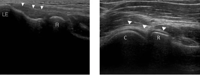
(4) 대한정형외과초음파학회지: 제 5 권 제 2 호 2012. 이전의 총신전건에서 관찰되는 부종성 저 반사 소견 ② 국소적 또는 광범위하게 관찰되는 불규칙적인 미 세섬유양 (fibrillar) 형태 소견 ③ 부분적 또는 전체 적인 파열 소견 등이다.11-13). 면서 요골 골두를 확인하는 동적 검사(dynamic study)가 가능하므로 더 효율적이다.8,9) 2. 외측방 검사(lateral aspect). 2) 요골 측부 인대. 주관절의 외측 방에서 초음파 검사의 목표가 되는 주된 구조물은 총신전건(common extensor tendon)과 요골 측부 인대(radial collateral ligament)인데, 이를 위해서는 주관절은 신전하고 손은 회내위와 회외위 중간 위치 정도의 자세에서 검사하 는 것이 관찰에 용이하다.10). 요골 측부 인대는 총신전건 심부에서 확인되는데 외상과 에서 요골 골두 쪽으로 주행하는 얇은 미세 섬유양 구조물을 확인하면 되며, 손상이 있는 경우 더 확실하게 관찰되는 경향이 있어 진단에 용이하다 (Fig. 5).12). 1) 총신전건. 3. 내측방 검사(medial aspect). 총신전건의 외상과 부착 부는 비교적 균일한 과반 사(hyperechoic) 소견이며 요골-소두(radiocapitellar) 관절을 가로 질러 주행하게 되는데, 초 음파 영상에서도 요골-소두 관절이 총신전건 반사 와 함께 보이도록 변환기를 위치시킬 때 나타나는 것이 비교적 정확한 총신전건 영상이라고 할 수 있 다(Fig. 4). 해부학적으로 단 요 수근 신전근(extensor carpi radialis brevis), 총수지 신전근(common extensor digitorum), 소 수 지 신 전 근 (extensor digiti minimi), 척 수근 신전근(extensor carpi ulnaris)들의 근 섬유를 받게 되나 초음 파 상으로 이를 구분할 수는 없다. 외측 방에서 가장 흔한 병리 소견은 외상과 염으로 명명하는 퇴행성 건 질환으로 초음파 상의 진단 기준은 ① 부착되기. 주관절 내측 검사를 위해서는 주관절은 신전한 상 태에서 전완 부는 완전 회외 상태에서 검사를 시행 한다. 내측 검사 시에 관찰하게 되는 주된 구조물은 총 굴곡건 (common flexor tendon), 척골 측부 인 대(ulnar collateral ligament) 등이다. 1) 총 굴곡건. 총 굴곡건은 원형 회내근(pronator teres)과 표 재성 굴곡근으로 구성되며 내상과에 부착하게 되는 데, 정상적인 초음파 소견은 과반사 소견을 보이는 삼각형의 구조물로 미세 섬유양 형태로 나타나며 외 측의 총 신전건 보다는 더 조밀하며 더 광범위한 부 착을 보인다(Fig. 6). 그렇지만 내상과 염에서 보이. Fig. 4. Fibrillar appearance of common extensor tendon (arrow head) is demonstrating in longitudinal image at the level of lateral epicondyle. LE: Lateral epicondyle, R: Radius. Fig. 5. Longitudinal image of radial collateral ligament image is demonstrating (arrow head). C: Capitellum, R: Radius. 126.
(5) 김철홍 외: 주관절의 초음파 검사. 골로부터 내상과로 주행함을 확인할 수 있다(Fig. 7). 만약 파열이 있는 경우에는 액성 반사를 동반한 저 반사의 비연속성이 보여 진단이 어렵지 않다.15). 는 총 굴곡건 질환의 초음파 소견은 외상과 염과 유 사한 판단기준으로 진단할 수 있다.14) 2) 척골 측부 인대. 척골 측부 인대는 전방 다발(anterior bundle), 후방 다발, 횡 다발 로 구성되나 초음파 상에서는 전 방 다발만이 관찰되는 경향이 있으며 요골 측부 인 대와 마찬가지로 종축 영상에서 관찰되며, 촘촘한 상대적으로 과반사의 미세 섬유양 구조물이 근위 척. 4. 후방 검사(posterior aspect). 주관절의 후방 검사를 위해서는 검사대에 손바닥 을 올려놓고 주관절을 90도 굴곡 시킨 자세를 유지 하게 한다. 이러한 자세는 후방 검사가 용이할 뿐만 아니라 관절의 잠김 작용(rocking motion)을 증가 시킴으로써 관절 액의 이동을 촉진하여 작은 유리. Fig. 6. Compact fibrillar appearance of common flexor tendon group (arrow head) is demonstrating in the longitudinal image at the level of medial epicondyle. ME: Medial epicondyle, Arrow: Medial joint space. Fig. 7. Fibers of ulnar collateral ligament (arrow heads) is showing in the longitudinal image of medial elbow joint. H: Humerus, U: Ulna. A. B. Fig. 8. (A) Transverse placement of probe just on cubital tunnel offers transverse image of ulnar nerve (arrow head). (B) Longitudinal placement of probe in cubital tunnel reveals the longitudinal image of ulnar nerve in cubital tunnel (arrow heads). CT: Cubital tunnel, U: Ulna. 127.
(6) 대한정형외과초음파학회지: 제 5 권 제 2 호 2012. elbow. Semin Musculoskelet Radiol. 2007;11:10516. 05. Barr LL, Babcock DS. Sonography of the normal elbow. Am J Roentgenol. 1991;157:793-8. 06. Lozano V, Alonso P. Sonographic detection of the distal bicep tendon rupture. J Ultrasound Med. 1995;14:389-91. 07. Miller T, Adler RS. Sonography of tears of the distal biceps tendon. Am J Roentgenol. 2000;175: 1081-6. 08. Martinoli C, Bianchi S, Zamorani MP, Zunzunegui JL, Derchi LE. Ultrasound of the elbow. Eur J Ultrasound. 2001;14:21-7. 09. Davidson RS, Markovitz RI, Dormans J, Drummond DS. Ultrasonographic evaluation of the elbow in infant sand young children after suspected trauma. J Bone Joint Surg Am. 1994; 76:1804-12. 10. Barr LL, Kirks DR. Ultrasonography of acute epitrochlear lymphadenitis. Pediatr Radiol. 1993;23:72-3. 11. Maffulli N, Regine R, Carrillo F, Capasso G, Minelli S. Tennis elbow:an ultrasonographic study in tennis players. Br J Sp Med. 1990;24: 151-4. 12. Jacobson JA, van Holsbeeck MT. Musculoskeletal ultrasonography. Orthop Clin North Am. 1998;29:13567. 13. Connell D, Burke F, Coombes P, et al. Sonographic examination of lateral epicondylitis. Am J Roentgenol. 2001;176:1763-77. 14. Ferrara MA, Marcelis S. Ultrasound of thee lbow. J Belge Radiol. 1997;80:122-3. 15. Vanderschueren G, Prasad A, van Holsbeeck M. Ultrasound of the elbow. Semin Musculoskel Radiol. 1998;2:223-35. 16. Chiou HJ, Chou YH, Cheng SP. Cubital tunnel syndrome: diagnosis by high-resolution ultrasonography. J Ultrasound Med.1998;17:643-8.. 체의 관찰에 용이한 것으로 되어 있다. 후방 검사에 서는 주로 상완 삼두건(triceps brachii muscle)과 주관(cubital tunnel)내에 위치한 척골 신경을 평가 하게 된다. 1) 상완 삼두건. 상완 삼두건의 경우 과반사의 촘촘한 선형(linear) 구조물이 척골 주두(olecranon process)에 부착하는 것을 확인하는 것으로 다소 용이하게 평가 할 수 있다. 2) 척골 신경. 변환기를 내상과 바로 위에 횡축으로 위치시키게 되면 내상과에 인접한 타원형의 구조물을 확인할 수 있으며(Fig. 8A) 이를 중심으로 변환기를 주관 (cubital tunnel)위에 종축으로 전환하면서 척골 신 경의 주관 내에서의 상태를 확인할 수 있다(Fig. 8B). 이와 관련된 비정상 소견 유무는 주관절에서 관찰되는 다른 신경인 정중 신경이나 요골 신경의 이상 소견에 비하여 다소 용이하게 판단할 수 있는 데, 주관증후군의 경우 척골 신경의 단면은 건강한 신경 혹은 반대 측보다 현저히 크기가 증가되어 있 다.16) 또한 변환기를 횡축으로 놓은 상태에서 주관 절을 점진적으로 굴곡 시키면서 주관에서의 탈구나 아탈구 유무도 관찰할 수도 있다.. 참고문헌 01. Radunovic G, Vlad V, Micu MC, et al. Ultrasound assessment of the elbow. Med Ultrason. 2012;14:141-6. 02. Martinoli C, Bianchi S, Giovagnorio F, Pugliese F. Ultrasound of the elbow. Skeletal Radiol. 2001;30:605-14. 03. Koski JM. Ultrasonography of the elbow joint. Rheumatol Int. 1990;10:91-4. 04. Tran N, Chow K. Ultrasonography of the. 128.
(7) 김철홍 외: 주관절의 초음파 검사. 국문초록 이 종설은 주관절에서의 초음파 검사 방법을 서술하였으며 주관절의 구조물들은 표층에 위치하기 때문에 초음파 사용이 용이하고 동적인 검사와 건 측과의 비교를 쉽게 할 수 있다는 장점으로 인하여 환자의 병변에 대한 많은 정보를 제공해 준다. 주관절에서 초음파 검사는 주관절 주변의 건, 근육, 인대 와 점액낭의 비정상적 형태뿐만 아니라 미세 골절(occult fracture), 주관(cubital tunnel) 내의 척골 신경의 압박 유무도 평가할 수 있다. 향후 주관절의 연부조직 이상을 진단하 는 수단으로서의 초음파의 가치는 점점 증가할 것이다. 색인단어: 주관절, 초음파 검사. 129.
(8)
수치


관련 문서
Muscle atrophy of the right thoracic limb, reduced biceps brachii muscle tendon tone, a tear of the medial glenohumeral ligament, and a rupture of the subscapularis
Distal biceps tendon ruptures result in loss of strength in forearm flexion of up to 30% and in forearm supination of up to 40%. 1) As these injuries often occur in active
Sonographic image shows short axis view of long head of biceps tendon (arrow), deltoid muscle, and subscapularis muscle.. D: deltoid,
Longitudinal ultrasonography finding of normal geun(筋) curved arrow: skin(epidermis and dermis), arrow heads: achilles tendon, SO: soleus muscle, FHL:.. flexor
Coronal reformatted CT image showing thickening and stenosis of the distal common bile duct (arrow) and dilatation of proximal biliary treeF. Coronal reformatted CT image showing
Transverse real time sonoelastography view and B mode image of distal Achilles tendon in healthy asymptomatic young adult, and it shows distal Achilles tendon arrows with
Purpose: This study aimed to investigate the impact of resistance on the muscle activities of the long and short heads of the biceps brachii, according to the elbow angle
Purpose: This study aimed to investigate the impact of resistance on the muscle activities of the long and short heads of the biceps brachii, according to the elbow
