Pulmonary Cryptococcosis Mimicking Primary Lung Cancer with Multiple Lung Metastases
5
0
0
전체 글
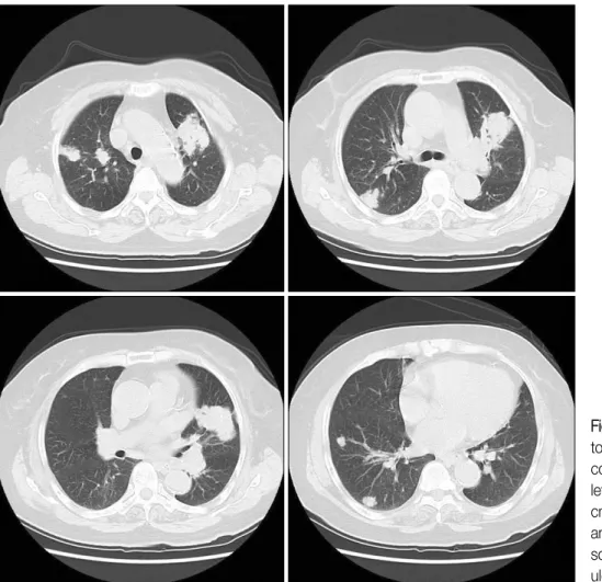
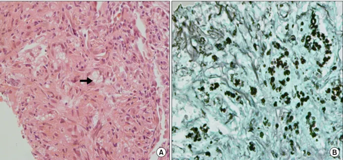
(2) Tuberculosis and Respiratory Diseases Vol. 73. No. 3, Sep. 2012. Figure 1. Simple chest radiograph shows a large irregular mass-like lesion in the left upper lobe with multiple opacities in both lungs.. left upper lobe (about 10 cm in longest diameter) and multiple nodular consolidations with small nodules, suggesting malignancy or granulomatous inflammation (Figure 2). Pulmonary function was within normal range. Considering old age and multiple pulmonary nodules leaded us to rule out the possibility of primary lung cancer with lung to lung metastases. There was no endobronchial lesion on fiberoptic bronchoscopic examination. Microbiologic studies with bronchial washing fluid revealed no bacterial, fungal, or mycobacterial pathogen. For confirmative diagnosis, percutaneous CT-guided biopsy was performed at the mass in left upper lobe. Microscopic findings revealed granulomatous inflammation with multiple yeast forms, morphologically suggestive of cryptococcosis (Figure 3). Serum cryptococcal antigen (Ag) was reactive. Lum-. Figure 2. Chest computed tomography reveals a large consolidative mass in the left upper lobe (about 10 cm in longest diameter) and multiple nodular consolidations with small nodules in both lungs.. 183.
(3) YS Kim et al: Consolidative lung mass with multiple pulmonary nodules. Figure 3. Histologic findings of percutaneous computed tomography-guided biopsy from mass in the left upper lobe. (A) Many yeast-form fungal organisms (arrow) are observed on the hematoxylin and eosin stain (×200). (B) Multiple fungal colonies with some budding yeasts, strongly stained by Gomori's methenamine-silver stain (×200).. Figure 4. After treatment with oral fluconazole for 4 months, chest radiographs shows significant decrease in size of the consolidative pulmonary mass in the left upper lobe and multiple nodular consolidations with small nodules in both lungs.. 184.
(4) Tuberculosis and Respiratory Diseases Vol. 73. No. 3, Sep. 2012. bar puncture revealed normal cerebrospinal fluid (CSF) opening pressure of 10 cm H2O. CSF microscopy showed no cells and no bacteria or fungus. CSF cryptococcal Ag was non-reactive. The patient was treated with oral fluconazole 400 mg daily according to the recent guidelines. Four months later, she became well without any symptom or sensation of weakness. Follow-up imaging studies showed a marked improvement of previously noted lesions (Figure 4).. Discussion Cryptococcosis is an invasive fungal infection, caused predominantly by Cryptococcus neoformans or Cryptococcus gattii1. It is an opportunistic infection that was recently estimated at 1 million cases and >600,000 deaths annually2. Cryptococcosis is more common in immune compromised patients. The infection is most often associated with human immunodeficiency virus infection. The other predisposing factors are patients with hematologic malignancies, chronic corticosteroid or immunosuppressive drugs, recipients of solid organ transplants. Cryptococcal infection is acquired by inhalation of aerosolized particles. The clinical presentation of cryptococcal infection varies along a spectrum from asymptomatic pulmonary colonization to severe pneumonia with respiratory failure and life threatening 3 meningitis . In immunocompromised patients, pulmonary cryptococcosis can be severe and rapidly progressive. However, the disease in immunocompetent patients may be asymptomatic and tends to remain confined to the lung as pulmonary nodules, although some patients develop serious meningitis and disseminated in4,5 fection . Symptoms are nonspecific and characterized by cough, fever, dyspnea, chest pain, hemoptysis, malaise, 6 or weight loss . Many cases are discovered incidentally during the work-up of an abnormal chest radiograph. The most common CT finding in immunocompetent patients is pulmonary nodule, well defined with smooth margin. Multiple nodules are more common than solitary nodule. Consolidation, lymphadenopathy, pleural. effusion, and cavitation can occur in the immunocompromised patients7-9. Diagnosis of pulmonary cryptococcosis requires direct evidence of the presence of cryptococcus in sputum, bronchial washing or bronchoalveolar lavage fluid or lung tissue. Cryptococcal Ag titer can be measured in serum or CSF. Although high titer suggests invasive disease, negative titer does not rule out the diagnosis. The changes in cryptococcal Ag titer reliably reflect the 10 response to therapy . The treatment of cryptococcosis depends on the immune status of the host and the site of infection. Pulmonary cryptococcosis in the immunocompetent patient sometimes resolves spontaneously. However, recent guidelines suggest treating even those who are asymptomatic, because dissemination of infection to central nervous system (CNS) in healthy host can occur and low toxicity therapy with fluconazole is availalbe. Mild to moderate disease limited to the respiratory system is usually treated with oral fluconazole in both immunocompetent and immunocompromised hosts (400 mg per day for 6∼12 months). Patients with severe disease, CNS involvement, or any other evidence of dissemination are treated with intravenous amphotericin B and flucytosine during induction phase, which is followed by prolonged therapy with oral fluconazole during consolidation phase. Immunocompromised patients remain on lifelong oral fluconazole until their immune impairment resolves or improves11. Although there are many case reports of pulmonary cryptococcosis in immunocompetent hosts, it is very rare that a huge consolidative mass like our case diagnosed as pulmonary cryptococcosis in a review of liter12 atures . In conclusion, we presented a case of 74-year-old patient with pulmonary cryptococcosis mimicking primary lung cancer with multiple lung to lung metastases. Although more common in immunocompromised patients, pulmonary cryptococcosis can occur in immunocompetent patients and should be considered on the differential diagnosis for nodular or mass-like lesions in chest radiograph.. 185.
(5) YS Kim et al: Consolidative lung mass with multiple pulmonary nodules. References 1. Brizendine KD, Baddley JW, Pappas PG. Pulmonary cryptococcosis. Semin Respir Crit Care Med 2011;32: 727-34. 2. Park BJ, Wannemuehler KA, Marston BJ, Govender N, Pappas PG, Chiller TM. Estimation of the current global burden of cryptococcal meningitis among persons living with HIV/AIDS. AIDS 2009;23:525-30. 3. Hung MS, Tsai YH, Lee CH, Yang CT. Pulmonary cryptococcosis: clinical, radiographical and serological markers of dissemination. Respirology 2008;13:247-51. 4. Nadrous HF, Antonios VS, Terrell CL, Ryu JH. Pulmonary cryptococcosis in nonimmunocompromised patients. Chest 2003;124:2143-7. 5. Goldman JD, Vollmer ME, Luks AM. Cryptococcosis in the immunocompetent patient. Respir Care 2010;55: 1499-503. 6. Mylonakis E, Muse VV, Mino-Kenudson M. Case records of the Massachusetts General Hospital. Case 28-2011. A 74-year-old man with pemphigus vulgaris and lung nodules. N Engl J Med 2011;365:1043-50. 7. Lindell RM, Hartman TE, Nadrous HF, Ryu JH. Pulmon-. 186. 8.. 9.. 10.. 11.. 12.. ary cryptococcosis: CT findings in immunocompetent patients. Radiology 2005;236:326-31. Song KD, Lee KS, Chung MP, Kwon OJ, Kim TS, Yi CA, et al. Pulmonary cryptococcosis: imaging findings in 23 non-AIDS patients. Korean J Radiol 2010;11:40716. Choe YH, Moon H, Park SJ, Kim SR, Han HJ, Lee KS, et al. Pulmonary cryptococcosis in asymptomatic immunocompetent hosts. Scand J Infect Dis 2009;41: 602-7. Chang WC, Tzao C, Hsu HH, Lee SC, Huang KL, Tung HJ, et al. Pulmonary cryptococcosis: comparison of clinical and radiographic characteristics in immunocompetent and immunocompromised patients. Chest 2006;129:333-40. Perfect JR, Dismukes WE, Dromer F, Goldman DL, Graybill JR, Hamill RJ, et al. Clinical practice guidelines for the management of cryptococcal disease: 2010 update by the infectious diseases society of america. Clin Infect Dis 2010;50:291-322. Garrett L, Marr K, West S, Allada G. 74-year-old man from the pacific northwest with fever and a lung mass. Chest 2011;140:814-7..
(6)
수치
관련 문서
A diagnosis of pulmonary siderosis is based on a patient history of iron inhalation, on chest radiographic findings, and on accumulation of iron oxide in macrophages within
The size of pre- viously described mass sh- adow of pulmonary arterio- venous malformation in the left lower lobe decreased on chest radiograph (A) and chest CT
Conventional chest CT scan reveals solitary 3.5 cm sized lung mass in left upper lobe without lynphadenopathy.. The distal porton of the mass shows
Simple chest radiograph shows old pulmonary tuberculous lesion in right upper lobe(small ar- rowheads) with newly appeared nodular increas- ed density (l arge
One day later increase in size of the mass with suspected pulmonary hemorrhage or infilteration a rouncl the mass was found in left lower
A lobulated mass narrowing right upper lobe bron chus , poststenotic pneumonitis , and thickening of the posterior wall of the right main bronchus.. Narrowing and
Chest CT showed diffuse bronchial wall thickening involving the left main bronchus, the left upper lobar bronchus and the lingular divisional bronchus of the left upper lobe..
Positron emission tomogra- phy/computed tomography (PET/CT) scan revealed an in- creased fluorodeoxyglucose (FDG) uptake in the mass involving the right upper lobe, which
