Received:November 30, 2018, Revised:December 31, 2018, Accepted:January 22, 2019 Corresponding to:Yun Jong Lee http://orcid.org/0000-0001-7615-8611
Division of Rheumatology, Department of Internal Medicine, Seoul National University Bundang Hospital, 82 Gumi-ro 173beon-gil, Bundang-gu, Seongnam 13620, Korea. E-mail:[email protected]
Copyright ⓒ 2019 by The Korean College of Rheumatology. All rights reserved.
This is an Open Access article, which permits unrestricted non-commerical use, distribution, and reproduction in any medium, provided the original work is properly cited.
Follicular Bronchiolitis Mimicking Lung Cancer in a Patient with Primary Sjögren’s Syndrome
Seunghwan Shin1, You-Jung Ha1,4, Eun Ha Kang1,4, Yeong Wook Song2,3,4, Yun Jong Lee1,4
1Division of Rheumatology, Department of Internal Medicine, Seoul National University Bundang Hospital, Seongnam, 2Department of Internal Medicine, Seoul National University Hospital, Seoul, 3WCU Department of Molecular Medicine and Biopharmaceutical Sciences, Medical Research Institute, Seoul National University College of Medicine, Seoul, 4Department of Internal Medicine, Seoul National University College of Medicine, Seoul, Korea
Follicular bronchiolitis (FB) is an uncommon pulmonary lymphoproliferative disorder that is characterized by the presence of peribronchiolar hyperplastic lymphoid follicles with reactive germinal centers. FB could be associated with systemic illnesses including immunodeficiency, infection, and autoimmune diseases. In Korea, a single case of FB with rheumatoid arthritis was recently described but there has been no report on FB associated with other rheumatic diseases. Herein, we describe the first case of FB presenting nodular ground-glass opacities (GGO), which mimicked lung cancer, in patients with primary Sjögren’s syndrome (SS). The differential diagnosis of nodular GGO lesions should include FB although it is a rare manifestation in SS patients. (J Rheum Dis 2019;26:206-210)
Key Words. Sjögren‘s syndrome, Lung neoplasms, Lung diseases, interstitial, Bronchiolitis
INTRODUCTION
Sjögren’s syndrome (SS) is a systemic autoimmune dis- ease characterized by the lymphocytic infiltration of the exocrine glands which often results in dryness of the mouth and eyes. Systemic manifestations occur in 30% to 40% of patients with SS [1]. SS can affect the lungs in 10%
to 20% of the patients and its pulmonary involvement in- cludes airway abnormalities, interstitial lung disease (ILD), and lymphoproliferative disorders [2].
Follicular bronchiolitis (FB) is a rare small airway dis- ease, and it is pathologically characterized by the pres- ence of hyperplastic lymphoid follicles with germinal cen- ters in the walls of small airways [3]. Radiographically, the most common findings of FB are bilateral cen- trilobular small nodules that are 1 to 3-mm in diameter and are often associated with bilateral patch ground-glass opacities (GGO) on chest computed tomography (CT) scans [4]. FB may occur without associated diseases
(idiopathic) or as a secondary form associated with other diseases including autoimmune disorders such as rheu- matoid arthritis (RA). In addition, FB has been consid- ered a pulmonary manifestation of SS since 1985 after Fortoul et al. [5] reported pathologically proven FB in three patients with SS.
In 1993, Lee et al. [6] reported the first Korean case of idiopathic FB and a single case of FB secondary to RA was recently published in 2016 [7]. However, there has been no FB case associated with SS in Korea until now. Herein, we describe a specific case of FB, presenting a newly de- veloped nodular GGO in a SS patient with ILD, and pro- vide the literature review of Korean FB cases.
CASE REPORT
A 67-year-old non-smoking female patient had been di- agnosed as having primary SS with ILD for seven years, based on her having dry mouth, recurrent parotid gland
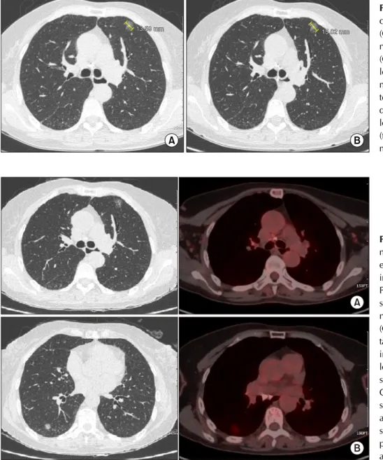
Figure 2. Chest computed to- mography (CT) and positron emission tomography (PET)-CT images. (A) Matched CT and PET/CT scan slices demon- strated a 15 mm-sized lesion of nodular ground-glass opacities (GGO) without pathological up- take of 18F-fluorodeoxyglucose in the anterior segment of the left upper lobe. (B) Chest CT scan images revealed a new GGO nodule in the superior segment of the right lower lobe and the corresponding PET-CT slice showed the lesion with hy- permetabolism (maximal stand- ardized uptake value 2.1).
Figure 1. Serial follow-ups of chest computed tomography (CT). (A) Chest CT showed a nodular ground-glass opacity (GGO) lesion in the left upper lobe, 31 months prior to ad- mission. (B) Seven months prior to admission, the longitudinal diameter of the nodular GGO lesion was slightly increased (from 13.5 to 15.0 mm over 24 months).
swelling, hyposalivation, positive anti-Ro/SSA, and focal sialadenitis. Thirty-one months ago, chest high reso- lution CT (HRCT) scans showed mild diffuse reticular opacities and traction bronchiectasis in the peripheral lower lobe of both lungs and a nodular GGO in the ante- rior segment of the left upper lobe (LUL). She visited our Rheumatology clinic 24 months ago and was treated with mizoribine, artificial saliva, and tears. The patient had an uneventful course during the follow-up, and her serial pulmonary function tests (PFTs) were normal over the re- cent 24 months. Seven months prior to admission, in the follow-up chest HRCT scan, the longitudinal diameter of the nodular GGO lesion had slightly increased (from 13.5 to 15.0 mm over 24 months), and the possibility of mini-
mally invasive adenocarcinoma could not be excluded (Figure 1). Six month later, another chest HRCT showed a newly developed GGO nodule (the diameter of 11 mm) in the superior segment of the right lower lobe (RLL;
Figure 2), but, the LUL nodular lesion was not changed.
Under the suspicion of lung cancer, further work-up was performed. Her complete blood count, renal, and liver function tests were within normal limits and C-reactive protein and complement levels were also normal. The er- ythrocyte sedimentation rate was 64 mm/h, and there was no evidence of monoclonal gammopathy. Serum tu- mor markers such as neuron-specific enolase, cytoker- atin-19 fragments, carcinoembryonic antigen, and squ- amous cell carcinoma antigen were within normal ranges.
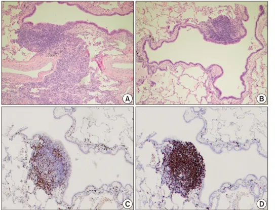
Figure 3. Pathology of lung bi- opsy specimen. In hematoxylin and eosin stained section, peri- bronchiolar lymphoid hyper- plasia with polymorphous lym- phoid populations was ob- served (A, ×100). Additionally, the destruction of peribronchial alveolar septa was not shown (B, ×100). Immunohistochem- istry study for CD3 (C) and CD20 (D) showed that most mononuclear cells were non- malignant B cells in the peri- bronchial follicular lymphoid hyperplasia (×40).
There were no malignant cells in the bronchoscopy and endobronchial transbronchial needle aspiration. Whole- body positron emission tomography/CT demonstrated a mild hypermetabolic focus corresponding to the RLL le- sion, but no uptake of 18F-fluorodeoxyglucose in the LUL lesion (Figure 2). The brain magnetic resonance imaging revealed no evidence of metastasis. Under the impression of adenocarcinoma in situ, she underwent superior seg- mentectomy of the RLL with selective mediastinal lymph node dissection. However, pathological examination re- vealed peri-bronchiolar lymphoid hyperplasia, showing polymorphous lymphocytes and intact lung parenchymal architecture. Additionally, immunohistochemistry showed B cell dominant infiltration with some reactive follicles (Figure 3), and the assay for IgH gene rearrangement demonstrated a polyclonal pattern. Finally, she was diag- nosed as having FB associated with SS, but she was ad- vised for regular radiographic follow ups for the LUL lesions. Four months after the segmentectomy, the chest HRCT showed no new findings and that the size of the LUL lesion was stable.
DISCUSSION
Apart from this recent case, a literature search revealed only five Korean FB cases. Three of the cases were idio-
pathic, while the others were secondary to multicentric Castleman’s disease or RA [6-10]. Table 1 presents a summarized comparison of clinical features in our and the previously reported Korean cases of FB. They include four adult cases (male to female 2:2, the range of age from 37 to 56 years) and a single pediatric case. Most of the pre- vious cases had respiratory symptoms such as productive cough and dyspnea. However, the current case did not have any respiratory symptoms. Interestingly, among the four Korean adult FB cases, two patients underwent a sur- gical approach under the suspicion of lung malignancy similar to the current case.
Patients with connective tissue diseases, especially RA, systemic sclerosis, and inflammatory myopathies are known to have increased risk of lung cancer [11]. In pa- tients with SS, the incidence of lymphoma is much higher than the general population [1]. Moreover, a nationwide population-based study of Korean SS patients recently de- scribed that the most common cause of death was respira- tory disease and lung cancer had the highest incidence among death-related cancers [12]. The present case was followed up with a chest CT because she had shown a pul- monary nodule suspicious of minimally invasive ad- enocarcinoma in the LUL. During follow-up, the size of the LUL nodule was slightly increased and a newly devel- oped hypermetabolic lesion was noted in the RLL.
Table 1. Comparison of clinical features of Korean patients with follicular bronchiolitis
Lee et al. [6] Kim et al. [9] Hwangbo et al. [8] Lee et al. [7] Jeong et al. [10] Current case Age (yr)/
Gender
46/male 49/female 37/female 56/male 12/male 67/female
Respiratory symptoms
Dyspnea, cough, sputum
Cough, sputum Cough, sputum, weight loss
Dyspnea None None
Underlying disease
None None Multicentric
Castleman’s disease
Rheumatoid arthritis
None Sjögren’s
syndrome
Chest CT/
X-ray
Interstitial infiltration in the peripheral portion of both lower lung field
5×4 cm sized lobular, ill-defined marginal mass with internal calcification in the right upper lobe
Thickening of bronchovascular bundles, interlobular septal thickening, poorly defined centrilobular nodules in both lungs
linear and reticular densities and small patchy areas of GGOs in the subpleural area of both lungs
Migrating ill-defined, irregularly shaped, air-space occupying GGOs in the left lower lobe
11 mm sized GGO nodule in the superior segment of the right lower lobe
Treatment Erythromycin Right upper lobectomy
High dose prednisolone and/
or azathioprine
Surgical removal under the possibility of malignancy
Regular follow-up, only
Superior segmentectomy of the right lower lobe Outcome Improvement in
the lung function
Improvement in symptoms
No significant improvement in the lung lesions
No new lesions were found on the chest CT at the 10 months follow-up
Consolidation in left lung completely disappeared over 5 months
No new findings on follow up image
CT: computed tomography, GGO: ground-glass opacities.
Therefore, pathological evaluation was needed to confirm the diagnosis. Of the two separate lesions, the hyper- metabolic RLL lesion was surgically removed and sub- mitted for histopathological diagnosis. Finally, and un- expectedly, she was diagnosed as having FB.
Most patients with FB complain of dyspnea, productive cough, hemoptysis, or chest pain. Some patients may have fever or weight loss. In cases of FB secondary to con- nective tissue diseases, progressively worsening dyspnea is the main symptom [3]. Although PFT is often non- specific, PFT can reveal a restrictive, obstructive, or mixed pattern in FB patients. The changes of PFT could be mild although the abnormalities on chest radiography were severe [13]. Additionally, chest radiographic find- ings are often normal in FB, but may show hyperinflation, small nodules or reticular or reticulonodular infiltrates in a severe case. The most frequent findings of HRCT are small centrilobular nodules (1 to 3 mm) associated with GGOs [14]. The patient of the present case patient did not complain of any respiratory symptoms and showed normal PFTs. Also, she showed two discrete nodules
measuring over 10 mm in diameter on chest CT imaging.
In such clinical context, before the segmentectomy, we believed that she was more likely to have a malignant le- sion than a benign pathology.
Bronchiolitis is one of the most common airway dis- eases in SS patients and it may be isolated or associated with interstitial pneumonitis. Pathologically, different types of bronchiolitis have been described and they in- clude FB, chronic bronchiolitis, obliterative bronchiolitis, lymphocytic bronchiolitis, constrictive bronchiolitis as- sociated with bronchiolar destruction and panbronchiolitis [2]. Histopathologies of FB are characterized by nodular aggregates of peribronchial or peribronchiolar lymphoid cells with or without reactive germinal centers. The le- sions distribute along the bronchovascular bundles with mild degrees of lymphocytic or monocytic infiltration in the interlobular septum [13]. But, in the case of FB, the alveolar septa remains largely spared. On the other hand, extensive alveolar septal involvement favors lymphocytic interstitial pneumonia (LIP). LIP shows histological fea- tures similar to those of FB and can also occur in patients
with SS. In the present case, there was no extensive septal involvement of the B cell dominant lesion.
The lung involvement could contribute to an elevated risk of death in SS patients [12,15], but all pulmonary manifestations of SS do not require an active treatment [2]. In FB secondary to connective tissue diseases, the treatment is usually aimed at controlling the underlying primary disease. The course of FB associated with SS has been described as being mild and stable [2].
Glucocorticoids, rituximab, or macrolides has been pre- scribed for the treatment of severe cases, but these ther- apeutic approaches have a low level of evidence [2]. As described in Table 1, FB was spontaneously resolved in the Korean case with RA [8], but FB in the case with Castleman’s disease did not respond to high doses of pre- dnisolone and azathioprine [9]. In the present case, the GGO nodule in RLL was surgically removed and the other nodular lesion in LUL remained. While maintaining the conservative treatment, the LUL lesion was post-oper- atively stable for four months and no further lesion was identified. But the possibility of malignancy could not be ruled out. If the size of the LUL lesion had progressively increased in serial chest CT, the patient cannot help but repeat surgical resection. However, if the size change is minimal, glucocorticoid therapy might be considered un- der the likelihood of FB.
SUMMARY
Conclusively, this report is the first one about FB in a Korean patient with primary SS which mimicked lung cancer. Clinical pulmonary manifestations are various in patients with SS. FB can not be easily diagnosed, because FB is pathologically confirmed and has non-specific clin- ical features. Therefore, Clinicians should include FB as a differential diagnosis of nodular lesion in SS although it is a rare manifestation.
CONFLICT OF INTEREST
No potential conflict of interest relevant to this article was reported.
REFERENCES
1. Mariette X, Criswell LA. Primary Sjögren's Syndrome. N Engl J Med 2018;378:931-9.
2. Flament T, Bigot A, Chaigne B, Henique H, Diot E, Marchand-Adam S. Pulmonary manifestations of Sjögren's syndrome. Eur Respir Rev 2016;25:110-23.
3. Tashtoush B, Okafor NC, Ramirez JF, Smolley L. Follicular bronchiolitis: a literature review. J Clin Diagn Res 2015;9:
OE01-5.
4. Pipavath SJ, Lynch DA, Cool C, Brown KK, Newell JD.
Radiologic and pathologic features of bronchiolitis. AJR Am J Roentgenol 2005;185:354-63.
5. Fortoul TI, Cano-Valle F, Oliva E, Barrios R. Follicular bron- chiolitis in association with connective tissue diseases.
Lung 1985;163:305-14.
6. Lee YJ, Park JH, Cho GJ, Lee BC, Kim DS, Suh YL, et al. A case of follicular bronchitis/bronchiolitis. Korean J Med 1993;45:795-800.
7. Lee JH, Choi M, Oh SS, Choi MR, Yang HJ, Lee KM, et al.
Case of follicular bronchiolitis in rheumatoid arthritis. J Rheum Dis 2016;23:392-5.
8. Hwangbo Y, Cha SI, Lee YH, Lee SY, Seo H, Oh S, et al. A case of multicentric castleman's disease presenting with fol- licular bronchiolitis. Tuberc Respir Dis (Seoul) 2013;74:
23-7.
9. Kim MS, Lim SC, Kim YH, Na KJ, Kim KS, Kwon KY, et al.
A case report of localized form of follicular bronchitis/
bronchiolitis with fibrosis. Tuberc Respir Dis 1998;45:
191-6.
10. Jeong Y, Bang YH, Kim YK. Migrating persistent pulmonary consolidation in a child: a case of follicular bronchiolitis.
Pediatr Pulmonol 2017;52:E22-5.
11. Ysamat Marfá R, Benito Ysamat A, Espejo Pérez S, Blanco Negredo M, Roldán Molina R. Lung disease associated with connective tissue disease. Radiologia 2013;55:107-17.
12. Kim HJ, Kim KH, Hann HJ, Han S, Kim Y, Lee SH, et al.
Incidence, mortality, and causes of death in physician-diag- nosed primary Sjögren's syndrome in Korea: a nationwide, population-based study. Semin Arthritis Rheum 2017;47:
222-7.
13. Aerni MR, Vassallo R, Myers JL, Lindell RM, Ryu JH.
Follicular bronchiolitis in surgical lung biopsies: clinical im- plications in 12 patients. Respir Med 2008;102:307-12.
14. Howling SJ, Hansell DM, Wells AU, Nicholson AG, Flint JD, Müller NL. Follicular bronchiolitis: thin-section CT and his- tologic findings. Radiology 1999;212:637-42.
15. Palm O, Garen T, Berge Enger T, Jensen JL, Lund MB, Aaløkken TM, et al. Clinical pulmonary involvement in pri- mary Sjogren's syndrome: prevalence, quality of life and mortality--a retrospective study based on registry data.
Rheumatology (Oxford) 2013;52:173-9.