기관지 협착을 동반한 원발성 폐동맥 육종
-1예 보고-
Primary Pulmonary Angiosarcoma Presenting as Enbodronchial Stenosis
-One Case Report-
Primary pulmonary angiosarcomas are extremely rare tumors. The diagnosis is often delayed due to nonspecific symptoms, mimicking pulmonary embolism and require careful clinical evaluation to exclude metastasis from the heart, pericardium, and distant extrathoracic sites. Most diagnosis are made postmortem. We report a case of primary pulmonary angiosarcoma histopathologically confirmed postoperatively, which was clinically suspected endobronchial carcinoma with endobronchial obstruction with relavant literature review.
(Korean J Thorac Cardiovasc Surg 2003;36:789-793) Key words
:1. Hemangioma
2. Lung neoplasms
3. Sarcoma
Fig. 1. Pre-operative chest CT. A: The chest CT one year ago showed mild endobrobchial stenosis and fibrosis at bronchiole above 2 cm from left upper lobar bronchus. B: At admission, chest CT showed 1.5 cm sized solitary pulmonary nodue at left upper lobe and progressive endobronchial stenosis.
A B
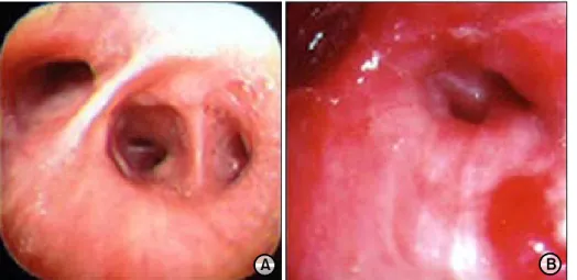
Fig. 2. Pre-operative bronchos- copy. A: The bronchoscopy one year ago was nonspecific find- ing. B: At admission, bronchos- copy shows bleeding from LB1+2 and near total obstruction on LB1+2 due to infiltrating mass but failed biopsy due to severe angulation.
A B
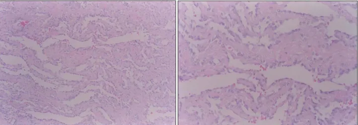
Fig. 3. The microscopic finding showed irregular anastomosing vascular channels lined by a single layer of enlarged endothelial cells permitting between collagen bundles (the vascular lumen are bloodless) (H-E××100, 400).
Fig. 4. The follow-up chest PA showed no evidence of the recurrent cancer but revealed the streaky density on LLL due to the radiation pneumonitis.
=국문 초록=
중심 단어