대 한 방 사 선 의 학 회 지 1992; 28(1) : 95~IOO
J.ournal of Korean Radiological Society, January, 1992
폐결핵과 공존하는 폐암의 CT진단
조선대학교 의과대학 진단방사선과학교실 김선주·김영숙·오재희·김은경·김영철 - Abstract-
CT Diagnosis of Primary Lung Cancer Coexisting with Pulmonary Tuberculosis
Sun Joo Kim, M.D., Young Sook Kim, M.D., Jae Hee Oh, M.D., Eun Kyoung Kim, M.D., Young Chul Kim M.D.
Department o[ Diagnostic Radiology. College o[ Medicine. Chosun University
When bronchogenic carcinoma is coexisting with pulrnonary tuberculosis. it is difficult to differentiate bronchogenic carcinoma from puJmonary tuberculosis radiologically. Thus. the object of this study is to define differential diagnosis of broríchogenic carcinoma by computed tomography. We analized CT scans of 27 patients with radiologic findings of pulmonary tuberculosis and mass of which twelve cases were p비monary tuberc비osis and fifteen cases were primary lung cancer. The location of parenchymal infiltration and the mass was the same in 60%(9115) of the primary lung cancer in cases and 83%(10112) of the pulmonary tuberculosis cases. The common location of the mass was the both upper lobes in 92 % (11/ (2) of the pulmonary tuberculosis cases and 53 % (8/ (5) of the primary lung cancer cases.
The common locations of the mediastinallymphadenopathy were 4R. 2R of the pulmonary tuberculosis cases and 4R. 10R of the primary lung cancer cases. In the feature of post enhanced lymph nodes‘ homogenous increased density was more frequent in primary lung cancer. Measurements of the maximum thickness part of the cavity wall was not a reliable indication of malignancy.
Index Words: Lung neoplasm. CT. 60.1211 Tuberculosis‘ Pulmonary.60.232
서 론
폐결핵과 폐암이 공존할 때 조기진단의 어려움으로 폐암 의 수술 시기를 놓치는 수가 종종있다(1). 특히 우리나라는 폐결핵의 유병률이 높아 이에 대한 연구가 더 필요한 실정 이다(2). 이에 저자들은 폐결핵을 앓고있는 환자에서 단순 흉부촬영상 종괴가 보일 때 종괴의 원인이 폐암에 의한 것 인지 폐결핵의 소견인지를 알고자 단순흉부 X 선 소견 및 흉부전산화단층촬영(이하 CT) 소견을 비교분석하여 다음과 같은 소견을 얻었기에 문헌고찰과 함께 보고하고자 한다.
대상 및 방법
1990년 1월부터 1991년 5월까지 조선대학교 부속병원을
내원한 환자중 과거 병력상 결핵치료를 한 환자에서 결핵약 에 잘 반응하지 않고 흉부 x-선사진상 결핵을 의심할 수 있 는 불규칙한 반점형 침윤음영과 종괴를 보여 흉부 CT를 시 행한 환자중 진단이 확진된 27례를 대상으로 하였다. 이중 폐결핵이 12례였으며 폐결핵과 폐암이 동반된 경우가 15례 였다. 환자의 연령분포는 31세에서 80세까지 였고 평균연령 은 56세였으며 남.녀비는 4 ’ l이었다. 폐암은 기관지경을 통한 생검법(13례), 객담 세포학 검사(2례), 경피적 세포조 직검사(2례), 기관지 세척물 세포학검사(1례)로 조직학 검 사상 확진된 15례였£며, 폐결핵은 객담(4례), 기관지 세척 물(1례)에서 세균학적으로 결핵균이 양성일때와 기관지 내 시경을 통한 조직생검(7례), 경피적 세포조직검사(1례), 경 부림프절 조직생검 (1례)으로 확진된 12례였다. 한 환자에서 두가지 이상의 진단법을 병행했던 경우도 있었다.
객담 결핵반응검사(AFB)는 모든 예에서 10회 이상 실시 하였으며 이중 양성소견을 보인 예는 결핵의 경우 4례 폐암 이 논문은 1991년 9월 25일 접수하여 1991년 12월 II일에 채택되었음
- 95-
대한방사선의학회지 1992; 28(1) : 95~IOO
의 경우 1례였다. 전산화단층촬영기기로 Hitachi 700을 사 용하였으며 폐첨부로 부터 횡격막까지 10mm간격으로 촬영 하였고, 모든 예에서 조영증강 전후촬영을 하였다. 분석기 준은 폐실질병변과 종괴의 위치, 종괴의 크기, 석회화 유무,
공동이 있을때 내면의 상태, 공동벽의 두께, 수면상 유무 및 림프절 이상소견으로 하였다. 종괴의 크기에 있어서는 단경 3.0cm을 기준으로 하였고 공동벽 두께에 있어서는 최대벽두 께를 측정하여 5mm미만, 5-15mm, 15mm이상으로 분류하 였다. 림프절은 단경 1. 0cm이상을 종대가 있다고 하였으며 1.0cm이하인 종대가 없는 경우도 중심부 저음영이 있을때는 이상소견으로 하였으며 럼프절 위치, 중심부 저음영 유무 및 균질성 조영증강 등을 분석하였다. 림프절의 위치는 미 국 흉곽협 회 (American Thoracic Society)의 구분에 따라 서 분류하였다.
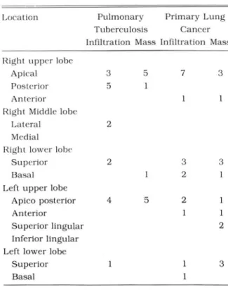
Table 1. Distribulion of Parenchymal Infiltration and Mass in the Pulmonary Tuberculosis. Primary Lung
Cancer
Location Pulmonary Primary Lung Tuberculosis Cancer Infiltration Mass Infiltration Mass Right. upper lobe
Apical 3 5 7 3
Posterior 5
Anterior Right Middle lobe
Lateral 2
Medial Right lowcr lobc
Superior 2 3 3
Basal 2
Left upper lobe
Apico posterior 4 5 2
Anterior
Superior lingular 2
Inferior lingular Left lower lobe
Superior 3
Basal
Table 2. Relation Between Parenchymal Infiltration and Mass
Same area Different area Pulmonary tuberculosis 10 2 Primary lung cancer
Squamous cell carcinoma 8 3
Adenocarcinoma 2
Small cell carcinoma 0
결 과
폐실질내 침윤성 병변의 위치는 폐결핵에서 우상엽 8예,
화상엽 4예의 순이었고, 폐암에서는 우상엽 8예, 우하엽 5 예 순이었다.
종괴의 위치는 폐결핵에서는 우상엽 6예, 좌상엽 5예, 우 하엽 l예 였으며 폐암에서는 우상엽 4예, 좌상엽 4예, 우하 엽 4예, 좌하엽 3예 였다(Table 1)
폐실질병변과 종괴의 위치가 같은 경우는 폐결핵이 10예 (83%), 폐암이 9예 (60%)였다.
폐암의 조직학적 검사에서 세포별 유형은 편평상피세포암 이 11예(73%)로 가장 많았으며 이중 8예가 침윤성 병변부 위와 같은 부위에 위치하였다 (Table 2).
종괴의 특징은 단경이 3.0cm 이상인 경우 폐결핵 6예, 폐 암 12예였으며 폐결핵에서 6예 모두 공동성 종괴였다. 종괴 내 석회화를 보이는 경우는 폐결핵에서 3예 모두 중심성 석 회화였고 폐암에서는 1예가 편심성 석회화였다. 공동성종괴 는 폐결핵, 폐암 각각 7례였으며 공동 내면이 평활한 경우 가 폐결핵 4예, 폐암 1예였고, 최대벽 두께가 15mm이상인 경우 결핵에서 4예(Fig. 1), 폐암에서 5예였으며, 4mm이하 인 경우 폐결핵 1예였다. 공동내 수변상형성은 폐결핵에서 2예 폐암에서 3예였다 (Table 3)
종격동 림프절의 이상소견은 폐결핵 5명 환자에서 8예, Table 3. Characteristics of Mass in Pulmonary Tuber
culosis and Primary Lung Cancer.
Characteristics Pulmonary Primary tuberculosis Lung Cancer Small mass(3.0cm less)
Solid 5 2
Cavity
Large Mass(larger than 3.0cm)
Solid 0 6
Cavity 6 6
Calcification
Concentric 3
Eccentric Cavity
Inner surface‘ sm~h 4
Inner surface: iFeguJar 3 6
Maximum thickness of cavity wall
more than 15mm 4 5
5 - 15mm 2 2
4mm or less
o
Air f1uid level 2 3
96 -
a
b
c
Fig. 1. a. Chest radiograph shows irregular infiltrations and smooth thickwalled cavitary mass in right upper lung field.
b. CT Scan shows small aggregated lymphnodes with central low density in right upper paratracheal area(arrow)
c. CT scan at the level of aortic arch shows large irregular thickwalled cavitary mass in posterior segment of right upper lobe with enhancing nodule(arrow). Small homogeneous enhancing lymphnodes in anterior area(arrows). Pathologica11y proven 잃 pulmonary tuber- culosis and subsided follow up chest after changed an- tituberculous regimen
김선주 외 : 폐결핵과 공존하는 폐암으I CT진단
a
b
Fig. 2. a. Chest .radiograph shows irregular patchy in- filtration and cavity(arrow) in right upper lung field.
b. CT scan at the level of aortic arch shows irregular lobulated thickwalled eccentric located cavitary mass in peripheral portion of anterior segment of right upper lobe with chest wall invasion. Pathologically proven as adenocarcinoma
폐암 10명에서 19예가 관찰되었다. 통일 림프절에 여러개의 림프질이 있을때는 l예로 간주하였다. 림프절의 위치는 폐 결핵의 경우 4R이 3예, 2R이 2예의 순이었으며, 폐암의 경 우 4R, 10R 이 각 4예씩, 5 , 6,7R 이 각각 3예의 순이 었다.
조영증강후 림프절이 중심부 저음영과 원형의 주변 조영증 강을 보이는 경우가 폐결핵 5예, 폐암 1예였으며, 불규칙한 저음영을 보이는 경우는 폐암 3예였으며, 균등한 조영증강 을 보이는 경우는 폐결핵 3예, 폐암 l4예였다 (Table 4). 한 환자에서 림프절의 위치에 따라 다른 조영증강을 보이는 경 우도 있었다.
97 -
대한방사선의학회지 1992 ; 28 ( 1) : 95~ 1 00
Fig. 3. a. Chest radiograph shows irregular infiltrations in right upper lung field and mass density in left parahilar area(arrow).
b. CT scan at the level of carina shows large lobulated mass with irregular centrallow density in left tracheobron.
chial area(arrow) and small multiple lymphnodes with centrallow densities in right tracheobronchial area(aπowhead).
Pathologically proven as small cell carcinoma.
Table 4 Characteristics and Location of Lymphnodes(Location by ATS. Mapping)
Pulmonary Tuberculosis Primary Lung Ca
Region Name
2R Upper paratracheal 2L
4R Lower paratracheal 4L
5 Aortopulmonry 6 Anterior 7 Subcarinal 8R Paraesophageal 9R Tracheobronchial lOR Peribronchial llR Intrapulmonary llL
14R Diaphragmatic 14L
Central Low density with Enhancing Rim
Homogeneous Central Low Irregular Homogeneous Enhancement density with low density enhancement
Enhancing Rim 2
2 3
’i qu qu
。ι
3
• A TS : American Thoracic Society
향상으로 감소의 추세에 있으나, 아직 선진국 수준에 미치 지 옷하고 있다(2). 반면에 폐암의 발생 빈도는 계속증가 추 세에 있다(3). 폐실질내에 반혼을 일으키는 만성폐질환의 과거 20년간 폐결핵의 유병율은 결핵퇴치사업, 경제여건 하나인 폐결핵이 폐암과 관련이 있다는 보고들이 있다(4
고 찰
- 98 -
6J. Mcquarre등어1(7) 의하면 결핵병변 부위에 폐암의 발 생빈도가 높다고 하였으며, Auerbach등은 (8) 조직반흔에 서 발생한 폐암의 원인중 폐경색 다음으로 폐결핵이 원인이 된다고 하였다. 폐실질내 반흔이 비전형상피 세포의 증식과 화생(metaplasia)을 자극하고 염증성 변화를 가진 기관지 점막에 세포화생이 변태 (atypism)를 일으켜 결국 변태는 암 세포(carcinoma in situ)로 되며, 침습성암(i nvasive car- cinoma)으로 진행될 수 있다(3, 10). Auerbach는(8) 반흔 암에서는 조직학적으로 기관지 선암의 빈도가 높다고하였으 나, Ting등어1( 5) 의하면 결핵과 병발된 폐암에서는 편평상 피세포암이 더 많다고 하였다. 폐결핵의 폐실질변화는 상엽 의 폐첨분절이나 후분절에 잘 생기는데 이는 서있는 상태 (upright)에서 이 부분의 산소분압이 가장높아 마이코 박테 리아의 독성(mycobacterial virulence)이 증가되고, 림프액 순환의 저하로 clearance mechanism이 장애되기 때문이라 고 한다(11, 12). 폐결핵에서 폐실질내 종괴를 보이는 경우 는 드물지만, 종괴가 있을때 대부분 상엽을 침범하며, 폐실 질내 병변과 같은 부위를 침범한다고 한다(11). 이때 폐암 과 구별을 요한다. 종괴의 석회화는 결핵성일때 빈번하고,
주로 미만성 석회화를 보이며, 편심성 석회화는 악성을 시 사한다고 한다(3, 13, 14). 폐암에서의 석회화는 석회화된 기 존 결핵성 병변근처에 암이 발생한 경우이거나, 종양괴사 부분에 영양장해로 인한 석회화, 종양 자체내에서 석회화를 일으키는 경우이다(1 5).
폐결핵 환자의 10%정도가 기관지 결핵을 동반하는데, 기 관지 결핵의 경우 염증성 병변이 기관지 점막으로 확산되어 기관지 협착이나 폐쇄를 유발하여 폐실질내 무기폐 소견을 주로 보인다고 보고되였다(16). 폐암의 경우 기관지 폐색에 의한 폐쇄성 폐염이나 무기폐소견을 보이며 침윤성 병변이 상엽의 전 분절이나 하엽의 기저분절을 침범했을때는 폐암 의 가능성을 시사해준다고 한다(3, 5).
종 격 동 림 프 절 종 대 의 원 인 은 주 로 악 성 림 프 종, sar-
coidosis, 폐암의 림프절 전이나 폐결핵에 의한 결핵성 임파
선염, 그외 전이암이 있다 (17). 결핵에 의한 경우는 소아에 빈번하며 주로 편측성으로 침범하고, 우상기관 우측과 하기 관 우측럼프절을 잘 침범하며 중심부 저음영의 소견과 임파 절 석회화의 빈도가 높다(18, 19). 폐암의 림프절 전이는 림 프절내 균등한 조영증강이 빈번히 관찰된다고 한다. 침범위 치는 상기관 우측, 하기관 우측, 기관분지하부를 많이 침범 한다(2이.
CT상 기관지 변형소견은 기관지 협착, 폐쇄, 변형퉁이며 기관지 협착이나 폐쇄가 기관지내 병변인지 외부종괴에 의 한 압박인지 정확히 구별하기는 곤란하나, 불규칙한 기관지 내벽의 비후는 기관지암을 강력히 시사해준다(21, 22). 그러 나, 기관지 결핵의 경우 종격동 럼프절 결핵이 기관지벽을 미란시키면서 기관지 내강으로 돌출되어 있는 상태에서는
김선주 외 : 폐결핵과 공존하는 폐암의 CT진단
폐암과 구별하기가 어렵다(16).
폐실질내 공동성 종괴는 기관지와 교통이 되어 발생되며 단순흉부 x-선보다 CT에서 발견률이 높다 (23). 폐결핵의 경우 건락성 괴사 (caseous necrosis)로 인한 다발성 공동이 흔하며 공동내면은 평활하고 최대벽 두게가 앓으며 (15mm 이하) 공동이 중심성인 경우가 많다. 폐암의 경우 혈관폐색 에 의한 액화성 괴사가 주 원인이며 단발성이 많고 공동내 벽이 불규칙하며 최대벽이 두껍고(15mm 이상) 공동이 편심 성인 경우가 많다(24, 25) (Fig_ 2).
공동내 수연상은 폐결핵의 경우 20% 정도로 나타나며 (26, 27) 폐암의 경우는 드울지만 (3) 공동내 수변상 존재 여 부는 악성과 양성을 구별하는데는 별 도움이 안된다고 한다 (28).
결론적으로 폐결핵을 앓고 있던 환자에서 흉부 x-선상 종괴가 있을 때 폐실질 병변과 종괴가 다른곳에 위치하거나 (Fig.3), 종괴가 하엽에 위치하였을 때는 폐암을 의심할 수 있으며 폐암의 림프절 전이는 주로 균질성 증가음영을 보였 다.
CT는 공동내면의 상태와 최대공동벽두께를 정확히 젤수 있으므로 통상의 공동벽 최대 벽 두께로 악성과 양성을 구멸 하는 기준은 재고를 해야 하며 앞으로 더 많은 연구가 있어 야할것으로사료된다.
참 고문 헌
1. Tunell WP. Koh YC. Adkin PC Washington DC. The dilemma of coincident active pulmonary tuber- culosis and carcinoma of the lung. J Thorac car- diovasc Surg 1971:62:563-567
2 보건사회부, 대한결핵협회 제 5차 결핵 실태조사 결과보 고, 1985
3 박연원, 김태선, 우영훈, 김상준, 전병희, 서정헥. 폐암에 대한 전산화 단층촬영 소견. 대한방사선의학회지 1985 ; 21 : 564-572
4. Cambell RE. Hughes FA. The development ofbron- chogenic carcinoma in patients with pulmonary tuberculosis. J Thorac cardiovasc Surg 1960:
40:98-101
5. Ting YM. Church WR. Ravikrishnan KP. Lung car- cinoma superimposed on pulmonary tuberculosis Radiology 1976: 119:307-312
6. Wofford JL. Webb WR. Stauss HK. Tuberculous scarnng 밍ld primary lung cancer. Arch Surg 1962:
85:928-935
7. Mcquarrie DG. Nicoloff DM. Van Nostrand D‘ Rao K. Hum phrey EW‘ Tuberculosis and carcinoma of the lung. Dis. Chest 1968:54:427-436
- QQ -
대한방사선의학회지 1992; 28(1) : 95~IOO
8. Auerbach 0‘ Gorfinkel L. Parks VR. Scar cancer of Lyons HA. Tuberculous mediastinallymphadenitis the lung increase over a 21 year period. Cancer in the adult. Radiology 1978:126:365-368 1979:43:636-642 19. 1m JG. Song KS. Kang HS. Yeon KM. Mediastinal 9. Greenberg SO. Jenkins OE. Bahar 0 et al. Coex- tuberculous Iymphadenitis CT manifestation
istence of carcinoma and tuberculosis of the lung. Radiology 1987: 164: 115-119
Am Rev Respir Ois 1964:90:67-76 20 이혜련, 황정원, 성규보 등. 종격동 임파절의 결핵성 임파 10. Farber E. Chemical carcinogenesis. N Engl J Med 선염 및 원발성 폐암 전이의 CT소견. 대한방사선의학회지
1981:305’1379-1389 1987 : 25 ‘ 911-916
11. WoodringJH. Vandiviere HM. Fried AM. Oillon ML. 21. Berkmen YM. Bronchial obslruction in unresolved Williams TO. Melvin IG. Uptate: The radiolographic pneumonia and its differentiation from bron- features of pulmonary tuberculosis AJR 1986: chogenic carcinoma. Radiology 1972:105‘309-313
146:497-506 22. Webb WR. Gamsu G. Speckman JM. Computed
12. Goodwin RA. Oesprez RM. Apical localization of tomography of the pulmonary hilum in patients pulmonary tuberculosis. chronic pulmonary with bronchogenic carcinoma. J Comput Assist histoplasmosis. and progressive massive fibrosis of Tomogr 1983‘7:219-225
the lung. Chest 1983;83 ’ 801~05 23 안우현, 조승환, 이준배, 김병수. 단순흉부사진상 공동을 13 최규옥 원형 폐종괴의 전산화단층촬영소견. 대한방사선의 보이지 않는 활동성 폐결핵의 흉부전산화 단층촬영 소견.
학회지 1984 : 20 : 804-819 대한방사선의학회지 1989: 25: 241-246
14. Shin MS. HO KJ. Computed tomographic evaluation 24. Woodring JH. Fried AM. Chuang VP. Solitary of solitary pulmonary nodule in chest roen- cavities of the lung.: Oiagnostic implications of cavi- tgenogram. J comput Assist Tomogr 1982;6: ty wall thickness. AJR 1980:135:1269-1271
947-954 25. 이관섭, 박기순, 최우석, 윤 엽, 임재훈. 단일공동성 폐
15. Mahoney MC. Shipley RT. Corcoran HL. Oickson 병변의 방사선학적 고찰. 대한방사선의학회지 1989 : 25 BA. Oemonstration of calcification in carcinoma of 410-415
the lung. AJR 1990:154:255-258 26. Cohen JR. Amorosa JK. Smith PR. The air-f1uid 16. Albert RK. Endobronchial tuberculosis progressing level in cavitary pulmonary tuberculosis. Radiology
to bronchial stenosis. Chest 1976:70:537-539 1978‘127:315-316
17 박정미, 임정기, 홍주희, 한만청. 종격동 림프절 질환의 27. Maka띠 uola O. Fluid levels in pulmonary tuber-
전산화단충촬영 소견에 대한 분석. 대한방사선의학회지 culosis cavities in a rural population ofNigeria AJR
1988: 24: 767-774 1983;14‘519-529
18. Amorosa JK. Smith PR. Cohen JR. Ramsey C. 28 박용희, 박석희. 흉부화상진단. 수문사 1990: 132-134