High Dose Involved Field Radiation Therapy as Salvage for Loco-Regional Recurrence of Non-Small Cell Lung Cancer
Sun Hyun Bae,
1Yong Chan Ahn,
1Heerim Nam,
4Hee Chul Park,
1Hong Ryull Pyo,
1Young Mog Shim,
2Jhingook Kim,
2Kwhanmien Kim,
2Jin Seok Ahn,
3Myung-Ju Ahn,
3and Keunchil Park
3Departments of 1Radiation Oncology, 2Thoracic Surgery, and
3Medicine (Division of Hematooncology), Samsung Medical Center, Sungkyunkwan University School of Medicine, Seoul;
4Department of Radiation Oncology, Kangbuk Samsung Hospital, Sungkyunkwan University School of Medicine, Seoul, Korea.
Received: August 31, 2011 Revised: November 29, 2011 Accepted: December 15, 2011
Corresponding author: Dr. Yong Chan Ahn, Department of Radiation Oncology, Samsung Medical Center,
Sungkyunkwan University School of Medicine, 50 Irwon-dong, Gangnam-gu,
Seoul 135-710, Korea.
Tel: 82-2-3410-2602, Fax: 82-2-3410-2619 E-mail: [email protected]
∙ The authors have no financial conflicts of interest.
© Copyright:
Yonsei University College of Medicine 2012 This is an Open Access article distributed under the terms of the Creative Commons Attribution Non- Commercial License (http://creativecommons.org/
licenses/by-nc/3.0) which permits unrestricted non- commercial use, distribution, and reproduction in any medium, provided the original work is properly cited.
Purpose: To determine the effectiveness of salvage radiation therapy (RT) in pa- tients with loco-regional recurrences (LRR) following initial complete resection of non-small cell lung cancer (NSCLC) and assess prognostic factors affecting sur- vivals. Materials and Methods: Between 1994 and 2007, 64 patients with LRR after surgery of NSCLC were treated with high dose RT alone (78.1%) or concur- rent chemo-radiation therapy (CCRT, 21.9%) at Samsung Medical Center. Twen- ty-nine patients (45.3%) had local recurrence, 26 patients (40.6%) had regional re- currence and 9 patients (14.1%) had recurrence of both components. The median RT dose was 54 Gy (range, 44-66 Gy). The radiation target volume included the recurrent lesions only. Results: The median follow-up time from the start of RT in survivors was 32.0 months. The rates of in-field failure free survival, intra-thoracic failure free survival and extra-thoracic failure free survival at 2 years were 52.3%, 33.9% and 59.4%, respectively. The median survival after RT was 18.5 months, and 2-year overall survival (OS) rate was 47.9%. On both univariate and multivar- iate analysis, the interval from surgery till recurrence and CCRT were significant prognostic factors for OS. Conclusion: The current study demonstrates that in- volved field salvage RT is effective for LRR of NSCLC following surgery.
Key Words: Concurrent chemo-radiation therapy, locoregional recurrence, non- small cell lung cancer, radiation therapy, salvage treatment
INTRODUCTION
In the management of non-small cell lung cancer (NSCLC), surgery is the treat-
ment of choice with the highest curative potential for the patients with stage I/II
and selective stage III disease.
1Unfortunately, however, a significant proportion of
the patients, following curative surgery, develop recurrence or metastasis during
their clinical course. Distant metastases (DM) are more common than loco-region-
al recurrences (LRR) after complete surgical resection, and the frequency of LRR
as the first site of failure ranges from 10-20% for stage I, and up to 50% for stage
III patients.
2-6The overall LRR in the mediastinum, the bronchial stump and the
patients (14.1%), lobectomy in 53 (82.8%), and wedge re- section in 2 (3.1%). The pathologic stages at the time of surgery were I in 38 patients (59.4%), II in 16 (25.0%) and III in 10 (15.6%). Following the initial surgery, adjuvant chemotherapy was given in 11 patients (17.2%), and none received adjuvant RT. Among 10 patients with pathologic stage III disease, 4 were recommended to receive postoper- ative RT because of pN2 disease. However, postoperative RT was not given to these 4 patients due to either postoper- ative morbidity (3 patients) or patient’s refusal (1 patient).
There were 53 males (82.8%) and 11 females (17.2%), and the age at diagnosis of LRR ranged from 45 to 80 years (median 66 years). Eastern Cooperative Oncology Group performance scores were 0 in 10 (15.6%), 1 in 44 (68.8%), 2 in 9 (14.1%) and 3 in 1 (1.5%). Squamous cell carcinoma (39 patients, 60.9%) and adenocarcinoma (17 patients, 26.6%) were the most common histologic types. The medi- an time from the date of initial surgery to LRR was 10 months (range, 1-72 months). The sites of LRR were local recurrence only at the bronchial stump or the chest wall in 29 patients (45.3%), regional recurrence only at the medias- tinal lymph node in 26 (40.6%), and combined local and re- gional recurrence in the bronchial stump and the mediasti- nal lymph node in 9 (14.1%). All patients were re-staged as if the patients had been newly diagnosed according to the sixth Edition of the AJCC Cancer Staging and the stages were I in 3 patients (4.7%), II in 27 (42.2%) and III in 34 (53.1%).
23Salvage RT was given using 6-15 MV photon beams from linear accelerators. Three-dimensional conformal RT after individual CT-based simulation was done in all pa- tients. The radiation target volume included the visible re- current lesions on axial CT images with 1-2 cm margins ac- cording to normal tissue constraints, and no attention was given to include the regional lymphatics electively. Three or four beam arrangements were typically used usually to adequately cover the target volumes and to minimize the dose to the normal tissues (e.g. lung, spinal cord and esoph- agus). All patients received high dose RT for curative pur- pose. Four different fractionation schedules were employed considering the expected morbidity, based on the target vol- ume, the patients’ performance, and the concurrent chemo- therapy. In 14 patients who received concurrent chemother- apy with salvage RT (CCRT), daily 1.8 or 2.0 Gy was used:
54-63 Gy by 1.8 Gy per fraction in 4 patients (6.3%); and 44-66 Gy by 2 Gy per fraction in 10 (15.6%). In the remain- ing patients receiving salvage RT alone, higher daily doses chest wall occurs with the incidence of 30% among all re-
currences without clinical evidence of DM.
7There have been a few reports on aggressive repeat sur- gery with the curative aim for the patients with resectable LRR or the second primary cancer.
8-11Even with the prom- ising survival outcomes, reoperation, however may be lim- ited only to those with adequate cardio-pulmonary func- tional reserves. There have been several reports on salvage radiation therapy (RT) as an alternative to repeated surgery, as RT can be widely applied.
6,12-22The survival outcomes were quite variable of 10-40% at 2-year because 1970’s and 1980’s and most studies had only a small number of pa- tients, with diverse RT dose schedules being used. Among these, few studies reported that post-resection recurrent NSCLC patients treated with salvage RT have survival comparable to that of newly diagnosed patients treated with radical RT.
14,17,22Therefore, salvage RT would be actively considered in patients with inoperable disease.
We report herein our experience with high dose salvage RT for patients with LRR after curative resection for NSCLC to determine the effectiveness of salvage RT and assess prognostic factors affecting survivals.
MATERIALS AND METHODS
We retrospectively reviewed the hospital charts of the pa- tients with completely resected NSCLC between 1994 and 2007 at Samsung Medical Center, and found 64 patients with LRR without evidence of DM and inoperable status, assessed by a trained surgeon, who were given high dose salvage RT. The clinical assessments at the time of diagnos- ing LRR included complete history and physical examina- tion, complete blood cell count, chemistry panel, simple chest X-ray, computed tomography (CT) of the chest and the upper abdomen, bronchoscopy (when clinically indicat- ed), fluoro-deoxy-glucose (FDG) positron emission tomog- raphy-computed tomography (PET-CT) scan, magnetic res- onance imaging of brain, and whole body bone scan. In 46 patients, biopsy seemed technically difficult or very risky, and, therefore, the clinical diagnosis of LRR was made based either on high FDG uptake on PET-CT scan without other possible explainable causes (28 patients, 43.8%), or on evi- dently progressive lesion by at least two consecutive chest CT scans (18 patients, 28.1%). Biopsy confirmation of the recurrent lesion was possible in 18 patients (28.1%).
The initial types of surgery were pneumonectomy in 9
post completion of salvage RT.
24Radiation pneumonitis and esophagitis were assessed by clinical symptoms, corre- lated with the radiographic findings, in the absence of other cause of symptoms, and estimated by the Radiation Thera- py Oncology Group Acute and Late Lung Morbidity Scor- ing Criteria.
As only the involved lesions were the targets for salvage RT in this study, the further recurrences following salvage RT were classified as in-field failure (IFF), intra-thoracic failure (ITF) and extra-thoracic failure (ETF). ITF was de- fined as any loco-regional recurrence regardless of the cur- rent RT volume and included IFF and recurrence at the su- praclavicular fossa or the pleura-pericardial seeding. ETF was any recurrence outside ITF.
Survival rates were estimated with the Kaplan-Meier method, and the comparisons between the groups were de- termined using the log-rank test.
25Multivariate analysis was performed to assess the relationships between the outcomes and the possible prognostic variables using the Cox propor- tional hazards model. Statistical analyses were performed using SAS software (SAS for Windows, version 9.0, SAS Institute, Cary, NC, USA).
RESULTS
Radiographic tumor response
The radiographic tumor responses were evaluable by chest CT in all patients. Complete response was achieved in 4 pa- tients (6.2%), partial response in 40 (62.5%) and stable dis- eases in 12 (18.8%). Eight patients (12.5%) showed pro- gressive disease: increased size of treated tumor in 3; new lesions in 5.
Patterns of failure
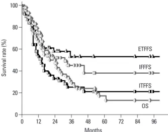
Three-fourth of the patients (48/64, 75.0%) experienced further recurrence following salvage RT: 29 (45.3%) had IFF; 40 (62.5%) had ITF; and 22 (34.4%) had ETF. The rates of IFF-free survival, ITF-free survival, and ETF-free survival at 2 years were 52.3%, 33.9%, and 59.4%, respec- tively (Fig. 1). On univariate analysis, there was no statisti- cally significant factor affecting the IFF-free survival. On both univariate analysis and multivariate Cox regression analysis, the radiographic tumor response to salvage RT proved to be the most important prognostic factor for ITF- free survival and ETF-free survival (p<0.05). The signifi- cances of the prognostic factors are summarized in Table 2.
were used to shorten the overall treatment duration: 50-60 Gy by 2.5 Gy per fraction in 3 (4.7%); and 40-66 Gy by 3 Gy per fraction in 47 (73.4%). The total RT doses were converted into biologically equivalent dose (BED) for the comparison purpose. When α/β was assumed to be 10 Gy, the median total BED was 70.2 Gy
10(51.5-85.8 Gy
10).
The chemotherapy was concurrently added as radiosensi- tizer, and the regimens had changed with time. During 1990’s, two patients received cisplatin (100 mg/m
2) plus et- oposide (50 mg/m
2) every four weeks. During 2000’s, 12 patients received paclitaxel (50 mg/m
2) plus cisplatin (25 mg/m
2) weekly. The median number of chemotherapy cy- cles was 6 cycles (2-6 cycles). The decision to add chemo- therapy to RT was individually made based on performance status of patients. The characteristics of the patients are summarized in Table 1.
Radiographic tumor response after salvage RT was eval- uated using the RECIST criteria by chest CT taken 1 month Table 1. Patients’ Characteristics at Diagnosis of Post-Resec- tion Recurrent NSCLC
Characteristics No. of patients (%)
Age Median 66 (45-80) yrs
<70 yrs 40 (62.5)
≥70 yrs 24 (37.5)
Sex
Male 53 (82.8)
Female 11 (17.2)
Disease-free interval Median 10 (1-72) months
<1 yr 33 (51.6)
≥1 yr 31 (48.4)
Recurrence site
Local 29 (45.3)
Regional 26 (40.6)
Loco-regional 9 (14.1)
Recurrent stage
I 3 (4.7)
II 27 (42.2)
III 34 (53.1)
BED Median 70.2 (51.5-85.8) Gy10
≤70.2 Gy10 33 (51.6)
>70.2 Gy10 31 (48.4) Concurrent chemotherapy
Yes 14 (21.9)
No 50 (78.1)
NSCLC, non-small cell lung cancer; BED, biologically equivalent dose.
Disease-free interval, time interval from initial surgery till postoperative recurrence. Recurrent stage, restaging according to the sixth edition of the AJCC Cancer Staging.
Survival
The median follow-up time from the start of salvage RT in survivors was 32.0 months (range, 2-102 months). The me- dian survival was 18.5 months and the overall survival (OS) rates at 2- and 3-years were 47.9% and 29.5%, respec- tively (Fig. 1). On univariate analysis, the factors that favor- ably correlated with the OS with statistical significance were the initial surgery type other than pneumonectomy (p=0.027), the disease-free interval till postoperative LRR longer than 1 year (p=0.025), the BED
10higher than 70.2 Gy
10(p=0.025), the application of CCRT (p=0.029), and the radiographic tumor response other than progressive dis- ease (p=0.017) (Table 2). Fig. 2 shows difference in the OS according to the BED
10and the treatment modality. Multi- variate Cox regression analysis of the possible prognostic factors on the OS revealed that the disease-free interval till
Table 2. 2-Year Survival Rates according to Clinico-Pathological Parameters on Univariate Analysis
Characteristics ITFFS p value OS p value
Age NS NS
<70 yrs 37.0 57.8
≥70 yrs 24.7 31.7
Sex NS NS
Male 36.3 46.6
Female 22.7 54.5
Surgical method NS 0.027
Pneumonectomy 57.1 29.6
Others* 31.5 50.9
Disease-free interval 0.044 0.025
<1 yr 24.5 31.6
≥1 yr 42.4 65.5
Recurrent site NS NS
Local 42.8 41.3
Regional 22.1 58.1
Loco-regional 41.6 40.0
Recurrent stage NS NS
I 33.0 0.0
II 53.2 48.1
III 53.9 47.9
Biologically equivalent dose NS 0.025
≤70.2 Gy10 24.5 37.3
>70.2 Gy10 42.6 59.2
Concurrent chemotherapy NS 0.029
Yes 25.7 65.2
No 36.9 43.5
Tumor response 0.003 0.017
CR+PR+SD 35.3 49.6
PD 33.3 37.5
ITFFS, intra-thoracic failure-free survival; OS, overall survival; CR, complete response; PR, partial response; SD, stable disease; PD, progressive disease.
Disease-free interval, time interval from initial surgery till postoperative recurrence.
Recurrent stage, restaging according to the sixth edition of the AJCC Cancer Staging.
*Includes wedge resection and lobectomy.
Fig. 1. In-field failure free survival (circle, IFFFS), intra-thoracic failure free survival (square, ITFFS), extra-thoracic failure free survival (triangle, ETFFS) and overall survival (diamond, OS) rates with salvage treatment in loco-re- gional recurrence of resected non-small cell lung cancer.
0 20 40 60 80 100
Survival rate (%)
0 12 24 36 48 60 72 84 96
ETFFS IFFFS ITFFS OS
Months
Treatment-related toxicity
All patients tolerated the salvage RT course well and there was no incidence of treatment interruption due to acute tox- postoperative LRR longer than 1 year (hazard ratio=0.372,
p=0.004) and the application of CCRT (hazard ratio=0.353, p=0.038) were favorable factors (Table 3).
Table 3. Multivariate Cox Regression Analysis of Risk Factors Affecting Overall Survival
Characteristics Hazard ratio (95% confidence limits) p value
Age ≥70 yrs 1.539 (0.782-3.029) 0.212
Sex (female) 0.724 (0.312-1.680) 0.452
ECOG performance score (2-3) 1.506 (0.647-3.506) 0.342
Disease-free interval ≥1 yr 0.372 (0.190-0.731) 0.004
Radiographic tumor response (progressive disease) 1.573 (0.984-2.514) 0.058
Use of concurrent chemotherapy 0.353 (0.132-0.944) 0.038
ECOG, Eastern Cooperative Oncology Group.
Disease-free interval, time interval from initial surgery till postoperative recurrence.
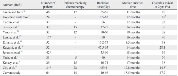
Table 4. Results of Salvage Radiation Therapy for Loco-Regional Recurrent NSCLC
Authors (Ref.) Number of
patients Patients receiving
chemotherapy Radiation
dose (Gy) Median survival
time Overall survival
at 2 yrs (%)
Green and Kern12 46 17 25-65 11 months 10
Kopelson and Choi13 24 - 18.5-62 12 months 10†
Curran, et al.14 37 - 56 12 months 22
Shaw, et al.15 37 15 12-75 14 months 30
Yano, et al.16 32 12 50-60 19 months 38
Leung, et al.17 17* - 60 16 months 40
Emami, et al.18 52 - 16.2-75 8.5 months 18
Kagami, et al.19 32 - 47.5-65 14 months 28.1
Jeremic, et al.20 42* - 55-60 18 months 36
Tada, et al.21 31 5 60 14 months 30
Kelsey, et al.6 29 15 46-74 17 months 38
Cai, et al.22 34* 22 >59.4 19.8 months 14.8†
Current study 64 14 40-66 18.5 months 47.9
NSCLC, non-small cell lung cancer.
*Includes only those treated with curative aim.
†5-years overall survival rate.
Fig. 2. Overall survival rate according to biologically equivalent dose (BED, A) and concurrent chemo-radiation therapy (CCRT, B). BED more than 70.2 Gy10
(p=0.018) and CCRT (p=0.029) were statistically significant prognostic factors in univariate analysis.
0 0
20 20
40 40
60 60
80 80
100 100
Survival rate (%) Survival rate (%)
0 12 24 36 48 60 72 84 96 0 12 24 36 48 60 72 84 96
BED>70.2 Gy10
CCRT (+)
BED≤70.2 Gy10
CCRT (-)
Months Months
A B
the elective regional nodal irradiation. However, recent clin- ical data on RT only to the involved sites in primary NSCLC showed no correlation between RT field size and outcomes, and low incidence of nodal failure at the unirradiated lym- phatics (6.4%).
31,32Tada, et al.
21reported patterns of failure after salvage RT in relation to the RT field in patients with LRR after surgery. They individualized the radiation target volume from only recurrent lesion to elective hilar and me- diastinum and found that a narrow RT field did not cause frequent marginal relapse. In our study, only recurrent sites were treated without elective regional lymph node irradia- tion, and 45.3% of patients had IFF and 62.5% had ITF, which is comparable with other series (50-60%). This im- plies that high dose RT which is focused only to the in- volved lesion would be effective in the salvage setting. A few factors might have contributed to the rationales of ap- plying the minimal RT volume: the accurate re-staging by PET-CT; absence of recurrence at other sites after a certain disease-free interval; possible changes in the lymphatic cir- culation following initial surgery; the use of high conformal radiation dose with the addition of chemotherapy; and im- proved RT technique.
Most studies did not report prognostic factors associated with the survival, because of small sample size. Until now, known prognostic factors are as follows; gender, disease- free interval, recurrent site, initial stage, recurrent stage, ra- diation dose and CCRT.
6,15,16,20,22We also found some prog- nostic factors on survival; the initial surgical method, the interval till the postoperative recurrence, the radiographic tumor response, radiation dose and the addition of chemo- therapy. With respect to the radiation dose, Jeremic, et al.
20showed that there was a significant difference in the median survival time (18 vs. 7 months) and the 5-year survival rates (14 vs. 0%) (p=0.000) between high-dose and low-dose RT groups. This concurred with our results, which showed that the low dose group with BED ≤70.2 Gy
10had 2-years sur- vival rates of 36.2% and high dose group with BED >70.2 Gy
10had 59.2% (p=0.018). In addition, it is thought that post-resection recurrent NSCLC patients treated with sal- vage RT have similar survivals to primary NSCLC patients treated with radical RT.
22Considering that the currently rec- ommended dose for primary NSCLC is 60-70 Gy with conventional fractionation, we suggest higher dose than BED >70.2 Gy
10for salvage treatment. However, further study for the dose escalation may be needed. With respect to the addition of chemotherapy, a few reports on salvage RT combined with chemotherapy have been published. Shaw, et icity. Forty-three patients (82.3%) experienced acute radia-
tion-induced esophagitis with grade 1 or 2. However, there was no grade 3 or higher acute esophagitis. Six patients (9.4%) had grade 3 radiation pneumonitis and were given corticosteroids, and one (1.6%) was hospitalized for grade 4 radiation pneumonitis. All patients with radiation pneu- monitis were given RT alone without concurrent chemo- therapy. There was no incidence of severe chronic toxicity or treatment-related death.
DISCUSSION
Before the wide clinical use of PET-CT in the oncology practice, the diagnosis of local or regional recurrence de- pended mainly on the histopathologic confirmation (bron- choscopic or mediastinoscopic biopsy) when an unusual and new soft tissue lesion shows up on the follow-up CT scans.
If the lesion suspected of recurrence was located at an inac- cessible position by either bronchoscopy or mediastinosco- py, the usual way of confirming the recurrence used to be re- peated CT scans at a few months’ interval to verify that the lesion had the evidence of progression, which might lead to the delay in the start of salvage therapy. Several studies re- cently reported the diagnostic accuracy of PET-CT for medi- astinal lymph node staging. The overall sensitivity, specifici- ty, positive and negative predictive values, and accuracy of PET-CT were reported as 45-86%, 64-100%, 47-100%, 86- 95% and 67-90%, respectively.
26-30The proportions of the diagnostic methods of biopsy, PET-CT, and repeated CT were 28.1%, 43.8%, and 28.1%, respectively, and the 2-year OS rates were 50.0%, 59.6% and 27.7%, respectively, in the current study. Though not statistically significant, the pa- tients who had to wait a few months before initiating sal- vage RT and were mostly diagnosed in the earlier study pe- riod, showed relatively poorer survival. Even though the decision based on PET-CT alone may have the risk of over- estimation of recurrence, its positive contribution to the im- proved outcomes is worthy of mention.
The role of elective regional lymph node irradiation in
salvage RT is unclear. For many years, the standard target
volume of radical RT for primary NSCLC usually included
regional nodes electively based on high incidence of hilar
and mediastinal lymph node metastasis. Hence, consider-
ation of elective regional lymph node irradiation in salvage
treatment setting seems instinctively logical, and most stud-
ies, including the latest study by Kelsey, et al.,
6employed
benefits of salvage RT for LRR of NSCLC (Table 4). The reported median survival ranged from 11 to 19.8 months, and the 2-year OS rates did 10-40%. To our knowledge, the present study, which included 64 patients, is the largest se- ries in the literature with favorable outcomes when com- pared to the previous reports: the median survival time was 18.5 months; and the 2-year OS rate was 47.9%. Favorable outcomes could have been possible because of relatively less tumor burden in the patients with LRR using improved diagnostic tools, and accordingly relatively smaller radia- tion target volume while delivering high conformal radia- tion dose. Based on these findings, the patients with LRR following surgical resection alone could be effectively sal- vaged by high dose RT.
Our study has some limitations. First, this study was a ret- rospectively analysis. It had heterogeneous patient’s group and radiation dose. The results may be affected by selection biases. Second, biopsy confirmation of the recurrent lesion was possible in 18 patients (28.1%). Because most patients were diagnosed with imaging method, it might have the risk of overestimation of recurrence and affect the result of sal- vage treatment. Third, we evaluated radiographic tumor re- sponse by chest CT taken 1 month post completion of salvage RT. This might be too early, since response can sometimes be further prolonged, thus leading to under-estimation of re- sponse to salvage RT. To confirm the effectiveness of in- volved field RT without elective regional lymph node irradi- ation and usefulness of CCRT, prospective trial including large number of patients might be needed.
In conclusion, the current study showed favorable surviv- al rates when compared to other published studies of sal- vage RT. The involved field RT without elective regional lymph node irradiation seemed to be effective as a salvage treatment for LRR after complete resection of NSCLC. Ag- gressive treatment such as CCRT is strongly encouraged for improvement of salvage treatment efficacy as long as patients can tolerate such treatment.
REFERENCES
1. Pearson FG. Non-small cell lung cancer: role of surgery for stages I-III. Chest 1999;116(6 Suppl):500S-3S.
2. Matthews MJ, Kanhouwa S, Pickren J, Robinette D. Frequency of residual and metastatic tumor in patients undergoing curative sur- gical resection for lung cancer. Cancer Chemother Rep 3 1973;4:63-7.
3. Lafitte JJ, Ribet ME, Prévost BM, Gosselin BH, Copin MC, Brichet AH. Postresection irradiation for T2 N0 M0 non-small cell
al.
15and Kelsey, et al.
6found no survival improvement by the addition of systemic chemotherapy to RT, whereas Cai, et al.
22observed that the addition of chemotherapy to RT proved prognostic factor significantly for the progression- free survival compared with RT alone (p=0.022), although the difference in the OS did not reach a statistical signifi- cance. On the other hand, our study showed that significant- ly better 2-year survival was achieved with CCRT when compared with RT alone (65.2% vs. 43.5%, p=0.029 by univariate analysis and p=0.038 by multivariate analysis). It should be noted, however, that the patients’ age in the cur- rent study was younger and the average RT dose was high- er in those who received CCRT than those who received RT alone.
It is generally thought that patients with bronchial stump recurrence only do better than patients with non-stump re- currence.
33There were 14 patients with bronchial stump only recurrence in the current study. However, there was no difference in survivals between bronchial stump recurrence only and other LRR. We speculate that there are several rea- sons for this difference. First, 3 patients in this study ac- counted for true stage I and other 11 patients had more ex- tensive bronchial component of disease. Law, et al.
34reported that patients with bronchial stump recurrence only had better survival than patients with more extensive bron- chial stump component of disease. In addition, the median time from the date of initial surgery to LRR was 6 months (range, 3-9 months) in these 3 patients. Considering the fact that the interval from surgery till recurrence is a significant prognostic factor, these adverse factors might negatively af- fect the survival in patients with bronchial stump recurrence only. Second, among other LRR, 14 patients had only single lymph node metastasis in the current study. Rea, et al.
35re- ported that the involvement of main bronchial nodes has a prognostic significance similar to that of N2 single station and should be considered as an early N2 disease. Recently, it is thought that pathologic N2 disease has heterogeneous na- ture and necessitates sub-classification: the best outcome in the case of single nodal chain.
36Since proximity of bronchi- al stump recurrence to main bronchus is classified as cT 3 or 4 and advanced T stage above 3 has similar survival to pN1, patients with single N2 lymph node recurrence might have similar survival to the patients with more extensive bronchi- al stump recurrence. Consequently, further study is needed to determine the prognosis of bronchial stump recurrence.
Several studies that were based on rather small number
of patients, usually in a single institution have shown the
21. Tada T, Fukuda H, Nakagawa K, Matsui K, Hosono M, Takada Y, et al. Non-small cell lung cancer: radiation therapy for locoregion- al recurrence after complete resection. Int J Clin Oncol 2005;
10:425-8.
22. Cai XW, Xu LY, Wang L, Hayman JA, Chang AC, Pickens A, et al. Comparative survival in patients with postresection recurrent versus newly diagnosed non-small-cell lung cancer treated with radiotherapy. Int J Radiat Oncol Biol Phys 2010;76:1100-5.
23. American Joint Committee on Cancer (AJCC). In: AJCC Cancer Staging Manual. 6th ed. Springer-Verlag New York; 2002. p.167-84.
24. Eisenhauer EA, Therasse P, Bogaerts J, Schwartz LH, Sargent D, Ford R, et al. New response evaluation criteria in solid tumours:
revised RECIST guideline (version 1.1). Eur J Cancer 2009;45:
228-47.
25. Kaplan EL, Meier P. Nonparametric estimation from incomplete observations. J Am Stat Assoc 1958;53:457-81.
26. Cerfolio RJ, Bryant AS, Ojha B, Eloubeidi M. Improving the in- accuracies of clinical staging of patients with NSCLC: a prospec- tive trial. Ann Thorac Surg 2005;80:1207-13.
27. Kim BT, Lee KS, Shim SS, Choi JY, Kwon OJ, Kim H, et al.
Stage T1 non-small cell lung cancer: preoperative mediastinal nodal staging with integrated FDG PET/CT--a prospective study.
Radiology 2006;241:501-9.
28. Lee BE, von Haag D, Lown T, Lau D, Calhoun R, Follette D. Ad- vances in positron emission tomography technology have in- creased the need for surgical staging in non-small cell lung cancer.
J Thorac Cardiovasc Surg 2007;133:746-52.
29. Melek H, Gunluoglu MZ, Demir A, Akin H, Olcmen A, Dincer SI. Role of positron emission tomography in mediastinal lymphat- ic staging of non-small cell lung cancer. Eur J Cardiothorac Surg 2008;33:294-9.
30. Billé A, Pelosi E, Skanjeti A, Arena V, Errico L, Borasio P, et al.
Preoperative intrathoracic lymph node staging in patients with non-small-cell lung cancer: accuracy of integrated positron emis- sion tomography and computed tomography. Eur J Cardiothorac Surg 2009;36:440-5.
31. Dosoretz DE, Galmarini D, Rubenstein JH, Katin MJ, Blitzer PH, Salenius SA, et al. Local control in medically inoperable lung can- cer: an analysis of its importance in outcome and factors determin- ing the probability of tumor eradication. Int J Radiat Oncol Biol Phys 1993;27:507-16.
32. Rosenzweig KE, Sim SE, Mychalczak B, Braban LE, Schindel- heim R, Leibel SA. Elective nodal irradiation in the treatment of non-small-cell lung cancer with three-dimensional conformal ra- diation therapy. Int J Radiat Oncol Biol Phys 2001;50:681-5.
33. Jeremic B, Bamberg M. External beam radiation therapy for bron- chial stump recurrence of non-small-cell lung cancer after com- plete resection. Radiother Oncol 2002;64:251-7.
34. Law MR, Henk JM, Lennox SC, Hodson ME. Value of radiother- apy for tumour on the bronchial stump after resection for bronchi- al carcinoma. Thorax 1982;37:496-9.
35. Rea F, Marulli G, Callegaro D, Zuin A, Gobbi T, Loy M, et al.
Prognostic significance of main bronchial lymph nodes involve- ment in non-small cell lung carcinoma: N1 or N2? Lung Cancer 2004;45:215-20.
36. Zheng H, Wang LM, Bao F, Jiang GN, Xie HK, Ding JA, et al.
Re-appraisal of N2 disease by lymphatic drainage pattern for non- small-cell lung cancers: by terms of nodal stations, zones, chains, and a composite. Lung Cancer 2011;74:497-503.
carcinoma: a prospective, randomized study. Ann Thorac Surg 1996;62:830-4.
4. al-Kattan K, Sepsas E, Fountain SW, Townsend ER. Disease re- currence after resection for stage I lung cancer. Eur J Cardiothorac Surg 1997;12:380-4.
5. Weisenburger TH. Effects of postoperative mediastinal radiation on completely resected stage II and stage III epidermoid cancer of the lung. LCSG 773. Chest 1994;106(6 Suppl):297S-301S.
6. Kelsey CR, Clough RW, Marks LB. Local recurrence following initial resection of NSCLC: salvage is possible with radiation therapy. Cancer J 2006;12:283-8.
7. Martini N, Bains MS, Burt ME, Zakowski MF, McCormack P, Rusch VW, et al. Incidence of local recurrence and second prima- ry tumors in resected stage I lung cancer. J Thorac Cardiovasc Surg 1995;109:120-9.
8. Gabler A, Liebig S. Reoperation for bronchial carcinoma. Thorax 1980;35:668-70.
9. Watanabe Y, Shimizu J, Oda M, Tatsuzawa Y, Hayashi Y, Iwa T.
Second surgical intervention for recurrent and second primary bronchogenic carcinomas. Scand J Thorac Cardiovasc Surg 1992;26:73-8.
10. Voltolini L, Paladini P, Luzzi L, Ghiribelli C, Di Bisceglie M, Gotti G. Iterative surgical resections for local recurrent and second primary bronchogenic carcinoma. Eur J Cardiothorac Surg 2000;18:529-34.
11. Yamaoka N, Uchiyama Y, Nakamura A, Muraoka M, Kondou M, Yamauchi H, et al. [Reoperation for lung cancer: indications and operation methods in cardiopulmonary function]. Kyobu Geka 1995;48:18-23.
12. Green N, Kern W. The clinical course and treatment results of pa- tients with postresection locally recurrent lung cancer. Cancer 1978;42:2478-82.
13. Kopelson G, Choi NC. Radiation therapy for postoperative local- regionally recurrent lung cancer. Int J Radiat Oncol Biol Phys 1980;6:1503-6.
14. Curran WJ Jr, Herbert SH, Stafford PM, Sandler HM, Rosenthal SA, McKenna WG, et al. Should patients with post-resection lo- coregional recurrence of lung cancer receive aggressive therapy?
Int J Radiat Oncol Biol Phys 1992;24:25-30.
15. Shaw EG, Brindle JS, Creagan ET, Foote RL, Trastek VF, Buskirk SJ. Locally recurrent non-small-cell lung cancer after complete surgical resection. Mayo Clin Proc 1992;67:1129-33.
16. Yano T, Hara N, Ichinose Y, Asoh H, Yokoyama H, Ohta M, et al.
Local recurrence after complete resection for non-small-cell carci- noma of the lung. Significance of local control by radiation treat- ment. J Thorac Cardiovasc Surg 1994;107:8-12.
17. Leung J, Ball D, Worotniuk T, Laidlaw C. Survival following ra- diotherapy for post-surgical locoregional recurrence of non-small cell lung cancer. Lung Cancer 1995;13:121-7.
18. Emami B, Graham MV, Deedy M, Shapiro S, Kucik N. Radiation therapy for intrathoracic recurrence of non-small cell lung cancer.
Am J Clin Oncol 1997;20:46-50.
19. Kagami Y, Nishio M, Narimatsu N, Mjoujin M, Sakurai T, Har- eyama M, et al. Radiotherapy for locoregional recurrent tumors after resection of non-small cell lung cancer. Lung Cancer 1998;20:31-5.
20. Jeremic B, Shibamoto Y, Milicic B, Milisavljevic S, Nikolic N, Dagovic A, et al. External beam radiation therapy alone for loco- regional recurrence of non-small-cell lung cancer after complete resection. Lung Cancer 1999;23:135-42.