Introduction
Idiopathic nonspecific interstitial pneumonia (NSIP) is one of the varieties of idiopathic interstitial pneumonias (IIP).
NSIP used to be considered as not an independent disease entity but provisional diagnosis. However, now, it is recog- nized as a distinct entity with clinical features that differentiate it from other interstitial pneumonias
1,2. Diagnosis of idiopathic NSIP can be done via multidisciplinary approach in which the clinical, radiologic and pathologic findings are discussed together and other causes are excluded.
Korean Guidelines for Diagnosis and
Management of Interstitial Lung Diseases:
Part 3. Idiopathic Nonspecific Interstitial Pneumonia
Jongmin Lee, M.D.
1, Yong Hyun Kim, M.D.
2, Ji Young Kang, M.D., Ph.D.
1,
Yangjin Jegal, M.D., Ph.D.
3and So Young Park, M.D., Ph.D.
4, on behalf of Korean Interstitial Lung Diseases Study Group
1
Division of Pulmonology and Allergy, Department of Internal Medicine, Seoul St. Mary’s Hospital, College of Medicine, The Catholic University School of Medicine, Seoul,
2Division of Allergy and Pulmonology, Department of Internal Medicine, Bucheon St. Mary’s Hospital, The Catholic University School of Medicine, Bucheon,
3Division of Pulmonary and Critical Care Medicine, Department of Internal Medicine, Ulsan University Hospital, Ulsan University College of Medicine, Ulsan,
4
Department of Pulmonary and Critical Care Medicine, Chungnam National University Hospital, Daejeon, Korea
Idiopathic nonspecific interstitial pneumonia (NSIP) is one of the varieties of idiopathic interstitial pneumonias.
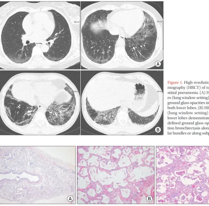
Diagnosis of idiopathic NSIP can be done via multidisciplinary approach in which the clinical, radiologic, and pathologic findings were discussed together and exclude other causes. Clinical manifestations include subacute or chronic dyspnea and cough that last an average of 6 months, most of which occur in non-smoking, middle-aged women. The common findings in thoracic high-resolution computed tomography in NSIP are bilateral reticular opacities, traction bronchiectasis, reduced volume of the lobes, and ground-glass opacity in the lower lungs. These lesions can involve diffuse bilateral lungs or subpleural area. Unlike usual interstitial pneumonia, honeycombing is sparse or absent.
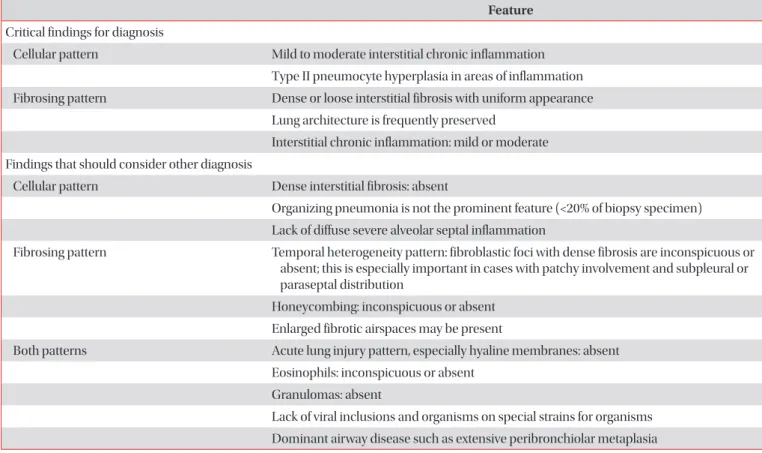
Pathology shows diffuse interstitial inflammation and fibrosis which are temporally homogeneous, namely NSIP pattern. Idiopathic NSIP is usually treated with steroid only or combination with immunosuppressive agents such as azathioprine, cyclophosphamide, cyclosporine, and mycophenolate mofetil. Prognosis of idiopathic NSIP is better than idiopathic pulmonary fibrosis. Many studies have reported a 5-year survival rate of more than 70%.
Keywords: Lung Diseases, Interstitial; Idiopathic Interstitial Pneumonias; Guideline
Address for correspondence: Yong Hyun Kim, M.D.
Division of Allergy and Pulmonology, Department of Internal Medicine, Bucheon St. Mary’s Hospital, College of Medicine, The Catholic University of Korea, 327 Sosa-ro, Wonmi-gu, Bucheon 14647, Korea
Phone: 82-32-340-7039, Fax: 82-32-340-2669 E-mail: [email protected]
Received: Dec. 21, 2018 Revised: Apr. 1, 2019 Accepted: Apr. 23, 2019 Published online: May. 31, 2019
cc