115
©The Korean Society of Food Science and Technology
LPS로 유도된 RAW264.7세포주에서 황금뿌리 물추출물의 항염증활성
이예은
1· 박홍진
1· 박충범
1· 황승미
1,2,*
1진안홍삼연구소, 2전북대학교 식품공학과
Anti-inflammatory activity of Scutellaria Baicalensis root extract in
lipopolysaccharide-induced RAW 264.7 cells
Ye Eun Lee1, Hong Jin Park1, Chung-berm Park1, and Seung-mi Hwang1,2,*
1Department of Efficacy Study, Institute of Jinan red ginseng 2Department of Food Science and Technology, Chonbuk National University
Abstract Scutellaria baicalensis has been used as a traditional medicine for diarrhea, dysentery, hypertension, hemorrhaging,
insomnia, inflammation, and respiratory infections. This study examined the anti-inflammatory effect of Scutellaria baicalensis water extract (SWE) in lipopolysaccharide (LPS)-induced RAW 264.7 cells. To evaluate the anti-inflammatory effect of SWE, RAW 264.7 macrophages were stimulated with LPS to induce the production of inflammation-related factors, which were measured by western blotting. In RAW 264.7 cells, SWE inhibited the production of nitric oxide (NO) without causing cell toxicity. SWE also reduced the expression of inducible NO synthase and cyclooxygenase-2 protein, as well as the production of pro-inflammatory cytokines (such as tumor necrosis factor-α). The phosphorylation levels of the mitogen-activated protein kinase family members, such as JNK and p38, were also reduced by SWE. Thus, SWE could be used as a potential anti-inflammatory agent.
Keywords: anti-inflammatory, Scutellaria baicalensis, RAW 264.7
서
론
외부항원으로 손상된 세포나 조직의 제거를 위해 필수적인 과 정인 염증반응은 과도하게 되면 종양의 성장을 돕거나 주변 조 직 손상과 질병의 악화를 초래한다(Park 등, 2014). 이 반응에는 각종 사이토카인과 단백질 등 다양한 면역계 세포와 매개물질들 이 관여한다. Lipopolysaccharide (LPS)는 그람음성균의 세포벽에 서 유도된 화합물로 염증성 사이토카인의 강력한 유도제로 알려 져있다. LPS로 염증이 유발된 대식세포 표면에서는 Toll-like receptor 4 (TLR4)가 자극되어 하부 세포 신호전달경로인 Mitogen-activated protein kinases (MAPKs) 활성화를 유도하게 된다. MAPKs는 여러 가지 외부자극에 의해 세포의 성장, 사멸, 분화 등에 관여하는데 extracellular signal-regulated kinase (ERK), c-Jun N-terminal kinase (JNK), p38 kinase로 구성되어 있으며, 전사조 절인자들을 인산화함으로써 다양한 유전자들의 발현을 조절한다 (Kang 등, 2013). 이렇게 활성화된 MAPKs 신호전달경로를 통해 tumor necrosis factor-α (TNF-α) 같은 전염증성 사이토카인이 생 성되며, 프로스타글란딘 생합성 속도조절 단계 효소인 cyclo-oxygenase-2 (COX-2)가 발현된다. 또한 L-arginine에서 inducible nitric oxide synthase (iNOS)에 의해 nitric oxide (NO)와 같은 염증인자가 생성된다(Kim과 Kim, 2015; Matthay 등, 2003). NO 생성의 증가는 류마티스관절염, 폐혈성쇼크, 조직손상 등 과 같은 질병을 유발하게 된다. 다양한 염증성 질환에서 주된 치 료제인 스테로이드와 비스테로이드성 항염증제가 염증반응의 주 요 매개체인 프로스타글란딘의 생합성을 억제하는 치료제로 사 용되어지고 있으나 이 제제들은 항염증 효과도 있지만 위염, 신 장염 및 심장질환과 같은 부작용 발생 우려로 장기간 사용이 어 렵다. 이처럼 부작용이 적은 천연물에서 유효성분을 얻어 개발하 려는 노력이 지속되고 있다(Park과 Kim, 2011).
황금(Scutellaria baicalensis Georgi)은 꿀풀과에 속하며 우리나 라 각처의 산지에 나고 밭에서 재배되는 다년생 초본이다. 한방 에서는 열을 내리고 해독의 목적으로 자주 사용되는 약재로써 약 용부위인 황금 뿌리에는 다양한 폴리페놀과 주요 활성물질인 플 라보노이드를 함유하고 있다(Zhao 등, 2019). 대표적인 플라보노 이드로 baicalin, baicalein 그리고 wogonin 등과 같은 화합물을 함 유하고 있으며, 특히 지표물질이자 활성성분인 baicalin는 항종양, 간보호, 항균 및 항바이러스, 항산화, 활성산소종 제거, 경련억제 등의 효능으로 알려져있다(Wang 등, 2018; Qing 등, 2016). 황금 추출물은 마우스 대식세포주에서 NO, IL-1β와 COX-2 발현수준 을 감소시키며 IL-6, IL-10, IL-17, IL-12p40, IP-10, KC, VEGF 생성증가를 유의하게 억제시킴으로써 항염효능을 보인다(Park 등, 2014; Yoon 등, 2011). 또한 지표성분인 baicalin의 경우 LPS로 자 극된 대식세포에서 TNF-α, IL-1β 및 IL-6을 포함한 NO, PGE2 및 전 염증성 사이토카인의 생성을 유의하게 억제했으며 전사 및 번역 수준에서도 LPS로 유도된 iNOS 및 COX-2 발현과 대식세 포에서 NF-κB의 핵 전좌를 현저하게 억제했다는 연구결과가 보 고된 바 있다(Kuo 등, 2020). 하지만 MAPK 경로 관련 면역매개 *Corresponding author: Seung-mi Hwang, Institute of Jinan red
gin-seng, Jinan, Jeonbuk 55442, Republic of Korea Tel: +82-63-432-0942
Fax: +82-63-432-0910 E-mail: [email protected]
Received December 23, 2020; revised February 23, 2021; accepted February 28, 2021
인자들에 대한 연구는 부족한 것으로 판단되었다. 따라서 본 연 구에서는 LPS로 염증반응을 일으켜 활성화된 RAW264.7세포에 서 황금 물 추출물의 iNOS, COX-2, 그리고 TNF-α와 같은 염증 성 사이토카인에 미치는 영향을 조사하고 더 나아가 염증반응의 주요 신호전달 분자인 MAPKs의 인산화 조사를 통해 염증 매개 물질에 대한 염증 완화 효과를 나타내는지 연구하여 황금의 항 염증 효과를 조사하고자 하였고 유의한 연구결과를 얻었기에 이 에 보고하는 바이다.
재료 및 방법
황금 물추출물 제조 본 실험에 사용한 황금(진안당, 진안)은 전라북도 임실군에서 2018년도 재배한 1년산 황금 뿌리로 분말화된 제품을 구입하였 다. 시료를 10배의 증류수로 혼합하여 80oC의 항온수조(MultiPurpose Extration Water Bath, WEB-6, Daihan Scientific Co. Ltd., Wonju, Korea)에서 3시간 동안 진탕하며 2회 반복 열수 추출을 하였고, 얻어진 용액을 합하여 여과하였다. 그 후 회전 감 압 농축하여 용매를 증발시켜 추출물을 얻었고 이를 동결 건조 하여 분말 형태의 황금 물추출물(SWE)을 얻었다. 분말형태의 SWE를 증류수로 녹여 시험농도로 만든 후 0.45 μm syringe filter (25HP045AN Syringe Filters, Advantec, Tokyo, Japan)로 여과하 여 본 연구에 사용하였다.
SWE의 정성 및 정량 분석
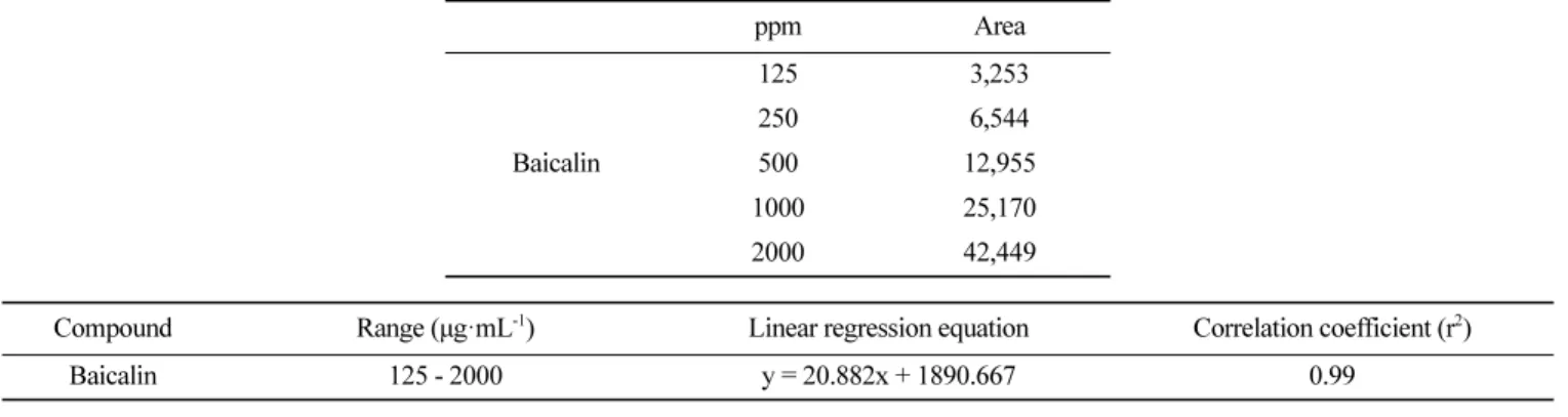
HPLC-UV 분석법에 적용하여 황금 지표성분인 baicalin의 함량 분석을 하였다(Park 등, 2014). Baicalin 표준용액은 baicalin standard (572667, Sigma-Aldrich Co., MO, USA) 1 mg을 50% 에 탄올 0.5 mL로 용해시켜 2000 µg/mL으로 조제한 것을 50% 에탄 올을 가해 1000, 500, 250, 125 µg/mL으로 희석시켜 표준용액으로 사용하였고, 분말형태의 SWE 10 mg을 10 mL 메탄올에 용해하여 0.45 μm syringe filter (Advantec)를 통하여 여과한 후 그 여액 10 μL를 HPLC (Fast-HPLC, Agilent 1260 Infinity, CA, USA)에 injection하였다. 컬럼은 CAPCELL PAK C18 (4.6 mm i.d.×250 mm, 5 μm, Shiseido, Tokyo, Japan)을 이용하였고 이동상의 용매로 는 1% acetic acid (solvent A)와 acetonitrile:methanol=7:3, 1% acetic acid (solvent B)를 사용하였다. 컬럼 온도는 40oC, 검출기는 다파장검출기(Multi Wavelength Detector), UV 조건은 275 nm, 유 량은 1.0 mL/min이며, 이동상 용매 조건은 Table 1 조건과 같이 분 석하였다. Baicalin에 대해서 각각 275 nm에서 125, 250, 500, 1000 그리고 2000 µg/mL의 농도로 분석하여 크로마토그램을 얻고 이로 부터 평균면적을 얻어 검량곡선을 그려 정량하였다. 세포배양 세포 배양을 위해 RAW264.7 세포는 5% CO2 및 37oC에서
95% 이상의 습도를 유지한 배양기(MCO-19AIC, SANYO, Tokyo, Japan)에서 10% FBS, 100 U/mL penicillin, 100 μg/mL streptomycin 을 포함하는 DMEM 배지에서 배양하였다.
MTT assay
RAW264.7세포에 대한 SWE의 세포독성을 MTT시약을 이용하 여 측정하였다(Mosmann, 1983). RAW264.7세포를 10% FBS, 100 IU/mL penicillin, 100 μg/mL streptomycin을 포함하는 DMEM 배 지를 이용하여 5×10⁵ cell/mL로 96-well plate에 접종하고 24시간
안정화하였다. 이후 세포에 SWE를 농도별(3.9, 7.8, 15.6, 31.2,
62.5, 125, 250, 500, 1000, 2000, 4000 μg/mL)로 24시간 처리하 였다. 세포배양액을 제거하고 배지로 희석한 MTT시약(0.05 mg/ mL, 298-93-1, Duchefa Biochemie, Haarlem, Netherland)을 100 μL씩 처리한 후 빛이 차단된 37oC 환경에서 2시간 반응시켰다. 이후 배지를 제거하고 100 μL의 DMSO를 이용해 formazan을 용 해시켜주고 37oC 환경에서 30분간 반응시킨다. 반응이 끝난 후
micro plate reader (SynergyTM HTX Multi-Mode Microplate Reader, Biotek, CA, USA)로 570 nm에서의 흡광도를 측정하였고 대조군과 비교하여 세포 생존률 %로 나타내었다.
NO assay
RAW264.7세포로부터 생성된 NO 함량은 Griess 원리를 기반으 로 만들어진 NO Plus Detection kit (21023, iNtRON, SungNam, Korea)를 이용하여 측정하였다. RAW264.7세포를 10% FBS, 100 IU/mL penicillin, 100 μg/mL streptomycin을 포함하는 DMEM 배 지를 이용하여 5×10⁵ cell/mL로 조절한 후 96-well plate에 접종
하고 37oC, 5% CO
2의 습윤배양기에서 24시간 안정화하였다. 안
정화된 세포에 LPS (1 μg/mL)와 SWE를 농도별(31.2, 62.5, 125, 250, 500, 1000, 2000 μg/mL)로 동시처리하여 24시간 배양하였다. 이후 세포배양 상등액 100 μL와 Griess시약을 혼합하여 96-well plate에서 20분동안 반응시킨 후 micro plate reader (SynergyTM HTX Multi-Mode Microplate Reader, Biotek, CA, USA)로 540 nm에서 흡광도를 측정하고 NO standard의 농도별 표준곡선을 이 용하여 배양액의 NO 농도를 결정하였다.
Western blot analysis
RAW264.7세포를 10% FBS, 100 IU/mL penicillin, 100 μg/mL streptomycin을 포함하는 DMEM 배지를 이용하여 5×10⁵ cell/mL 로 100 π dish에 분주하고 37oC, 5% CO 2의 습윤배양기에서 24 시간 안정화하였다. 안정화된 세포에 LPS (1 μg/mL)와 SWE를 농 도별(31.2, 62.5, 125, 250, 500 μg/mL)로 동시처리한 후 24시간 배양하였다. 반응이 종료된 후 배지를 제거하고 PBS로 2회 세척 한 후 0.25% trypsin-EDTA를 처리하여 세포를 수확하고 용출 완 충용액(RIPA buffer, 1% protease inhibitor cocktail, 1% phosphatase Table 1. HPLC condition for the analysis of baicalin from Scutellaria baicalensis
HPLC system Fast-HPLC, Agilent 1260 Infinity Column OSAKA SODA CAPCELL PAK C18 Column Temp 40oC
Detector UV 275 nm Flow rate 1.0 mL/min Inj. vol. 10 μL
Mobile phase Solvent A-1% acetic acid
Solvent B-ACN:MeOH=7:3 (v/v), 1% acetic acid
Gradient elution
Time (min) Solvent (%A)
0-10 75-68 10-20 68-55 20-24 55-55 24-35 55-52 35-40 52-75 40-45 75-75
inhibitor cocktail3)을 가해준 뒤 4oC에서 2시간 동안 단백질을 추 출하였다. 이후 30분 동안 4oC, 125×g에서 원심분리한 상층액을
따서 단백질 정량에 사용하였다. 정량된 40 μg의 단백질을 10% sodium dodecyl sulfatepolyacrylamide gel electrophoresis (SDS-PAGE)로 분리하고 PVDF membrane으로 전이시켰다. 전이된 membrane은 Tris-buffered saline & Tween 20 (TBST: Tris 24.7 mM, potassium chloride 2.7 mM, sodium chloride 137 mM, 0.2% Tween 20)에 용해된 5% skim milk에 1시간 동안 실온에서 blocking한 후 목적 단백질에 대한 1차 항체(iNOS, COX-2, TNF-α, JNK, p38, ERK, p-JNK, p-p38, p-ERK 그리고 β-actin)를 4oC 에서 overnight 반응시켰다. TBST로 3회 washing하고, HRP-conjugated secondary antibody로 3시간 동안 실온에서 반응시킨 후 다시 TBST로 3회 washing하였다. ECL kit (ECL plus western blotting substrate, Thermo, MA, USA)를 사용하여 목적단 백질을 검출하고 필름으로 현상하여 분석하였다.
통계처리
실험결과는 3회 이상 실시하여 평균±표준편차(mean±SD)로 표 기하였다. 통계적 분석은 SPSS (PASWstatistics 18, SPSS Inc., IBM Corporation, Armonk, NY, USA) 분석프로그램의 One-way ANOVA, Duncan test에 준하였고 p<0.05 수준에서 유의성을 검 정하였다.
결과 및 고찰
SWE 추출 수율 및 지표물질
황금 50 g을 10배수의 증류수로 열수추출해 15.21 g의 추출물 을 얻었으며 수율은 30.42%로 나타났다. 또한 SWE에 함유되어 있는 baicalin을 정량하기 위해 HPLC를 이용하여 baicalin standard 125, 250, 500, 1000, 그리고 2000 µg/mL의 농도로 제조하여 기 기분석을 하였고. 농도에 따른 검량곡선을 그려 직선식을 구하였 다(Table 2). SWE 함유된 baicalin 정량 분석 결과 100.030± 2.922 mg/g으로 분석되었다(Table 3). SWE의 세포 독성평가 염증반응에서 중추적인 역할을 하는 대식세포는 여러 자극원 이나 다른 세포의 사이토카인에 의해 활성화된다(Iontcheva 등, 2004; Li 등, 2015). 그러므로 본 연구에서는 SWE의 항염증 효 과를 확인하기 위하여 먼저 대식세포에 독성을 미치지 않는 농 도를 결정하기 위하여 MTT assay를 수행하였다. RAW264.7 세 포주에 SWE를 농도별(3.9, 7.8, 15.6, 31.2, 62.5, 125, 250, 500, 1000, 2000, 4000 μg/mL)로 처리한 결과 cell viability가 SWE 1000 μg/mL 이하의 농도에서 96.9% 이상의 세포생존율을 가지며 2000 μg/mL와 4000 μg/mL에서 각각 92.9%와 84.2%로 약간의 세 포독성이 보였다. 따라서 RAW264.7 세포만 배양한 대조군에 비 하여 유의한 세포독성이 없는 농도는 SWE 1000 μg/mL 이하로 설정하였다(Fig. 1). 따라서 SWE는 고농도를 제외한 나머지 농도 에서 독성을 보이지 않으므로 항염증 소재로서의 활용 가능성을 가지며 추후 진행되는 효능 실험 결과는 SWE의 세포독성에 의 한 세포생존율 감소가 아닌 SWE의 약리활성으로 사료된다. SWE의 NO 생성 억제 효과 LPS나 다양한 병원체 유래 인자에 의해 활성화된 대식세포는 외래인자 탐식을 위해 NO나 pro-inflammatory 사이토카인과 같은 염증 매개인자를 유리함으로써 외래인자를 제거하는 중요한 역 할을 담당한다(Kim, 2013). 박테리아를 제거하는 기능을 하는 NO 는 과도하게 생성되면 염증을 일으켜 조직손상, 유전자변이, 신 경손상을 유발한다(Jung 등, 2010). 본 연구에서는 LPS에 의해 RAW264.7 세포로부터 생성되는 NO에 대한 SWE의 억제효과를 세포배양액으로부터 Griess assay 방법에 의해 측정하였다. NO의 생성함량은 RAW264.7 세포만 배양한 대조군에 비해 LPS (1 μg/ mL)를 처리한 군에서 NO의 농도는 2.3 μM에서 36.0 μM로 현저 히 증가되었다. SWE를 농도별(31.2, 62.5, 125, 250, 500, 1000 μg/mL)로 처리한 실험군은 농도의존적으로 NO 생성이 억제되는 것을 관찰할 수 있었으며 500 μg/mL에서 7.5 μM로 유의한 억제 를 보였다(Fig. 2). 이 같은 결과는 SWE가 LPS에 의해 유발된 마우스 대식세포주에서 과도한 NO 생성을 억제함으로써 염증을 완화할 수 있는 효능이 있음을 의미한다. 기존 연구는 황금 열수 추출물이 마우스 대식세포의 NO 생성을 억제한다고 보고했으며 이는 본 연구결과와 일치한다(Park 등, 2014). SWE의 염증관련 사이토카인 단백질 발현 조절 효과 RAW264.7 세포에서의 SWE의 NO 생성억제효능을 검증하였 고 LPS 자극에 의해 NO를 생성하는 것으로 알려져있는 iNOS와 Table 2. Measurement of linearity for baicalin
ppm Area Baicalin 125 3,253 250 6,544 500 12,955 1000 25,170 2000 42,449
Compound Range (μg·mL-1) Linear regression equation Correlation coefficient (r2)
Baicalin 125 - 2000 y = 20.882x + 1890.667 0.99
Table 3. Quantitative analysis of baicalin from Scutellaria baicalensis
Sample mg·L-1 (ppm) Area±SD Ave mg·g-1±SD
Scutellaria baicalensis
염증단계에서 중요한 역할을 하는 전 염증성 사이토카인인 COX-2, 그리고 LPS 자극의 주요 매개체로 선천면역반응에 있어서 중 요한 역할을 하는 TNF-α의 단백질 발현 수준의 조사를 통해 SWE 의 항염증효과를 조사하였다(Lee 등, 2003). 안정화된 RAW264.7 세포에 LPS (1 μg/mL)와 SWE를 농도별(31.2, 62.5, 125, 250, 500 μg/mL)로 24시간 처리한 후 western blot을 통해 단백질 발현 을 확인하였고 β-actin으로 이들 단백질의 정량화를 나타내었다. 실험결과, 대식세포에 LPS를 처리함에 따라 iNOS, COX-2 그리 고 TNF-α 단백질 발현이 증가하는 것을 확인하였다. 증가된 iNOS 와 COX-2 발현은 SWE를 처리함에 따라 농도의존적으로 이들 단백질 발현을 억제하였으며, 특히 SWE 250 μg/mL 농도에서는 iNOS와 COX-2 발현을 공시험군과 비슷한 수준까지 억제함을 알 수 있었다. 또한, TNF-α의 경우 SWE 250 μg/mL 이상의 농도에 서는 발현이 관찰되지 않았고 나머지농도에서는 농도간의 단백 질 발현 차이는 크게 나지는 않지만 LPS만 단독처리한 군에 비 해 하향 조절되었다. 특히 SWE 62.5 μg/mL에서 공시험군과 비 슷한 수준으로 단백질 발현을 억제함을 알 수 있었다(Fig. 3). SWE의 MAPKs 활성화에 미치는 영향 SWE의 염증 매개물질 기전을 탐색하기 위해서 MAPKs 경로 를 조사하였다. MAPK는 세포분화 및 성장의 조절뿐만 아니라 사이토카인과 스트레스에 대한 세포내 반응을 조절하는 중요한 역할을 한다(Park 등, 2018). MAPKs는 ERK, JNK, p-38로 구성 되어 있으며, 이들을 통한 신호경로들은 LPS로 염증이 유발된 마 우스 대식세포인 RAW264.7에서 iNOS 및 전염증성 세포활성물 질 발현에 관여한다(Kim 등, 2011). RAW264.7 세포에 LPS Fig. 1. Effects of Scutellaria baicalensis water extract (SWE) on cell viability in RAW 264.7 cells. RAW 264.7 cells were plated at a concentration of 5×10⁵ cell/mL in 96-well plate. Following 24 h of stabilization, cell were cultured in the presence of SWE and LPS (1 μg/mL) for 24 h under serum-free conditions. The cell viabilities were determined via MTT assay. Each determination was made in triplicate and the data were expressed as means±SD.
Fig. 2. Effects of Scutellaria baicalensis water extract (SWE) on NO production in LPS-stimulated RAW 264.7 cells. RAW 264.7 cells were plated at a concentration of 5×10⁵ cell/mL in 96-well plate. Following 24 h of stabilization, cell were cultured in the presence of SWE and LPS (1 μg/mL) for 24 h under serum-free conditions. The NO production were determined by Griess reaction. Each determination was made in triplicate and the data were expressed as means±SD. *p<0.05, **p<0.01 and ***p<0.001, compared with LPS stimulated control group.
Fig. 3. Effects of Scutellaria baicalensis water extract (SWE) on expression of iNOS and COX-2 protein and production of pro-inflammatory cytokines in LPS-stimulated RAW 264.7 cells. (A) RAW 264.7 cells were treated with SWE (31.2-500 μg/mL) and LPS (1 μg/ mL) for 24 h. Equal amounts of whole cell extract were separated on SDS-PAGE gels, and iNOS, COX-2 and TNF-α were detected by western blot. β-actin was used as a control. (B) iNOS, COX-2, TNF-α to β-actin ratio were calculated based on the relative level of each protein after the determination of the intensity of each band using an imaging densitometer. Data are mean±SD of three independent experiments. *p<0.05, **p<0.01 and ***p<0.001, compared with LPS stimulated control group.
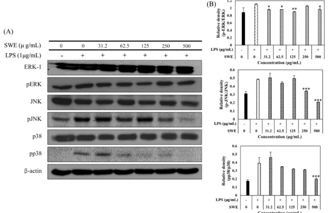
Fig. 4. Inhibition of mitogen-activated protein kinases (MAPKs) phosphorylation by Scutellaria baicalensis water extract (SWE) in LPS-stimulated RAW 264.7 cells. (A) RAW 264.7 cells were plated at a concentration of 5×10⁵ cell/mL in 100 π dish. Following 24h of stabilization, cells were cultured in the presence of SWE and LPS (1 μg/mL) for 24 h under serum-free conditions. Equal amounts of whole cell extract were separated on SDS-PAGE gels, and ERK/pERK, p38/pp38, JNK/pJNK and β-actin were detected by western blot. (B) ERK/pERK, p38/pp38, JNK/pJNK ratio was calculated based on the relative level of each protein after the determination of the intensity of each band using an imaging densitometer. Data are mean±SD of three independent experiments. *p<0.05, **p<0.01 and ***p<0.001, compared with LPS stimulated control group.
(1 μg/mL)와 SWE를 농도별(31.2, 62.5, 125, 250, 500 μg/mL)로 처리하여 western blot을 수행하였고 β-actin으로 이들 단백질의 정량화를 확인하였다. 실험결과, LPS로 자극한 세포에서 phospho-ERK, phospho-JNK와 phospho-p38의 발현이 현저히 증가하였고 SWE를 전처리한 세포에서 phospho-JNK와 phospho-p38 발현이 감소하는 것으로 보아 SWE가 JNK와 p38의 인산화를 억제하는 것을 알 수 있고 ERK의 인산화에는 영향을 끼치지 않는 것으로 확인하였다(Fig. 4) 이와 같은 결과는 SWE가 MAPKs 경로에서 JNK 및 p38 인산화 억제를 통해 염증성 사이토카인의 발현을 억 제하여 항염증 작용을 하고 있음을 나타낸다.
요
약
Scutellaria baicalensis water extract (SWE)는 지질 다당류 LPS 로 유도된 RAW 264.7 세포에서 NO 및 전 염증성 사이토카인인 TNF-α의 생성을 세포 독성을 유발하지 않고 유의하게 억제하였 다. 또한, SWE는 iNOS 및 COX-2의 단백질발현을 농도의존적으 로 감소시켰으며, ERK, JNK, p38과 같은 MAPKs 계열의 인산화 발현 수준을 조사한 결과 JNK와 p38의 발현 수준을 감소시켰다. 이는 SWE가 p38 인산화를 억제함으로써 iNOS, COX, 그리고 TNF-α와 같은 전 염증성 사이토카인의 발현을 감소시키며 결론 적으로 NO의 생성을 억제시킨다는 결과를 도출할 수 있었다. 본 연구는 항염증 효능 검증뿐 아니라 염증대사기전의 주요인자를 탐색함으로써 황금의 기능성 소재로써의 가능성을 시사한다.
감사의 글
본 연구는 농림축산식품부가 지원하는 2015년 농촌자원복합산 업화지원사업 향토건강식품명품화사업으로 수행된 연구결과입니다.References
Iontcheva I, Amar S, Zawawi KH, Kantarci A, Van Dyke TE. Role for moesin in lipopolysaccharide-stimulated signal transduction. Infect. Immun. 72: 2312-2320 (2004)
Kuo SW, Su WL, Chou TC. Baicalin improves the survival in endo-toxic mice and inhibits the inflammatory responses in LPS-treated RAW 264.7 macrophages. Eur. J. Inflamm. 18: 1-12 (2020) Jung JY, Lee JR, Byun SH, Jung JW, Kim YH, Kim SC. Inhibitory
effect of Dioscorea bulbifera MeOH extract on pro-inflammatory mediator In vitro and In vivo. J. Physiol. Path. Kor. Med. 24: 310-318 (2010)
Kang SW. Role of Reactive Oxygen Species in Cell Death Pathways. Hanyang Medical Reviews 33: 77-82 (2013)
Kim TY. Effect of Gagam-Dangueumja through regulation of MAPK on LPS-induced inflammation in Raw 264.7 cells. Kor. J. Orient. Intern. Med. 34: 339-348 (2013)
Kim MS, Jeong J, Lee HY, Ju YS, Bae GS, Seo SW, Cho IJ, Park SJ, Song HJ. The anti-inflammatory effect of Achyranthes japon-ica on lipopolysaccharide-induced inflammatory activity in murine macrophages. Kor. J. Herbology. 26: 51-57 (2011)
Kim MK, Kim DY. Anti-inflammatory effect of barley leaf ethanol extract in LPS-stimulated RAW264.7 macrophage. Kor. J. Food Preserv. 22: 735-743 (2015)
Lee AK, Sung SH, Kim YC, Kim SG. Inhibition of lipopolysaccha-ride-inducible nitric oxide synthase, TNF-α and COX-2 express-tion by sauchinone effects on I-Ba phosphorylaexpress-tion, C/EBP and AP-1 activation. Br. J. Pharmacol. 139: 11-20 (2003)
Li Y, Wu Q, Deng Y, Lv H, Qiu J, Chi G, Feng H. D(-)-Salicin inhibits the LPS-induced inflammation in RAW264.7 cells and mouse models. Int. Immunopharmacol. 26: 286-94 (2015)
Matthay MA, Zimmerman GA, Esmon C, Bhattacharya J, Coller B, Doerschuk CM, Floros J, Gimbrone Jr MA, Hoffman E, Hub-mayr RD, Leppert M, Matalon S, Munford R, Parsons P, Slutsky AS, Tracey KJ, Ward P, Gail DB, Harabin A. Future research directions in acute lung injury: summary of a National Heart, Lung, and Blood Institute working group. Am. J. Respir. Crit. Care Med. 167: 1027-1035 (2003)
Mosmann T. Rapid colorimetric assay for cellular growth and sur-vival: application to proliferation and cytotoxicity assays. J. Immunol. Methods. 65: 55-63 (1983)
Park HJ, Kim SM, Kwon HJ, Lee HT, Kim BW, Kim TH, Kim MM. Anti-inflammatory effect of Scutellaria baicalensis hot water extracts containing baicalin on modulation of the immune system in Raw264. 7 cells. J. Life Sci. 24: 219-226 (2014)
Park JS, Kim MH. Anti-inflammatory effects of ricd bran ethanol extract in murine macrophage raw 264.7 cells. Yakhak Hoeji 55: 451-461 (2011)
Park JW, Kwon OK, Ryu HW, Paik JH, Paryanto I, Yuniato P, Choi SH, Oh SR, Ahn KS. Anti-inflammatory effects of Passiflora foetida L. in LPS-stimulated RAW264.7 macrophages. Int. J. Mol. Med. 41: 3709-3716 (2018)
Wang ZL, Wang S, Kuang Y, Hu ZM, Qiao X, Ye M. A comprehen-sive review on phytochemistry, pharmacology, and flavonoid bio-synthesis of Scutellaria baicalensis. Pharm. Biol. 56: 465-484 (2018)
Qing Z, Chen XY, Martin C. Scutellaria baicalensis, the golden herb from the garden of Chinese medicinal plants. Sci. Bull. 61: 1391-1398 (2016)
Zhao T, Tang H, Xie L, Zheng Y, Ma Z, Sun Q, Li X. Scutellaria baicalensis Georgi. (Lamiaceae): a review of its traditional uses, botany, phytochemistry, pharmacology and toxicology. J. Pharm. Pharmacol. 71: 1353-1369 (2019)