• 검색 결과가 없습니다.

Inhibitory Activity of Cordyceps bassiana Extract on LPS-induced Inflammationin RAW 264.7 Cells by Suppressing NF-κB Activation

N/A
N/A
Protected

Academic year: 2022

Share "Inhibitory Activity of Cordyceps bassiana Extract on LPS-induced Inflammationin RAW 264.7 Cells by Suppressing NF-κB Activation"

Copied!
7
0
0

로드 중.... (전체 텍스트 보기)

전체 글

(1)

23(3) : 162-168 (2017)

https://doi.org/10.20307/nps.2017.23.3.162

162

Inhibitory Activity of Cordyceps bassiana Extract on LPS-induced Inflammation in RAW 264.7 Cells by Suppressing NF- κB Activation

Deok Hyo Yoon

1,†

, Changwoo Han

2,†

, Yuanying Fang

2

, Shankariah Gundeti

2

, In-Sook Han Lee

3

, Won O Song

4

, Ki-Chul Hwang

5

, Tae Woong Kim

1

, Gi-Ho Sung

5

*, and Haeil Park

2

*

1

Department of Biochemistry, Kangwon National University, Chunchon 200-701, Republic of Korea

2

College of Pharmacy, Kangwon National University, Chunchon 200-701, Republic of Korea

3

Department of Science Education, Kangwon National University, Chunchon 200-701, Republic of Korea

4

Department of Food Science and Human Nutrition, Michigan State University, East Lansing, MI48824, USA

5

Institute for Bio-Medical Convergence, Catholic Kwandong University, Incheon 404-834, Republic of Korea

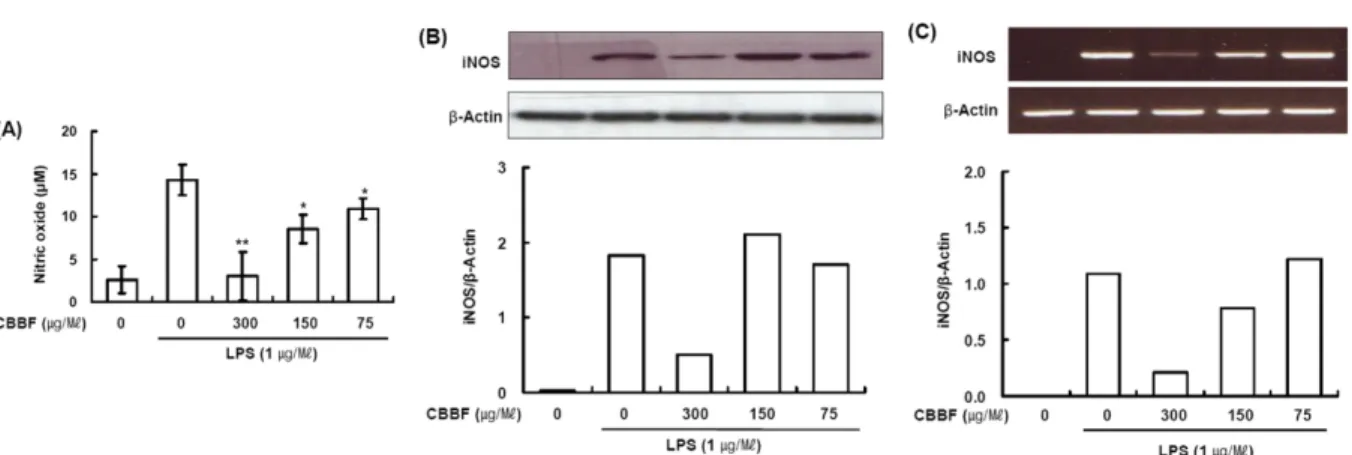
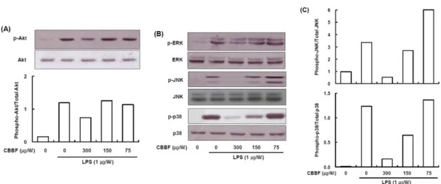
Abstract − Cordyceps bassiana has long been used as an oriental medicine and reported to possess diverse biological activities. The fruiting bodies of Cordyceps bassiana was extracted with ethanol and then further fractionated with n-hexane, ethyl acetate, n-butanol and water. The butanol fraction from Cordyceps bassiana (CBBF) exhibited the most effective in anti-inflammatory activity in RAW 264.7 macrophages and the roles of CBBF on the anti-inflammation cascade in LPS-stimulated RAW 264.7 cells were studied. To investigate the mechanism by which CBBF inhibits NO, iNOS and COX-2, the activation of IκB and MAPKs in LPS-activated macrophage were examined. Our present results demonstrated that CBBF inhibits NO production and iNOS expression in LPS-stimulated RAW 264.7 macrophage cells, and these effects were mediated through the inhibition of IκB-α, JNK and p38 phosphorylation. Also, CBBF suppressed activation of MAPKs including p38 and SAPK/JNK. Furthermore, CBBF significantly suppressed LPS-induced intracellular ROS generation. Its inhibition on iNOS expression, together with its antioxidant activity, may support its anti-inflammatory activity.

Thus Cordyceps bassiana can be used as a useful medicinal food or drug for further studies.

Keywords − Cordyceps bassiana, Oriental ethnopharmacology, n-butanol fraction, Anti-inflammatory activity

Introduction

Parasitic Cordyceps fungi (winter worm summer grass) is a parasitic complex of fungus and caterpillar, which has been used for medicinal purposes for centuries particularly in Asia.

1

Cordyceps is a genus of ascomycete fungi that include about 400 described species.

2

Some Cordyceps species are sources of biochemicals with biological and phamacological properties, such as 3'- deoxyadenosine (cordycepin), cordycepic acid, and Cordyceps polysaccharide, etc. It is commonly used to treat conditions such as asthenia after severe illness, hyperglycemia, hyperlipidemia, hyposexuality, respiratory

disease, renal dysfunction, renal failure, arrhythmias, and other heart disease and liver disease.

The entomopathogenic fungus Beauveria bassiana is a globally distributed hyphomycete under intensive study as a biocontrol agent for a variety of pest insects.

Beauveria is presumed to be related to Cordyceps by morphological and physiological characters.

3

Schaerffenberg reported a possible clavicipitaceous sexual state for Beauveria bassiana,

4

and the new species Cordyceps bassiana was described by Li et al. on carpenterworm larva (Lepidoptera: Cossidae).

5

Recently, it was known that Beauveria bassiana is the anamorph (asexually reproducing form) of Cordyceps bassiana.

Recent studies have shown that extracts and isolated components from mushrooms suppress tissue injury of pathological processes associated with many inflammatory diseases.

6-8

Cordyceps species and their extracts have been recognized for the prevention and treatment of cancer, immunity, and several other diseases and the protective effects on human organs.

1,9

Although there are 400 species of Cordyceps, the

*Author for correspondence

Haeil Park, College of Pharmacy, Kangwon National University, Chunchon 200-701, Republic of Korea.

Tel: +82-33-250-6920 E-mail: [email protected]

Gi-Ho Sung, Institute for Bio-Medical Convergence, Catholic Kwandong University, Incheon 404-834, Republic of Korea.

Tel: +82-32-290-2772 E-mail: [email protected]

These authors contributed equally to this work

(2)

research into its pharmacological effect has focused only on two specis, C. sinensis and C. Militaris. Since C.

bassiana has been shown biocontrol agent for insects and other arthropod pests but not fully studied yet about other medicinal value and described with anti-inflammatory activity.

In this study, we aim to investigate the effect of C.

bassiana extracts on the NO production and iNOS gene and protein expression, and attempt to clarify its mechanism of action in LPS-stimulated RAW 264.7 macrophages cells.

Information from our research may provide an additional pharmacological background on its efficacies of C. bassiana.

Experimental

General experimental procedures − Dulbecco's modified Eagle's medium (DMEM), penicillin, streptomycin, and fetal bovine serum (FBS) were purchased from Invitrogen Co. (Grand Island, NY, USA). Lipopolysaccharide (LPS, Escherichia coli O11:B4) was obtained from Sigma Co.

(St. Louis, MO, USA). Mouse iNOS (#610328), phospho- p38 MAPK (#612280) antibody was obtained from BD Biosciences (San Jose, CA, USA). Akt (#9272), JNK (#9252), p38 (#9217), phospho-Akt (#9271), phospho- JNK (#9251) antibody were obtained from Cell signaling Technology, Inc. (Boston, MA, USA). COX-2 (#1746), I κB-α (#1643), phospho-IκB-α (#9242) were obtained from SANTA CRUZ Biotechnology, Inc. (Santa Cruz, CA, USA). β-Actin (A5441) and DPI were obtained from Sigma Co. (St. Louis, MO, USA). All other chemicals and solvents were purchased from Sigma unless indicated otherwise.

Extraction and fractionation from Cordyceps bassiana − Artificial cultured C. bassiana

2

were obtained from Mushtech Co. (Chuncheon, Korea) and authenticated by Dr. J.M. Sung (Dep. of Applied biology, Kangwon National University). A voucher specimen (EFCC #12988) was deposited in the Entomopathogenic Fungal Culture Collection (EFCC), Kangwon National University, Korea.

The fruiting bodies were dried at 50

o

C and crushed in a blender and the crude powder was extracted with ethanol at 80

o

C for 8 h. The extracts were evaporated at 60

o

C under pressure and suspended again in distilled water. The aqueous layer was mixed with n-hexane, n-butanol and ethyl acetate. The n-hexane layer was evaporated to dryness under pressure. n-Butanol, ethyl acetate and aqueous fractions were progressed following the same method.

MTT assay − The above extracts or fractions of C.

bassiana were dissolved in DMSO and diluted with medium. These were added to the cultures at the concen-

tration of 0~100 μg/ml each. We first test inhibition of cell proliferation by C. bassiana, measured by MTT (3- (4,5-dimethylthiazol-2-yl)-2,5-diphenyltetrazolium bromide) assay. Briefly, A549 cell line (5.0 × 10

3

cells/well) were cultured in the 96 well culture plate in the presence or absence of the extracts or fractions of C. bassiana. After 24 h incubation, 50 μl of the MTT solution, which was prepared by mixing 3 mg of the MTT powder with 1 ml of the PBS, was then added to each well. After incubation, the medium containing MTT solution was replaced with isopropanol for extraction of dye. After incubation for 30 min, absorbance was measured on micro plate reader at a test wavelength of 570 nm.

Macrophage cell culture − Murine macrophage cell line RAW 264.7 (American Type Culture Collection, Bethesda, USA) was cultured in DMEM including 2 mM L-glutamine, 100 U/ml penicillin, 100 μg/ml streptomycin and 10% FBS. Macrophages were cultured in 6-well plates (4 × 10

6

cells/well) at 37

o

C in 5% CO

2

/95% air. Cells were washed twice with fresh medium and stimulated with 1 μg/ml LPS.

Determination of NO concentration − RAW 264.7 cells (4 × 10

6

cells/well) were incubated for 16 h with 1 μg/ml LPS. The presence of nitrite, a stable oxidized product of NO, was determined in cell culture media by Griess reagent. Briefly, 100 μl of culture supernatant was removed and combined with 100 μl Griess reagent (mixture of equal volume of 1% sulfanilamide in 5% H

3

PO

4

and 0.1% N-(1-naphthyl) ethylenediamine dihydrochloride in H

2

O) in a 96-well plate, followed by spectrophotometric measurement at 550 nm. Nitrite concentrations in the supernatants were determined by comparison with a sodium nitrite standard curve.

Preparation of nuclear extracts − Nuclear extracts were prepared by a modified method of Wadsworth and Koop (1999).

10

Treated cells were washed, then scraped into 1.5 mL of ice-cold TBS (pH 7.9), and centrifuged at 12,000 × g for 30 s. The pellet was suspended in 10 mM HEPES, pH 7.9, with 10 mM KCl, 0.1 mM EDTA, 0.1 mM EGTA, 1 mM DTT, 0.5 mM PMSF, 5 μg/ml of leupeptin, aprotinin, and pepstatin, incubated on ice for 15 min and then vortexed for 10 s with 0.6% Nonidet P-40.

Nuclei were separated from cytosol by centrifugation at

12,000 × g for 60 s. The supernatant was removed, and

the pellet was suspended in 50 - 100 μl of 20 mM HEPES,

pH 7.9, with 25% glycerol, 0.4 M NaCl, 1 mM EDTA,

1 mM EGTA, 1 mM DTT, 0.5 mM PMSF, 10 μg/ml of

leupeptin, aprotinin and pepstatin. The samples were

incubated with rocking at 4

o

C for 15 min, and centrifuged

for 5 min at 12,000 × g. Protein concentration of the

(3)

supernatant was determined by BCA protein assay kit (Thermo Fisher Scientific Inc., USA).

Western blot analysis − Macrophages were incubated with or without LPS in the presence or absence of CBBF.

Cells were harvested, washed twice with ice-cold TBS, and resuspended in Lysis buffer 100 mM Tris, 5 mM EDTA, 50 mM NaCl, 50 mM β-glycerophosphate, 50 mM NaF, 0.5% NP-40, 1% sodium deoxycholate, 0.1 mM sodium orthovanadate and 1% PMSF. The cytosolic fraction was obtained from the supernatant of 12,000 × g centrifugation at 4

o

C for 20 min. Protein (30 - 50 μg per lane) were separated using 8 - 12% polyacrylamide SDS gels and then transferred electrophoretically to a polyvinylidene difluoride membrane. The membrane was blocked for 2 h at room temperature with TBS containing 0.1% Tween-20 and 5% fat-free dried milk and then incubated with antibodies. Immune complexes were performed using enhanced chemiluminescence method and exposed to X- ray film. Band intensity was measured by computer analysis using ‘Scion Image’ software.

RT-PCR analysis − Total RNA was prepared from RAW 264.7 cells using a Trizol Reagent kit (Invitrog Co.). One microgram of total RNA was converted to cDNA by treatment with 200 U of reverse transcriptase and 500 ng of oligo-dT primer in 50 mM Tris-HCl (pH

8.3), 75 mM KCl, 3 mM MgCl

2

, 10 mM DTT, and 1 mM dNTPs at 42

o

C for 1 h. The reaction was stopped by heating at 70

o

C for 15 min. Three microliters of the cDNA mixture was used for enzymatic amplification.

Polymerase chain reaction was performed in 50 mM KCl, 10 mM Tris-HCl (pH 8.3), 1.5 mM MgCl

2

, 0.2 mM dNTPs, 2.5 U of Taq DNA polymerase, and 0.1 μM of each primer for iNOS and COX-2. The amplification was performed in a DNA thermal cycler under the following condition: denaturation at 95

o

C for 5 min for the first cycle; 95

o

C for 45 s annealing at 55

o

C (iNOS) or 50

o

C (COX-2) for 30 s, and extension at 72

o

C for 45 s for 35 repetitive cycles. Final extension was performed at 72

o

C for 10 min. The PCR products were electrophoresed on a 1.5% agarose gel and stained with ethidium bromide. The primers were purchased from COSMO co, Ltd. The primers used were 5'-TCTTCGAAATCCCACCTGAC-3' (sense) and 5'-CCATGATGGTCACATTCTGC-3' (antisense) for the iNOS, 5'-TCCTTCGTTGCCGGTCCACA-3' (sense) and 5'-CGTCTCCGGAGTCCATCACA-3' (antisense) for the β-actin was used as an internal control.

Measurement of intracellular ROS − Intracellular ROS release analysis was performed with Confocal Laser Scanning Microscope (CLSM). RAW 264.7 cells were plated on a 25 mm coverslip at a density of 10

6

cells/ml.

Fig. 1. Effects of CBEE on NO production, iNOS and COX-2 protein expression in LPS-induced macrophages: (A) RAW 264.7 cells

(4 × 10

6

cells/well) were incubated for 16 h with 1 μg/ml LPS in the presence or absence of CBEE. RAW 264.7 cells were pretreated with

the indicated concentrations of CBEE for 30 min before incubation with LPS for 16 h. The culture supernatants were subsequently

isolated and analyzed for nitrite levels. Data are expressed as mean ± S.D.(n = 3). Asterisks indicate a significant difference from LPS

alone (* P < 0.05, ** P < 0.01 vs. LPS alone). (B) The cells were lysed, and the lysates were analyzed by immunoblotting used anti-iNOS

and anti-COX-2. The blot was stripped from the bound antibody and reprobed with anti-β-actin to confirm equal loading. (C~D) The

intensity of the bands was scanned and quantified by Scion Image software.

(4)

Confluent cells were made quiescent by placing in medium containing 0.5% FBS. They were treated with DCFH-DA for 30 min and washed twice with PBS. The samples were excited at 488 nm using an argon-krypton laser to observe the green fluorescence of the fluorescein and RAW 264.7 cells cultured in a 60 mm dish at 5 × 10

5

cells/dish were treated with LPS for 20 min. DCFH-DA (5 M) was added to the cultures and incubated for 30 min.

The cells were harvested and washed three times with PBS, and intracellular ROS was quantified by flow cytometry with excitation at 488 nm and emission at 525 nm.

Statistical analysis − Values were expressed as mean ± S.D. Statistical significance was determined using Student’s t-test. Values with P < 0.05 were considered significant.

Result and Discussion

The effects of C. bassiana ethanol extract (CBEE) on NO, iNOS and COX-2 expression in LPS-induced RAW 264.7 cells were explored. As shown in Fig. 1A., LPS caused an increase from basal level of 1.3 ± 0.05 μM to 16.3 ± 0.03 μM for 16 h and CBEE inhibited NO production in a dose-dependent manner. No significant effect on cell viability was observed at a test concentra- tion as determined by MTT assay (data not shown). As shown in Fig. 1B., treatment of LPS for 16 h markedly enhanced expression of iNOS and COX-2 protein, and co-treatment with CBEE showed a dose-dependent inhibition of LPS-induced iNOS (Fig. 1C) and COX-2 protein expression (Fig. 1D).

Further study on the effects of C. bassiana solvent fractions on NO, iNOS and COX-2 expression in LPS- induced RAW 264.7 cells was conducted. RAW 264.7 cells were treated with 150 μg/ml of n-hexane fraction (HF), n-butanol fraction (BF), ethyl acetate fraction (EAF) and aqueous fraction (AF) for 30min before exposure to LPS. CBBF was strongly inhibited LPS- induced NO production (Fig. 2) than the other solvents fractions. Collectively, the butanol sub-fraction of CBEE (CBBF) was clearly found to contain major anti- inflammatory principle(s) of C. bassiana.

Effects of C. bassiana butanol fractions (CBBF) on iNOS protein and mRNA expression was studied. As shown in Fig. 3B., the treatment of LPS (1 μg/ml) for 16h markedly enhanced expression of iNOS protein, and this effect was inhibited by the co-treatment of CBBF in a dose-dependent manner. To further characterize whether CBBF decrease NO production (Fig. 3A) via suppression of iNOS mRNA levels, the levels of iNOS mRNA in

cells treated with LPS in the presence of various concentration of CBBF were determined by RT-PCR.

RAW 264.7 cells expressed high levels of iNOS mRNA when stimulated with LPS (1 μg/ml) for 8 h, while the expression of iNOS mRNA was barely detectable in unstimulated cells. RT-PCR analysis of LPS-activated macrophages treated with CBBF showed the suppression of iNOS mRNA expression in a dose-dependent manner (Fig. 3C). These findings suggest that the inhibitory activity of iNOS by CBBF might be correlated to the suppression of iNOS gene and protein expression.

Effect of CBBF on LPS-induced phosphorylation of I κBα − was studied. The NF-κB is a transcription factor that modulated the expression of variety of genes involved in inflammatory responses, including iNOS, COX-2. The NF- κB/Rel transcription factors are present in the cytosol in an inactive state complexed with the inhibitory I κB proteins. Translocation of NF- κB to the nucleus is proceeded by the phosphorylation, ubiquitination, and proteolytic degradation of I κB-α, since nuclear translocation of NF- κB is directly linked to I κB-α degradation and phosphorylation.

11-13

To determine whether CBBF could affect degradation and phosphorylation of I κB-α, we determined IκB-α in RAW 264.7 cells after incubation for 30 min and 2 h with LPS and 0 up to 300 μg/ml CBBF. Western blot analysis of cell extracts with antibodies specific for I κB-α showed that CBBF inhibited LPS-mediated I κB-α phosphorylation in a dose-dependent manner (Fig. 4). These results suggested that CBBF might block LPS-induced nuclear Fig. 2. Effect of Cordyceps bassiana solvent fractions on NO production in LPS-induced macrophages. RAW 264.7 cells (4 × 10

6

cells/well) were incubated for 16 h with 1 μg/ml LPS in the presence or absence of CBEE. RAW 264.7 cells were pretreated with 150 μg/ml of various solvent fractions for 30 min before incubation with LPS for 16 h. HF, n-hexane fraction; BF, n-butanol fraction; EAF, ethyl acetate fraction; AF, aqueous fraction. The culture supernatants were subsequently isolated and analyzed for nitrite levels. Data are expressed as mean ± S.D.

(n = 3). Asterisks indicate a significant difference from LPS alone

(* P < 0.05, ** P < 0.01 vs. LPS alone).

(5)

translocation of NF- κB by suppression of IκB-α phosphorylation and degradation.

Effects of CBBF on LPS-induced phosphorylation of Akt and MAPKs − The expression of iNOS is regulated by pathways that involved Akt and MAPKs in macrophages. The Akt signal molecule is known to regulate NF- κB activation via IKK activation. Activation of IKK is mediated by phosphorylation through various upstream kinases such as NF- κB-inducing kinase, NK-

κB-activating kinase, and Akt, which are involved in cellular signaling in response to pro-inflammatory stimuli.

14,15

Therefore, the effects of CBBF on LPS- induced Akt phosphorylation were examined. To investigate whether Akt pathway was involved in the regulation of macrophage inflammation, we examined the phosphory- lation of Akt after stimulation of RAW 264.7 with LPS.

Phosphorylation of Akt was inhibited by 300 μg/ml CBBF. (Fig. 5A). The MAPKs play a critical role in the regulation of cell growth and differentiation and the control of cellular responses to cytokines and stresses.

Recently, a number of in vitro studies have shown that the production of inflammatory mediator is strongly affected by MAPKs, such as extracellular signal-regulated kinase (ERK) 1/2, c-jun N-terminal kinase (JNK) and p38 in macrophages.

16-18

In order to investigate whether the inhibition of inflammatory mediators by CBBF is mediated through the MAP kinase pathway, we examined the effect of CBBF on the LPS-induced phosphorylation of ERK 1/

2, SAPK/JNK and p38 MAP kinase in RAW 264.7 cells using Western blot analysis. As shown in Fig. 5B, CBBF suppressed LPS-induced activation of SAPK/JNK and p38 MAPK. Therefore, these results suggest that CBBF blocked SAPK/JNK and p38 phosphorylation of the MAPK pathways to suppress the LPS-stimulated RAW 264.7 cells.

CBBF suppresses ROS in the LPS-stimulated RAW 264.7 cells − The inflammatory responses of LPS in macrophages include the initial induction of ROS, which leads to the activation of MAPKs and NF- κB and to the Fig. 3. Effects of CBBF on NO production, iNOS protein and mRNA expression in LPS-induced macrophages. RAW 264.7 cells (4 × 10

6

cells/well) were incubated with 1 μg/ml LPS in the presence or absence of CBBF. (A) RAW 264.7 cells were pretreated with indicated concentrations of CBBF for 30 min before incubation with LPS for 16 h. The culture supernatants were subsequently isolated and analyzed for nitrite levels. Data are expressed as mean ± S.D. (n = 3). Asterisks indicate a significant difference from LPS alone (*P < 0.05, **P < 0.01 vs. LPS alone). (B) The cells were lysed, and the lysates were analyzed by immunoblotting used anti-iNOS. The blot was stripped from the bound antibody and reprobed with anti-β-actin to confirm equal loading. (C) After 8 h stimulation, total RNAs were obtained from RAW 264.7 cells using a Trizol reagent kit. The mRNA levels of iNOS was determined by RT-PCR analysis. Actin was used as an internal control.

Fig. 4. Effect of CBBF on LPS-induced phosphorylation of I κB-α.

Raw 264 Macrophages were incubated with LPS (1 μg/ml) without

or with different concentration of CBBF for 2 h. Cytosolic extracts

of the cells were subjected to Western blot analysis with anti-

phospho-IκBα (Ser-32/36) antibody and anti-IκBα.

(6)

induction of iNOS protein expression and NO produc- tion.

18,19

All macrophages express the multi-component enzyme, NADPH oxidase and generate super-oxide anion to various extents. Macrophages stimulated with LPS

generate ROS via the activation of a membrane-bound NADPH oxidase, and ROS plays an important role in NF- κB activation. A number of reports have shown that antioxidant and other molecules inhibited inflammatory gene expression and NO production by suppressing NF-B activation through the removal of ROS.

12,19,20,22

Thus, the cellular redox can affect NF- κB and MAPKs activation.

We examined the intracellular levels of ROS by confocal microscopy following treatment of RAW 264.7 cells with CBBF in the presence of 2,7-dihydrodichlorofluorescein diacetate (DCFH-DA). We determined the antioxidant activity of CBBF, and compared with diphenylene iodonium (DPI), which is widely used as an inhibitor of NADPH oxidase. As shown in Fig. 6, ROS level was markedly increased upon exposure to LPS alone, but treatment of CBBF (150 μg/ml) decreased ROS level in LPS-stimulated RAW 264.7 cells. These results suggest that the antioxidant activity of CBBF contributes to the suppression of NF- κB dependent iNOS expression.

In conclusion, this study suggests that butanol fraction from Cordyceps bassiana (CBBF) inhibits NO production, iNOS expression in LPS-stimulated RAW 264.7 macro- phage cells, and that these effects are mediated through the inhibition of I κB-α, JNK and p38 phosphorylation.

Furthermore, CBBF significantly suppressed LPS-induced intracellular ROS. Its inhibition on iNOS expression, together with its antioxidant activity, may support its anti- inflammatory and anti-angiogenic activities. These novel findings provide an additional pharmacological background on its efficacies of C. bassiana apart from the anti- inflammatory properties found in several phytochemicals Fig. 5. Effects of CBBF on Akt and MAPKs activation in RAW 264.7 macrophage cells: (4 × 10

6

cells/well) were treated with or without LPS (1 μg/ml), or with LPS plus different concentrations of CBBF. Control values were obtained in case of LPS and CBBF.

CBBF was pretreated for 30 min and then treated with LPS for 30 min. Western blot analysis was performed using a specific antibody raised against Akt (A) and MAPKs (B). (C) The intensity of the bands was scanned and quantified.

Fig. 6. Effect of CBBF on LPS-activated ROS production. RAW

264.7 cells were treated with LPS in the presence or absence of

CBBF (150 μg/ml). After 30 min incubation, cells were incubated

with DCFH2-DA (5 μM) for an additional 30 min. Cells were

washed twice with PBS, and the intracellular levels of ROS were

analyzed by confocal microscope. DCF fluorescence intensities

were determined from the same numbers of cells in randomly

selected area. DPI, NADPH oxidase inhibitor, 10 μM.

(7)

which were recently separated from the C. bassiana extraxt.

23

Characteristics of previously unidentified active constituents from CBBF that mediate the anti-inflamma- tory activity of C. bassiana are under investigation and will be reported in due time.

Acknowledgments

This research was supported by the BioGreen 21 Program (No. PJ011949), Rural Development Administra- tion, Republic of Korea. The authors thank to Institute of New Drug Development Research and Central Laboratory of Kangwon National University for the use of analytical instruments and bioassay facilities.

References

(1) Zhou, X.; Gong, Z.; Su, Y.; Lin, J.; Tang, K. J. Pharm. Pharmacol.

2009, 61, 279-291.

(2) Sung, G. H.; Hywel-Jones, N. L. ; Sung, J. M.; Luangsa-Ard, J. J.;

Shrestha, B.; Spatafora, J. W. Stud. Mycol. 2007, 57, 5-59.

(3) Priest, F. G.; Goodfellow, M. Applied Microbial Systematics (9eds.);

Kluwer Academic Publishers: Dordrecht. P, 2 000, pp 203-230.

(4) Schaeffenberg, B. Z. Pflanzenkrankh. Pflanzensch. 1955, 62, 544- 549.

(5) Li, Z.; Li, C.; Huang, B.; Fan, M. Chinese Science Bulletin 2001, 46, 751-753.

(6) Kim, K. M.; Kwon, Y. G.; Chung, H. T.; Yun, Y. G.; Pae, H. O.; Han, J. A.; Ha, K. S.; Kim, T. W.; Kim, Y. M. Toxicol. Appl. Pharmacol. 2003, 190, 1-8.

(7) Park, Y. M.; Won, J. H.; Kim, Y. H.; Choi, J. W.; Park, H. J.; Lee, K.

T. J. Ethnopharmacol. 2005, 101, 120-128.

(8) Kim, B. C.; Choi, J. W.; Hong, H. Y.; Lee, S. A.; Hong, S.; Park, E.

H.; Kim, S. J.; Lim, C. J. J. Ethnopharmacol. 2006, 106, 364-371.

(9) Paterson, R. R. Phytochemistry 2008, 69, 1469-1495.

(10) Wadsworth, T. L.; Koop, D. R. Biochem. Pharmacol. 1999, 57, 941-949.

(11) Makarov, S. S. Mol. Med. Today 2000, 6, 441-448.

(12) D'Acquisto, F.; May, M. J.; Ghosh, S. Mol. Interv. 2002, 2, 22-35.

(13) Moynagh, P. N. J. Cell Sci. 2005, 118, 4589-4592.

(14) Giri, S.; Rattan, R.; Singh, A. K.; Singh, I. J. Immunol. 2004, 173, 5196-5208.

(15) Islam, S.; Hassan, F.; Mu, M. M.; Ito, H.; Koide, N.; Mori, I.;

Yoshida, T.; Yokochi, T. Microbiol. Immunol. 2004, 48, 729-736.

(16) Chan, E. D.; Riches, D. W. Am. J. Physiol. Cell Physiol. 2001, 280, C441-C450.

(17) Hommes, D. W.; Peppelenbosch, M. P.; van Deventer, S. J. Gut 2003, 52, 144-151.

(18) Kim, S. H.; Johnson, V. J.; Shin, T. Y.; Sharma, R. P. Exp. Biol.

Med(Maywood). 2004, 229, 203-213.

(19) Bai, S. K.; Lee, S. J.; Na, H. J.; Ha, K. S.; Han, J. A.; Lee, H.;

Kwon, Y. G.; Chung, C. K.; Kim, Y. M. Exp. Mol. Med. 2005, 37, 323- 334.

(20) Kim, J. H.; Kim, D. H.; Baek, S. H.; Lee, H. J.; Kim, M. R.; Kwon, H. J.; Lee, C. H. Biochem. Pharmacol. 2006, 71, 1198-1205.

(21) Torres, M.; Forman, H. J. Biofactors 2003, 17, 287-296.

(22) Suh, S. J.; Chung, T. W.; Son, M. J.; Kim, S. H.; Moon, T. C.; Son, K. H.; Kim, H. P.; Chang, H. W.; Kim, C. H. Arch. Biochem. Biophys.

2006, 447, 136-146.

(23) Suh, W.; Nam, G.; Yang, W. S.; Sung, G. H.; Shim, S. H.; Cho, J. Y.

Biomol. Ther. 2017, 25, 165-170.

Received April 1, 2017

Revised May 5, 2017

Accepted May 5, 2017

수치

Fig. 1. Effects of CBEE on NO production, iNOS and COX-2 protein expression in LPS-induced macrophages: (A) RAW 264.7 cells (4 × 10 6  cells/well) were incubated for 16 h with 1 μg/ml LPS in the presence or absence of CBEE
Fig. 4. Effect of CBBF on LPS-induced phosphorylation of I κB-α.
Fig. 6. Effect of CBBF on LPS-activated ROS production. RAW 264.7 cells were treated with LPS in the presence or absence of CBBF (150 μg/ml)

참조

관련 문서

japonica alkaloids on LPS-stimulated NO production and iNOS mRNA and protein level in BV2 cells It has been relatively well known that LPS induced iNOS expression

Inhibitory Effect of Ethanol Extract of Monascus-fermented Red Yeast Rice on Proinflammatory iNOS and COX-2 Protein Expression in LPS-stimulated RAW 264.7 Macrophage Cells.. Ki

Inhibitory effects of ethanolic extract from Polyopes affinis (PAEE) on the protein expression of NF-κB p65, p-IκBα, p-p38, p-ERK, and p-JNK in LPS-induced RAW 264.7 cells..

Effect of Grateloupia crispata ethanol extract on LPS-induced iNOS, COX-2, and NF-κB p65 expression in RAW 246.7 cells.. The levels of iNOS, COX-2, and NF-κB p65 in cytosolic

Effects of Chondrus nipponicus Yendo ethanol extract on LPS-induced iNOS, COX-2, and NF-κB p65 expression in RAW 264.7 cells.. The level of NF-κB p65 in nuclear protein

Total RNA was prepared for the RT-PCR analysis of iNOS gene expression from RAW 264.7 macrophages stimulated with LPS (1 µg/ml) with/without different concentration (25, 50, 100

4 Inhibition of LPS-induced iNOS and COX-2 protein (A) and mRNA (B) expression by the addition of EtOAc fraction of fermented Bat Faeces to RAW 264.7 macrophage cells..

Effect of tuna oil (TO) on western blot analysis of NF- κB p65, iNOS, and COX-2 protein expression on LPS-induced inflam- matory responses in RAW 264.7 cells, and (B-D) relative unit