Vol. 40, No. 1, March 2014, 21-28 http://dx.doi.org/10.15230/SCSK.2014.40.1.21
β -hexosaminidase 분비 억제 및 각질형성세포 분화에 대한 두충(Eucommia ulmoides Oliver) 추출물의 효과
홍 인 기⋅김 은 지⋅석 지 현⋅김 보 현*⋅장 진 동*⋅조 기 정*⋅최 신 욱†
(주)래디안 기업부설연구소, *(주)소망화장품
(2013년 8월 8일 접수, 2013년 8월 15일 수정, 2013년 9월 29일 채택)
Effects of Eucommia ulmoides Oliver Extract on Inhibition of β-hexosaminidase and Keratinocyte Differentiation
In Kee Hong, Eun Ji Kim, Ji Hyun Seok, Bo Hyeon Kim*, Jin Dong Jang*, Gi Jung Joe*, and Shin Wook Choi†
RADIANT INC. R&D Center, 1143, G-tech village, Geodu-ri, Dongnae-myeon, Chuncheon, Kangwon 200-883, Korea
*SOMANG COSMETICS INC., 7 Lot, 116 Block, 687-14, Kozan-dong, Namdong-Gu, Incheon-City, Korea (Received August 8, 2013; Revised August 15, 2013; Accepted September 29, 2013)
요 약: 본 연구에서는 두충 추출물이 RBL-2H3 세포의 β-hexosaminidase의 분비 억제와 HaCaT kerati- nocytes 피부장벽의 회복과 관련한 filaggrin, transglutaminase-1 (TGase-1), cornified cell envelope (CE)의 발현에 미치는 영향에 관하여 연구하였다. β-hexosaminidase 방출 억제 능은 13% 효능을 확인하였 고, 피부장벽기능의 회복과 관련된 인자들은 발현과 활성의 정도가 매우 우수한 것을 확인하였다. 각질형성세포 의 분화를 판단할 수 있는 CE 측정에서는 두충추출물이 양성대조군보다 더 좋은 효능을 나타내기도 하였다. 따 라서 두충 추출물은 β-hexosaminidase 분비 억제에 효과가 있으며, 손상된 피부장벽강화에 영향을 미치는 각질형성세포의 분화 촉진에 효과가 있음을 확인하였다.
Abstract: In this study, Eucommia ulmoides Oliver extracts was studied in order to see any effects on the β-hex- osaminidase release suppression of RBL-2H3 cells and on the expression of filaggrin, transglutaminase-1 (TGase-1) and cornified cell envelope (CE) related to the recovery of HaCaT keratinocyte skin barrier. Results showed that Eucommia ulmoides Oliver extracts reduced β-hexosaminidase release in RBL-2H3 cells and increased the effects of Eucommia ulmoides Oliver extract on the expression of filaggrin, transglutaminase-1 (TGase-1) and cornified cell envelope (CE) in HaCaT keratinocytes. Taken together, these results suggested that Eucommia ulmoides Oliver extract may be appli- cable for keratinocyte differentiation.
Keywords: Eucommia ulmoides Oliver, anti-allergic, skin barrier, filaggrin, cornifide envelope
1. 서 론
1)
같은 물질에 노출되었을 때 피부 자극이나 알러지 반응이 일어나는 사람들도 있지만 아무런 증상이 나 타나지 않는 사람들도 있는데, 예전부터 화장품 업계 나 소비자들은 화장품 사용 후에 이러한 증상이 나타
† 주 저자 (e-mail: [email protected])
나는 피부를 ‘민감성 피부’라고 하였으며, 어느 서양 화장품회사가 실시한 설문조사에서는 소비자의 약 40%가 ‘민감성 피부’를 가지고 있다고 하였다[1].
민감성 피부가 급증한 원인으로는 사회생활의 변화 와 환경오염의 증가로 인한 피부 장벽 기능의 손상이 다. 피부 장벽이란, 우리 피부의 가장 바깥층으로 우 리가 볼 수 있는 피부 표면을 말한다. 외부의 자극을 막아주는 피부 장벽이 손상되고 약해지면 피부 면역
력이 떨어져 알러지나 발진, 습진 등이 생길 수 있으 며 거칠고 칙칙한 피부가 되기 쉽다. 이에 대해 각질 형성세포의 분화를 촉진시킴으로서 경표피수분손실 을 감소시켜 궁극적으로 피부 장벽이 강화되게 된다.
몇몇 연구자들은 질병의 발생 기전에 피부장벽의 손상이 최초의 사건이 되어 피부에서 수분이 함께 소 실되고 박테리아, 이스트 바이러스 등이 피부 보호막 을 통과하기 쉬워져 습진이나 알러지를 일으키는 것 [2-4]이라고 밝혔다.
알러지 반응은 염증성 질환의 한 종류로[5], Mast cell이 탈과립되어 일어나는데 이때 히스타민이 분비 되게 된다. 히스타민은 Mast cell에서 합성, 저장되고 급성 염증반응에 많은 영향을 끼치는 것으로 알려져 있으며, β-hexosaminidase는 히스타민과 Mast cell 내 에 함께 존재하는 효소로 탈과립에 의해 누출되는 히 스타민의 양과 비례하는 것으로 알려져 있어[6,7] 항 알러지 효과 확인의 지표로서 이용되고 있다.
두충(杜冲) (Eucommia ulmoides Oliver)은 두충나무 과에 속하는 낙엽교목으로서 우리나라 중부이남 지역 어디에서나 잘 서식하며 약용가치가 높아 전국적으로 많이 재배되는 약용식물 중 하나이다. 이를 이용한 차 는 항산화효과[8], 활성산소종의 소거작용[9], 항암효 과[10,11], 항당뇨효과[12] 뿐만 아니라 collagen 생성 을 촉진시키는 효과[13]까지 다방면에서 효능이 보고 되고 있다.
본 연구에서는 두충 추출물이 민감성 피부를 완화 시킬 수 있는 생리활성 효능이 있는지를 확인하기 위 하여 알러지의 증상을 완화 시키는데 중요한 역할을 하는 β-hexosaminidase의 분비를 측정하였다. 또한 cornified cell envelope (CE), transglutaminase-1 (TGase-1) 및 filaggrin의 발현 양을 확인하여 피부 장 벽기능을 보호 및 회복하는 효능을 나타내는지 확인 하여 두충이 천연물 소재의 민감성 피부 보호용 미용 제로서의 가능성을 살펴보고자 하였다.
2. 재료 및 방법
2.1. 추출물의 제조
건조된 두충(Eucommia ulmoides Oliver) 100 g을 분 쇄기를 이용하여 분쇄하였다. 분쇄된 두충 건조분말 에 2 L의 70% 에탄올을 이용하여 실온에서 5 h 추출
하였다. 추출된 두충 추출물은 여과지(Pore size : 60 µm)를 이용하여 여과하였다. 여과한 시료를 water bath 온도 47 ℃, 플라스크 회전속도 110 rpm, 40 mbar 의 조건하에서 회전감압농축기(LABOROTA 20, He- idolph, Germany)를 이용하여 농축하였다. 감압 농축된 농축물을 Deepfreezer에서 12 h 동결시킨 후 동결건조 기(FreeZone 4.5 µm, LABCONCO, USA)를 이용하여 -86 ℃, 0.12 bar 조건에서 72 h 동결건조를 실시하였다.
2.2. 세포주 및 배양
본 연구에서 사용한 세포 주는 비만세포인 RBL- 2H3과 각질형성세포인 HaCaT은 10% heat-inactivated fetal bovine serum (FBS)(HycloneⓇ, USA)과 1% pen- icillin-streptomycin (PEST)(HycloneⓇ, USA)이 첨가된 Dulbecco’s modified eagle medium (DMEM)(HycloneⓇ, USA) 배지를 이용하여 세포배양접시에 접종한 후 37 ℃, 5% 이산화탄소를 공급하는 배양기(Forma Direct Heat CO2 Incubator, 311, Thermo, USA) 내에서 배양하였다.
2.3. MTT assay를 통한 세포생존율 측정
배양을 완료한 세포의 생존율은 MTT (3-[4,5-dime- thylthiazole-2-yl]-2,5-diphenyl-tetrazolium bromide; Sigma) (Sigma, USA) 환원 방법을 이용하여 측정[14]하였다.
본 연구에 사용된 RBL-2H3, HaCaT 세포를 각각 10%
FBS와 1% PEST가 첨가된 DMEM 배지를 이용하여 1
× 105 cells/mL의 농도로 희석하여 96-well plate에 100 µL씩 분주한 뒤 24 h 배양하였다. 배양 후 배지를 모 두 제거하고, FBS가 포함되지 않은 배지를 이용하여 시료를 적당한 농도로 희석하여 세포에 처리한 후 다 시 24 h 동안 배양하였다. 이후 DMEM의 10분의 1에 해당하는 MTT 용액(5 mg/mL)을 가하고 37 ℃, 5%
CO2 incubator에서 4 h 추가 배양하여 MTT를 환원시 켜 formazan을 형성하도록 하였다. 그런 다음 formazan 이 유실되지 않도록 배지를 제거한 후 암조건에서 30 min 간 건조 후에 0.4 N acid isopropanol을 첨가하여 10 min 교반 후 ELISA reader (1420 Multipliable coun- ter, Perkin Elmer precisely, USA)를 이용하여 570 nm에 서 흡광 값을 측정하였다.
2.4. β-Hexosaminidase 분비 측정
β-Hexosaminidase release assay는 Choi 등[15]의 방
Name Sequence β-actin (Human) Forward
Reverse
5'-ACACTGTGCCCATCTACGAGGGG-3'
5'-ATGATGGAGTTGAAGGTAGTTTCGTGGAT-3' Filaggrin (Human) Forward
Reverse
5'-TGATGCAGTCTCCCTCTGTG-3' 5'-TGTTTCTCTTGGGCTCTTGG-3' TGase-11) (Human) Forward
Reverse
5'-GCAGTAGAGACAGCAGCAGCCCA-3' 5'-CTGTACTTCACACTCCTGGCCAA-3'
1) Transglutaminase-1
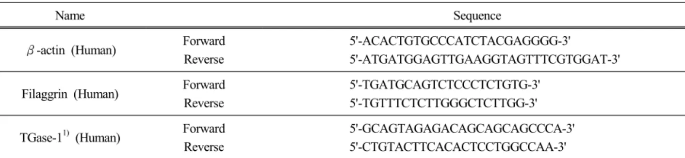
Table 1. PCR Primer Used in this Experiment
법을 이용하여 측정하였다. RBL-2H3 세포를 10%
FBS와 1% PEST와 0.5 µg/mL dinitrophenyl-Immuno- globulinE (DNP-IgE)(Sigma, USA)가 포함된 DMEM 배 지를 이용하여 24-well plate에 2 × 10⁵ cells/mL로 분주 한 뒤 37 ℃, 5% CO2 incubator에서 12 h 동안 배양하 였다. 상층액을 제거 후 Siraganian buffer (119 mM NaCl, 5 mM KCl, 5.6 mM glucose, 0.4 mM MgCl2, 25 mM PIPES, 1 mM CaCl2, 0.1% BSA, pH 7.2)로 2회 세 척한 다음 37 ℃, 5% CO2 incubator에서 10 min 간 반 응시킨 후 FBS가 제외된 DMEM 배지에 시료를 20 µg/mL로 처리하여 37 ℃, 5% incubator에서 30 min 동 안 반응시켰다. 이후 2 µg/mL DNP-HSA (Sigma, USA) 을 가하여 37 ℃, 5% incubator에서 2 h 반응시키고 ice bath에서 10 min 간 incubation하여 반응을 종결시켰다.
상층액 40 µL를 96-well plate에 옮기고 substrate buffer (4-p-nitrophenyl-N-acetyl-β-D-glucosaminide 2 mM, so- dium citrate, 0.05 M, pH 4.5) 40 µL를 넣고 37 ℃에서 1 h 동안 배양시킨 다음 각 well 당 stop solution 200 µL를 첨가하여 반응을 종결시키고 ELISA reader를 이 용하여 405 nm에서 흡광도를 측정하였다.
2.5. RT-PCR을 이용한 mRNA 발현 확인
장벽강화와 관련한 유전자의 mRNA 발현 양을 확 인하기 위한 실험으로 RT-PCR을 이용하였다. 각각의 세포를 10% FBS와 1% PEST가 포함된 DMEM 배지를 이용하여 24-well plate에 2 × 10⁵ cells/mL로 분주한 뒤 37 ℃, 5% CO2 incubator에서 24 h 동안 배양하였다.
상층액 제거 후 FBS가 제외된 DMEM 배지에 각 시료 를 처리하여 37 ℃, 5% CO2 배양기에서 24 h 배양 후 상층액을 제거하고 QIAzol lysis reagent (QIAGEN sci-
ences, USA)를 이용하여 매뉴얼에 따라 RNA를 추출 하였고, M-MLV reverse transcriptase (USB corporation, USA)를 이용하여 50 ℃에서 1 h, 95 ℃에서 5 min 동 안 반응시켜 cDNA를 합성하였다. 실험에 사용한 PCR primer는 Bioneer (Daejeon, Korea)에서 Table 1과 같이 디자인하여 사용하였다.
2.6. Western blot을 이용한 단백질 발현 확인
연구와 관련된 단백질들의 발현 양을 알아보기 위 하여 western blot 실험을 수행하였다. 각각의 세포를 10% FBS와 1% PEST가 포함된 DMEM 배지를 이용하 여 1 × 10⁶ cells/mL로 6-well plate에 분주한 뒤 24 h 동안 배양하여 상층액을 제거한 후에 추출물을 처리 하여 24 h 추가 배양한 세포를 이용하였다. 상층액이 제거된 plate에 RIPA Buffer (Theromo, USA)를 처리하 여 세포를 lysis 하고 원심분리(12,000 rpm, 10 min, 4
℃)하여 상층액의 단백질 농도를 측정한 후 동일양의 단백질(30 µg)로 희석하여 sample buffer (Theromo, USA)를 혼합하여 100 ℃에서 5 min 간 끓였다. 그 후 Mini PROTEANⓇ Tetra cell (552BR, Bio-Rad, USA)을 이용하여 SDS-PAGE 후 Mighty small transphor (Amersham Bio Science, USA)를 이용하여 단백질을 Transfer membranes (Millipore, USA)로 전이하였다. 비 특이적 단백질 결합부분은 0.1% Tween20 (Sigma, USA)과 5% 탈지분유를 포함한 Tris-Buffer saline (TBS)에 1 h 동안 반응하여 blocking하였다. 1차 anti- body (Filaggrin: Santa Cruz Biotechnology, USA)는 1 : 1,000 (v/v)으로 희석하여 상온에서 1 h 교반 후 TBST (TBS containing 0.1% Tween-20)로 5 min 동안 3회에 걸쳐 세척하였다. 그 다음 2차 antibody가 첨가된 용액
Figure 1. Effect of Eucommia ulmoides Oliver extracts on the cell viability in RBL-2H3 cells. (n = 3) ***p < 0.001 compared to control; one-way ANOVA, followed by Newman-Keuls Multiple Comparison test.
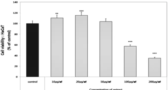
Figure 2. Effect of Eucommia ulmoides Oliver extracts on the cell viability in HaCaT cells. (n = 3) ***p < 0.001, **p
< 0.01 compared to control; one-way ANOVA, followed by Newman-Keuls Multiple Comparison test.
을 넣고 마찬가지로 상온에서 1 h 교반 후 TBST (TBS containing 0.1% Tween-20)로 5 min 동안 3회에 걸쳐 세척하고 SuperSignalⓇ West Pico Chemiluminescent Substrate (Thermo, USA)를 이용하여 발색 시키고 x-ray 필름(AGFA, Germany)에 감광시켜 밴드를 확인 하였다. 내부 표준 단백질로는 β-actin (Santa Cruz Biotechnology, USA)을 사용하였다.
2.7. Cornified envelope assay
각질 분화 유도능을 측정하기 위한 실험방법으로 Cornifide envelope assay를 수행하였다. HaCaT 세포를 24-well plate에 1 × 10⁵ cells/mL로 접종하고 37 ℃, 5%
CO2 incubator에서 24 h 동안 배양하였다. 배양 후 배 지를 모두 제거하고 혈청을 포함하지 않은 배지를 이 용하여 두충 추출물을 적정농도로 희석하여 well 당 처리하여 세포가 바닥 면적의 70 ~ 80% 정도 자랄 때 까지 약 5 days 간 배양하였다. 이 세포를 수확하여 PBS로 세척한 뒤 2% Sodium dodecyl Sulfate (SDS) (Sigma, USA)와 20 mM Dithiothreitol (DTT) (Sigma.
USA)를 포함한 10 mM Tris-HCl (pH 7.4)를 1 mL 넣고 3 min 동안 sonication을 수행한 후 10 min 간 100 ℃에 서 끓여 주었다. 일부를 취해 단백질 정량하여 평가 시 기준으로 잡는데 이용하고 나머지를 원심분리 하 여(1,200 rpm, 30 min, 4 ℃) 분리한 침전물은 다시 PBS에 현탁 시켜 340 nm에서 흡광도를 측정하였다.
2.8. Transglutaminase-1 assay
피부의 turn over 주기를 평가하기 위하여 trans-
glutaminase-1의 발현 양을 측정하였다. 먼저 96-well plate에 세포를 1 × 10⁵ cells/mL로 접종하고 37 ℃, 5%
CO2 incubator에서 24 h 동안 배양하였다. 배양 후 배 지를 모두 제거하고 혈청을 포함하지 않은 배지를 이 용하여 두충 추출물을 적정농도로 희석하여 well 당 처리하여 2 days 동안 배양한 후 배지를 제거하고 얼 렸다 녹이기를 3회 반복하였다. 그 후 에탄올과 아세 톤을 1 : 1 (v/v) 의 비율로 섞어 –20 ℃로 용액의 온도 를 낮춘 후 각각의 well에 100 µL씩 넣어 4 ℃에서 30 min 처리하고 제거한 후에 약 20 min 간 plate를 건조 시킨다. 건조된 플레이트에 1%의 BSA가 첨가된 PBS 를 100 µL 넣어 1 h 반응 후 제거하고, 여기에 TGase-1 antibody (Santa Cruz Biotechnology, USA)를 1 : 300 (v/v)의 비율로 희석하여 50 µL 씩 넣어 4 ℃ 냉장고에 서 12 h 동안 둔다. 이 후 PBST를 이용하여 3회 세척 하고 2차 antibody를 1 : 500 (v/v)의 비율로 희석하여 실온에서 1 h 반응 후 다시 3회 세척한다. 물기가 없도 록 잘 제거한 후에 SIGMAPASTTM OPD tablet (Sigma, USA)을 100 µL 넣고 ELISA reader를 이용하여 450 nm에서 흡광 값을 측정하였다.
3. 결과 및 고찰
3.1. MTT assay를 통한 세포생존율 측정
RBL-2H3 세포와 HaCaT 세포에 대한 두충 추출물 의 세포독성을 알아보기 위하여 MTT assay를 수행하 였다(Figure 1, Figure 2). 두충 추출물을 농도별로 처리 한 결과 RBL-2H3 세포에서는 20 µg/mL 이하의 농도
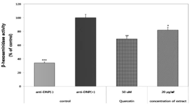
Figure 3. Effects of Eucommia ulmoides Oliver extracts on secretion of b-hexosaminidase. (n = 3) ***p < 0.001, **p <
0.01, *p < 0.1 compared to control; one-way ANOVA, followed by Newman-Keuls Multiple Comparison test.
Figure 4. Effects of Eucommia ulmoides Oliver extracts increased differentiation-related marker proteins in HaCaT cells. mRNA [A] and protein [B] levels of filaggrin. (1) Ca2 + 0.03 mM (2) Ca2 + 1.2 mM (3) Eucommia ulmoides Oliver 50 µg/mL.
에서 세포독성을 나타내지 않는 것으로 확인하였고, HaCaT cell의 경우 50 µg/mL의 농도에서 세포의 생존 율에 영향을 미치지 않음을 확인하였다. 따라서 본 연 구에서 세포에 처리하는 두충추출물의 농도를 RBL- 2H3 세포는 20 µg/mL, HaCaT 세포는 50 µg/mL으로 선정하여 실험을 수행하였다.
3.2. β-Hexosaminidase 분비 측정
항원과 항체가 비만세포에 반응하여 세포내에서 외 부로 분비되는 히스타민, 프로스타글란딘 등의 과립 의 양을 측정하는 것은 항 알레르기 효과를 확인하기 에 적합한 방법이다[16]. 따라서 본 연구에서는 세포 외로 분비된 β-Hexosaminidase 분비량을 측정하여 두 충 추출물의 항 알레르기 효능을 확인하였다(Figure 3). 추출물의 처리 농도는 세포 생존율 측정 결과에 따 라 세포에 영향을 미치지 않는 최고농도인 20 µg/mL 로 처리하였다. 그 결과 anti-DNP만 처리한 군을 기준 으로 양성대조군인 quercetin이 31%의 감소 효능을 보 인데 비해 두충 추출물은 18%가 감소하는 효능을 확
인하였다.
3.3. Filaggrin 발현 측정
포유동물 표피 과립층의 각질 초자질 과립에서 전 구단백질인 profilaggrin으로부터 합성되며, 보습인자 이자, TGase-1의 기질로 사용되는 단백질인 filag- grin[17-18]의 발현에 두충 추출물이 어떠한 영향을 미 치는지 확인하였다. RNA 발현 확인을 위해 수행한 RT-PCR 결과(Figure 4A) 두충 추출물을 처리하였을 경우 대조군인 0.03 mM Ca2+ 비하여 mRNA 발현이 증가되어 있는 것을 확인할 수 있었으며, 이와 마찬가 지의 결과로 단백질의 발현 또한 대조군에 비하여 증 가되어있음(Figure 4B)을 확인하였다.
3.4. Transglutaminase-1의 활성 및 발현 측정 Transglutaminase는 각질세포 형성에 관여되는 구조 단백질 사이를 연결하여 cornified cell envelop을 만드 는데 관여하는 효소로서 단백질 내의 글루타민과 리 신 사이를 연결하는 기능을 가지고 있는데, Transglu- taminase-1은 기저층에서부터 발현이 되기 시작하여 극세포층, 과립층으로 갈수록 점차 발현이 높아지는 효소이다[19]. 피부 장벽층인 cornified cell envelop를 만드는데 가장 중요한 효소인 TGase-1의 발현을 측정 한 결과 대조군에 비하여 31% 가량 발현이 증가되어 있다는 것을 알 수 있었으며(Figure 5A), RT-PCR 결과 (Figure 5B) 또한 단백질의 발현과 같은 경향의 결과를 나타내 두충추출물을 처리한 군에서 mRNA의 발현양 이 현저히 증가되어 있음을 확인할 수 있었다.
3.5. Cornified envelope assay 측정
각질형성세포는 분화 단계에서 특이한 단백질을 발 현시켜 표피의 기저층부터 각질층까지 분화가 이루어
Figure 5. Effects of Eucommia ulmoides Oliver extracts on [A] TGase-1 activity, [B] expression of TGase-1 mRNA in HaCaT cells. (1) Ca2 + 0.03 mM (2) Ca2 + 1.2 mM (3) Eucommia ulmoides Oliver 50 µg/mL (n = 4) ***p < 0.001, **p < 0.01 compared to control; one-way ANOVA, followed by Newman-Keuls Multiple Comparison test.
Figure 6. Effect of Eucommia ulmoides Oliver extracts on the proliferation and differentiation of HaCaT cells. (n = 3)
***p < 0.001, compared to control; one-way ANOVA, followed by Newman-Keuls Multiple Comparison test.
지도록 프로그램화 되어 있는데[20-21] 말기 분화과정 에서 출현하는 각화세포피(cornified cell envelope, CE) 는 filaggrin과 같은 구조 단백질을 전구물질로 하여 TGase-1의 매개에 의해 교차 결합되어 단단한 구조를 형성하게 된 것이다[22]. CE의 측정은 피부장벽의 보 호를 위한 최종단계인 각질형성세포의 분화를 판단할 수 있는 좋은 기준이 된다. 두충 추출물을 처리하여 CE를 측정한 결과(Figure 6) 대조군을 기준으로 보았 을 때 양성대조군보다 더 높은 효능(360%)을 나타내 어, 두충 추출물이 HaCaT 세포의 분화를 촉진하여 각 화세포피를 형성하는데 뛰어난 효능을 나타내는 것을 알 수 있었다.
4. 결 론
본 연구에서는 두충 추출물이 RBL-2H3 세포의 β -hexosaminidase의 분비 억제와 HaCaT keratinocytes 피 부장벽의 기능을 강화시키는 bio marker인 filaggrin, TGase-1, CE의 발현에 미치는 영향에 관하여 연구하 였다. 먼저 비만세포에서 알러지의 시초가 되는 물질 인 β-hexosaminidase의 방출을 확인한 결과, anti-DNP 만 처리한 군을 기준으로 두충 추출물은 13%가 감소 하는 효능을 확인하였다. 피부장벽기능의 회복과 관 련된 모든 bio marker들은 mRNA, protein, activity 모두 에서 뛰어난 효능을 확인하였는데, Filaggrin의 경우 mRNA와 protein 모두에서 대조군보다 높은 발현을 나 타내었으며, TGase-1은 activity와 mRNA 발현율 모두 높인다는 것을 확인하였다. 각질형성세포의 분화를 판단할 수 있는 CE 측정에서는 두충추출물이 양성대 조군보다 더 좋은 효능을 나타내기도 하였다. 따라서 두충 추출물은 β-hexosaminidase의 분비 억제에 효과 가 있고, 각질형성세포의 분화에 영향을 주어 분화를 촉진시키는 효과가 있음을 확인하였다.
Reference
1. C. H. Lee, Sensitive skin, The korean society for
skin barrier research, 11(2), 55 (2009).
2. P. M. Elias, L. C. Wood, and K. R. Feingold, Epidermal pathogenesis of inflammatory dermatoses, Am. J. Contact Dermatol., 10(3), 119 (1999).
3. J. P. Allam, T. Bieber, and N. Novak, Recent high-lights in the athophysiology of atopic eczema, Int. Arch. Allergy. Immunol., 136(2), 191 (2005).
4. H. Zhang, Immunopharmacological effect of Cordyceps sinensis, Zhong Xi Yi Jie He Za Zhi, 10(9), 570 (1990).
5. H. J. Cho, J. Y. Lee, and D. G. Kim, In vitro and in vivo Anti-inflammatory and Anti-allergic re- sponses caused by water extract of Mori Cortex, J.
Korean Oriental Pediatrics, 19(2), 175 (2005).
6. P. D. Moon, J. J. Na, H. J. Jeong, S. H. Hong, H.
R. Kim, J. O. Choi, S. H. Lee, J. Y. Shin, and H.
M. Kim. Inhibitory effect of Gamibojungikqitang ex- tract on mast cell-mediated allergicreaction in murine Model, J. Pharm. Sci., 8(1), 94 (2005).
7. M. Ennis, F. L. Pearce, and P. M. Weston, Some studies on the release of histamine from mast cells stimulated with polylysine, Br. J. Pharmacol., 70(2), 329 (1980).
8. G. C. Yen and C. L. Hsieh, Reactive oxygen species scavenging activity of Du-zhong (Eucommia ul- moides oliv.) and its active compounds, J. Agric.
Food Chem., 48(8), 3431 (2000).
9. C. L. Hsieh and G. C. Yen, Antioxidant actions of du-zhong (Eucommia ulmoides oliv.) toward oxida- tive damage in biomolecules, Life Sci., 66(15), 1387 (2000).
10. Y. F. Sasaki, A. Chiba, M. Murakami, K. Sekihashi, M. Tanaka, M. Takahoko, S. Moribayashi, C.
Kudou, Y. Hara, Y. Nakazawa, T. Nakamura, and S.
Onizuka, Antimutagenicity of Tochu tea (an aqueous extract of Eucommia ulmoides leaves): 2. Suppress- ing effect of Tochu tea on the urine mutagenicity af- ter ingestion of raw fish and cooked beef, Mutat.
Res., 371(3-4), 203 (1996).
11. T. Nakamura, Y. Nakazawa, S. Onizuka, A. Chiba, K. Sekihashi, A. Miura, N. Tasugahira, and Y. F.
Sasaki, Antimutagenicity of Tochu tea (an aqueous extract of Eucommia ulmoides leaves) 1. The clas- togen-suppressing effects of Tochu tea in CHO cell and mice, Mutat. Res., 388(1), 7 (1997).
12. J. Watanabe, J. Kawabata, H. Kurihara, and R. Niki, Isolation and identification of alpha-glucosidase in- hibitors from Tochu tea (Eucommia ulmoides), Biosci. Biotechnol. Biochem., 61(1), 177 (1997).
13. Y. Li, T. Sato, K. Metonri, K. Koike, Q. M. Che, and S. Takahashi, The promoting effects of geniposi- dic acid and aucubin in Eucommia ulmoides oliver leaves on collagen synthesis, Biol. Pharm. Bull.,
21(12), 1306 (1998).
14. S. P. Choi, M. Y. Kang, and S. H. Nam, Inhibitory activity of pigmented rice bran extract to the allergic inflammation in basophilic cell line and peritoneal mast cells, J. Korean Soc. Appl. Biol. Chem., 48(4), 315 (2005).
15. O. B. Choi, Anti-allergic Effects of Petasites japoni- cum, Korean J. Food & Nutr., 15(4), 382 (2002).
16. P. Yamada, M. Zarrouk, K. Kawasaki, and H. Isoda, Inhibitory effect of various Tunisian olive oils on chemical mediator release and cytokine production by basophilic cells, J. Ethnopharmacol., 116(2), 279 (2008).
17. B. A. Dale, Purification and characterization of a ba- sic protein from the stratum cormeum of mammalian epidermis, Biochem. Biophy. Acta., 491(1), 193 (1977).
18. R. L. Meek, J. D. Lonscale-Eccles, and B. A. Dale, Epidermal filaggrin is synthesized on a large mes- senger ribonucleic acid as a high-molecular-weight preecursor, Biochemistry, 22(21), 4867 (1983).
19. J. M. Yang, Mutations of transglutaminase 1 gene in lamellar ichthyisis, The JSBR., 2(1), 22 (2000).
20. A. C. Steven and P. M. Steinert, The protein compo- sition of cornified cell envelopes, J. Cell Sci.,
107(2), 693 (1994).
21. P. M. Steinert and L. N. Marekov, The proteins ela- fin, rilaggirn. keratin, intermediate rilaments, loricrin, and small prolinerich proteins 1 and 2 are isodipep-
tide cross-linked components of the human epikermal cornified cell envelope, J. Biol. Chem., 270(30), 17702 (1995).
22. R. Schmidt, U. Reichert, and M. Bouclier, Plasma
membrane transglutaminase and cornified envelope competence in vultured human keratinocytes, REBS Lett., 186(2), 201 (1985).
![Figure 5. Effects of Eucommia ulmoides Oliver extracts on [A] TGase-1 activity, [B] expression of TGase-1 mRNA in HaCaT cells](https://thumb-ap.123doks.com/thumbv2/123dokinfo/4729294.511452/6.892.165.730.131.406/figure-effects-eucommia-ulmoides-oliver-extracts-activity-expression.webp)
