Withaferin A의 다양한 항암 효과 및 분자생화학적 기전
Anti-cancer Effects and Molecular Mechanisms of Withaferin A
저자 (Authors)
우선민, 민경진, 권택규
Seon Min Woo, Kyoung-jin Min, Taeg Kyu Kwon
출처 (Source)
생명과학회지23(3), 2013.3, 462-469 (8 pages)
Journal of Life Science23(3), 2013.3, 462-469 (8 pages)
발행처 (Publisher)
한국생명과학회
Korean Society Of Life Science
URL
http://www.dbpia.co.kr/Article/NODE02127662APA Style 우선민, 민경진, 권택규 (2013). Withaferin A의 다양한 항암 효과 및 분자생화학적 기전. 생명과학 회지, 23(3), 462-469.
이용정보 (Accessed)
저작권 안내
DBpia에서 제공되는 모든 저작물의 저작권은 원저작자에게 있으며, 누리미디어는 각 저작물의 내용을 보증하거나 책임을 지지 않습니다.
이 자료를 원저작자와의 협의 없이 무단게재 할 경우, 저작권법 및 관련법령에 따라 민, 형사상의 책임을 질 수 있습니다.
Copyright Information
The copyright of all works provided by DBpia belongs to the original author(s). Nurimedia is not responsible for contents of each work. Nor does it guarantee the contents.
You might take civil and criminal liabilities according to copyright and other relevant laws if you publish the contents without consultation with the original author(s).
계명대학교 114.71.5.212
2016/03/25 15:18 (KST)
Anti-cancer Effects and Molecular Mechanisms of Withaferin A
Seon Min Woo
†, Kyoung-jin Min
†and Taeg Kyu Kwon*
Department of Immunology, School of Medicine, Keimyung University, Daegu 704-701, Korea
Received March 1, 2013 /Revised March 20, 2013 /Accepted March 20, 2013Withaferin A is a steroidal lactone purified from the Indian medicinal plant Withania somnifera. It ex- hibits a wide variety of activities, including anti-tumor, anti-inflammation, and immunomodulation properties. In this review, we focused on the anti-cancer effects of withaferin A. Withaferin A inhibits cell proliferation, metastasis, invasion, and angiogenesis in cancer cells. Furthermore, it sensitized irra- diation, tumor necrosis factor-related apoptosis-inducing ligand (TRAIL)-, and doxorubicin-mediated apoptosis. The results showed that multiple mechanisms were involved in withaferin A-mediated an- ti-cancer effects. First, withaferin A increased intracellular reactive oxygen species (ROS) production and induced ER stress- and mitochondria-mediated apoptosis. Second, withaferin A inhibited the sig- naling pathways (Jak/STAT, Akt, Notch, and c-Met), which are important in cell survival, pro- liferation, and metastasis. Third, it induced apoptosis and inhibited cancer cell migration through the up-regulation of prostate apoptosis protein-4 (Par-4). Finally, withaferin A up-regulated pro-apoptotic protein expression levels through the inhibition of proteasome activity. Our findings suggested that withaferin A is a potential, potent therapeutic agent.
Key words : Anti-cancer effect, apoptosis, sensitization, signaling pathway, withaferin A
†These authors contributed equally to this work
*Corresponding author
*Tel:+82-53-580-3882, Fax:+82-53-580-3795
*E-mail : [email protected]
This is an Open-Access article distributed under the terms of the Creative Commons Attribution Non-Commercial License (http://creativecommons.org/licenses/by-nc/3.0) which permits unrestricted non-commercial use, distribution, and reproduction in any medium, provided the original work is properly cited.
Journal of Life Science 2013 Vol. 23. No. 3. 462~469 DOI : http://dx.doi.org/10.5352/JLS.2013.23.3.462
서 론
Withania somnifera는 인도에서 전통적으로 사용되던 약재 로 다양한 류머티즘과 관절염에 사용되고, 건강과 장수를 위 해 노인과 임산부들에게 사용되었다. 이 후, 많은 연구자들이 Withania somnifera의 약물 효과를 다양한 분야에서 연구하여 염증 억제기전[23, 39]과 항스트레스 작용[2], 그리고 항산화제 로서의 역할 등이 있음을 확인하였다[4, 5]. 본 연구는 Withania somnifera에서 추출한 steroidal lactone인 withaferin A의 다양 한 항암제로써의 역할과 기존의 항암제들과의 병합처리에 의 한 암세포 사멸 증진효과, 그리고 이에 관여하는 세포 내 신호 전달 타깃에 대하여 중점적으로 논하고자 한다.
Withaferin A의 구조적 특징
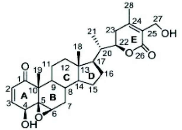
Withaferin A는 28개의 탄소로 이루어진 스테로이드 락톤 계(steroidal lactones)로, 스테로이드에 기본적으로 존재하는4 개의 사이클로 알칸 고리(cycloalkane ring) 구조로 이루어져, 3개의 사이클로헥산 고리(cyclohexane ring)와 1개의 사이클
로펜테인 고리(cyclopentane ring)로 이루어져 있다 (Fig. 1) [58]. Withaferin A는 타깃 단백질에 존재하는 시스테인(cys- teine) 잔기의 sulfhydryl과 같은 친핵성군(nucleophilic group)과 알킬화(alkylation) 작용을 하는 3가지 위치가 있다.
A ring의 C3, E ring의 epoxide 구조와 C24가 여기에 속한다.
이들 사이트는 타깃 단백질의 친핵성군과 작용하여 Michael addition thioalkylation 반응을 통하여 공유 결합하게 된다.
A 고리에 있는 C2와 C3의 이중결합은 withaferin A에 의해 나타나는 효과 중 세포사멸을 일으키는데 중요한 구조로 알려 져 있어, 이중결합을 없앤 withaferin A의 경우 세포에 대한 독성이 줄었다[12]. A 고리에 있는 ketone carbon은 with- aferin A의 기능 중 하나인 proteasome 억제효과에 중요하고
Fig. 1. Structure of withaferin A. Withaferin A (5,6-epoxy- 4,27-dihydroxy-1-oxowitha-2,24-dienolide), Molecular formula: C28H38O6), Molecular weight: 470.61.
- Review -
Journal of Life Science 2013, Vol. 23. No. 3 463
[55], A 고리의 C2와 C3의 이중 결합과 C5와 C6의 에폭시드 (epoxide) 그룹은 withaferin A 와 vimentin의 결합에도 중요 하게 관여한다[3]. 또한, B 고리에 있는 에폭시드 그룹 역시 withaferin A의 기능에 매우 중요한 역할을 하는데, mercap- toethanol을 withaferin A와 반응시키게 되면 B ring에 있는 에폭시드 그룹에 영향을 주어 암세포사멸 효과가 사라진다 [30]. 이와 동일하게 작용으로 강력한 환원제인 DTT와 N-ace- tyl cysteine (NAC)을 withaferin A와 함께 처리하면 with- aferin A에 의해 나타나던 항염증작용과 암세포사멸 작용이 나타나지 않는다[17, 41, 49].
Withaferin A의 항암작용
Withaferin A의 암세포의 세포성장 억제 및 세포사 멸 증진효과
Withaferin A의 항암작용 중 가장 많이 연구된 내용은 withaferin A에 의한 세포사멸에 대한 연구이다. Withaferin A는 대부분의 암세포들에서 apoptotic 세포사멸을 유도하는 것으로 알려져 있는데, 이러한 암세포로는 백혈병[35, 49], 전 립선 암[45], 폐암[8], 신장암[7, 52], 유방암[15, 46, 60], 자궁경 부암[32], 난소암 [61], 흑색종암[40] 그리고 췌장암[59] 들이 알려져 있다. 또한, withaferin A는 세포사멸 이외에 7,12-dimethylbenz[a]anthracene (DMBA)에 의한 oral carci- nogenesis를 유의적으로 억제한다. DMBA에 의한 종양의 생 성, 종양의 크기를 withaferin A 처리 군과 처리하지 않은 대조 군에서 비교하였을 때 withaferin A 처리 군에서 현저히 낮은 것을 확인하였다[26, 27]. 또한, withaferin A는 세포의 분열을 억제하기도 하는데 유방암에서 세포 주기 단백질인 cyclin-dependent kinase 1 (Cdk1), cell division cycle 25C (Cdc25C) 그리고 Cdc25B 의 발현을 억제하여 G2/M arrest를 유도함으로써 세포분열 억제효과를 나타내기도 한다[47].
Withaferin A의 암세포 전이(metastasis)와 침투 (invasion) 억제 효과
암세포의 경우 암이 처음으로 형성된 일차적인 장기 및 부 위에서 다른 이차적인 장기 및 부위로 이동하여 종양을 형성 하게 되는데, 이러한 것을 전이(metastasis)라고 한다. 암환자 의 치료에 있어서 전이에 의한 이차적인 종양을 형성하게 되 면 안 좋은 예후를 나타내게 된다. 따라서, 암세포의 전이를 억제하는 것이 종양의 치료에 중요하게 관여하게 된다.
Withaferin A는 이러한 암세포의 전이를 억제하는 효과가 있 다. 유방암에서 세포가 죽지 않는 저농도 withaferin A 는 세포 의 이동에 중요하게 관여하는 vimentin의 serine 56의 인산화 를 통한 분해를 유도하여 세포의 전이 및 침투를 억제하는 효과가 있다[51]. 또한 본 연구실에서 실험한 결과에 의하면 자궁 경부암세포인 Caski 세포에서 세포외 기질(extracellular matrix) 분해를 통해 세포의 전이에 중요하게 관여하는 PMA 유도된 matrix metalloproteinase (MMP)-9의 활성과 세포의
이동을 withaferin A 가 유의적으로 억제하는 것을 확인하였 다 (in press).
Withaferin A의 혈관생성(angiogenesis) 억제 효과 암세포가 계속적으로 분열하여 종양을 형성하는 과정에서 필요한 영양분을 공급받기 위하여 새로운 혈관생성은 필수적 이다. 혈관생성이 되지 않으면 산소와 영양분 공급이 제대로 이루어 지지 않아 암세포는 사멸하게 된다. 따라서, 이러한 혈관생성을 억제하는 것 또한 암세포사멸을 증진시키는 타깃 이 될 수 있다. Human umbilical vein endothelial cell (HUVEC)에 withaferin A를 처리하여 혈관생성효과를 확인한 결과 HUVEC 세포의 분열을 저해함으로써 새로운 혈관생성 을 억제하였다. 이러한 효과는 앞에서 언급하였던 암세포의 세포분열억제효과를 보이는 withaferin A 의 농도보다 현저하 게 낮은 농도에서 효과를 나타내었다[31].
Sensitizer로서의 withaferin A
Withaferin A의 방사선 (irradiation)에 의한 세포사 멸 증진 효과
방사선 요법은 가장 많이 사용되고 있는 항암요법이지만, 정상 조직에 상처를 입히고 장시간 치료 시 저항성이 생기며 염증을 유발하는 부작용이 나타나기 때문에 매우 제한적이다.
또한, 방사선치료만으로는 종양을 제거하는 것에 한계가 있어 최근에는 이러한 방사선요법의 효율성 및 효과를 증진시킬 수 있는 전략을 수립하는 연구가 많이 이루어지고 있다.
Withaferin A 역시 방사선 요법에 의한 항암효과를 증가시킬 수 있는 약물 중 하나로, 흑색종암 세포[18]와 에를리히 복수 암 세포[9, 44]에서 방사선에 의한 세포사멸증진효과가 알려져 있다. 하지만, withaferin A에 의한 방사선요법에 민감화 증가 기전은 잘 알려져 있지 않다. 본 연구실에서 최근 수행한 연구 에 의하면, 백혈병세포에서 withaferin A는 활성산소의 생성 증가와 JNK와 p38 MAPK의 인산화 증가, 그리고 항 세포 사 멸단백질인 Bcl-2의 발현을 억제를 통하여 세포사멸을 증가시 킨다[53].
Withaferin A의 TNF-related apoptosis-inducing ligand (TRAIL) 유도 세포사멸의 증진효과
TRAIL은 정상세포의 조직 및 세포에는 아무런 영향을 주지
않으면서 암세포에서만 특이적으로 세포사멸을 유도하는 획
기적인 항암제로 알려져 있다. 이는 암세포에는 TRAIL의 수
용체로 알려진 death receptor (DR)의 발현이 높지만, 정상세
포에서는 상대적으로 decoy receptor의 발현이 높아 정상세포
의 세포사멸을 유도하지 않는다[36]. 하지만, 많은 암세포에서
TRAIL 에 대한 저항성이 나타나고, TRAIL을 계속 처리하게
되면 저항성을 가지는 세포가 만들어지면서 TRAIL 단독처리
에 의한 세포사멸 효과는 미미한 경우가 나타난다. 이러한 문
제점을 해결하고자 세포에 독성을 유발하지 않는 저농도의
TRAIL과 다른 약물과의 병합 처리하여 TRAIL에 대한 민감성 을 증가시키는 연구가 진행되고 있다. Withaferin A는 TRAIL 과의 병합처리를 통하여 유의적으로 세포사멸을 증가시키는 약물 중 하나이다. 본 연구실에서 수행한 연구결과에 의하면 약물 단독에 의한 세포사멸유도효과를 보이지 않는 저농도의 TRAIL과 withaferin A를 처리하면 신장암세포의 세포사멸을 유도하는 것을 확인하였다[22]. Withaferin A에 의한 TRAIL 민감화 기전은 활성산소를 만들어 ER stress 관련 단백질인 C/EBP homologous protein (CHOP) 단백질 발현을 증가시키 고, 이를 통한 DR5의 증가가 관여한다. 또한 NF-κB의 활성을 억제함으로써 세포사멸 억제 단백질인 cellular FADD-like IL-1β-converting enzyme inhibitory protein (cFLIP) 발현 감소 도 withaferin A에 의한 TRAIL 민감화 증가에 관여한다[22].
Withaferin A의 doxorubicin 유도 세포사멸의 증진 효과
Doxorubicin은 Streptomyces peucetius에서 추출한 an- thracylin계 항암제로서, 그 작용기전은 topoisomerase II의 기 능을 억제하여 DNA 복제를 위해 DNA chain이 끊긴 후 다시 연결되는 것을 막음으로써 복제과정을 억제한다. 이러한 dox- orubicin은 FDA로부터 승인 받은 항암제로서 다양한 암세포 사멸을 유도한다. 하지만, doxorubicin은 농도 의존적으로 구 토, 신경장애, 심근 독성, 탈모, 그리고 구내염과 같은 부작용 을 초래한다. 따라서, 저농도의 doxorubicin을 이용한 항암전 략수립이 필요하여 다른 약물과의 병합처리를 통한 민감화 증가 연구가 진행되고 있다. Withaferin A는 저농도의 doxor- ubicin과 함께 병합 처리하면 세포사멸을 증가시킨다. 난소암 세포에서 doxorubicin을 withaferin A와 함께 병합 처리하면 활성산소가 증가하고 이를 통한 autophagy가 증가하여 세포 사멸을 유도한다[11]. 따라서, doxorubicin에 의한 약물 부작 용을 줄이기 위하여 저농도의 doxorubicin과 withaferin A과 의 병합처리가 효과적인 항암전략이 될 수 있다.
Withaferin A에 항암효과에 관여하는 신호전달체계 Withaferin A에 의한 활성산소 증가
활성산소는 항암제들에 의한 암세포 사멸증가 기전으로 잘 알려진 신호전달 물질 중 하나이다. Withaferin A는 백혈 병세포[24, 54], 신장암 세포[22], 흑색종 세포[28], 난소암 세 포[11]에서 활성산소를 증가시킴으로써 세포사멸을 유도하 는 것으로 알려져 있다. 하지만, withaferin A에 의한 암세포 사멸에 활성산소가 중요하게 관여하지만 활성산소를 만들어 내는 기전 및 관여하는 활성산소 형성 단백질에 대한 연구는 전무하다.
Withaferin A에 의한 미토콘드리아 세포사멸 관련 단백질 조절
미토콘드리아에서 세포사멸을 조절하는 단백질로는 Bcl-2
family가 잘 알려져 있다. Bcl-2 family는 크게 두 분류로 나누 어 세포사멸을 증가시키는 Bcl-2 family와 세포사멸을 억제하 는 Bcl-2 family가 있다. 먼저, 세포사멸을 억제하는 단백질로 는 Bcl-2, Bcl-xL, Bcl-w, 그리고 Mcl-1 등이 속한다. 세포사멸을 증가시키는 Bcl-2 family로는 Bax, Bak, 그리고 Bok/Mtd가 있 다. 이들은 미토콘드리아에 monomer로 존재하다가 세포사멸 유도 신호가 오게 되면 미토콘드리아에 pore를 형성함으로써 내부에 존재하는 cytochome C, Apaf-1, Smac/Diablo, 그리고 AIF 등을 유출한다. BH3 domain만을 갖고 있는 BH3 only protein으로 Bim, Noxa, Bad, Puma, 그리고 Bid 등이 속한다.
이들은 세포사멸 억제 단백질인 Bcl-2와 Bcl-xL 등과 결합하여 기능을 억제함으로써 세포사멸을 일으키게 한다. Withaferin A는 이러한 미토콘드리아에 존재하는 Bcl-2 family들을 조절 하여 암세포의 세포사멸을 조절한다. 유방암 세포에서 with- aferin A는 BH3 domain만 유일하게 함유한 단백질인 Bim의 발현을 증가시키고[46], 백혈병 세포에서는 Bax의 발현을 증 가시킴으로써 세포사멸을 유도한다[25]. Withaferin A에 의한 Bcl-2 단백질의 발현 감소를 통한 세포사멸은 신장암 세포[35, 54]와 흑색종[28]에서도 관찰된다.
Withaferin A에 의한 ER stress 유도
세포 소기관중 하나인 endoplasmic reticulum (ER)은 단백
질의 합성을 담당하는 소기관으로 단백질이 합성된 후 폴딩
(folding)과 조립(assembly), 당화(glycation) 및 이황화결합
(disulfide bond) 등을 통한 단백질의 구조를 결정한다. 또한
ER 중요한 기능은 세포 내 칼슘저장고로써 작용하여 칼슘농
도를 일정수준으로 유지하는데 중요하게 관여한다. 하지만,
구조적으로 망가진 단백질이 과다하게 만들어지거나, 칼슘이
고갈되면 ER 기능에 문제가 생겨 ER stress를 유도하고
unfoled protein response (UPR)을 일으킨다[50]. ER stress에
의한 UPR 반응은 주로 소포체 막에 존재하는 pancreatic ER
kinase (PKR)-like ER kinase (PERK), activating transcription
factor-6 (ATF-6)와 inositol-requiring enzyme 1 (IRE1)와 같은
ER stress 인식 단백질에 의해 조절된다. ER stress가 오게 되
면 PERK에 의한 eIF2a의 인산화가 일어나 세포 내 전반적인
단백질 합성을 억제하게 되고, ATF-6는 ER에서 골지로 이동
한 후, 단백질 가수분해를 통해 활성화되어 핵으로 이동함으
로써 단백질 폴딩에 중요한 ER chaperone과 X box-binding
protein-1 (XBP-1)의 발현을 조절한다[42]. IRE1은 RNAase 기
능이 있어 XBP-1을 mRNA 단계에서 절단함으로써 spliced
XBP-1을 형성하고, 이는 핵으로 이동하여 chaperone의 전사
과정을 조절한다[57]. 하지만, 이러한 ER stress가 과도하거나
오랫동안 지속될 경우 ER의 UPR이 충분하게 작용하지 못하
여 세포사멸을 일으킨다. 암세포에서는 이러한 ER stress에
의한 세포사멸이 유도되는 경우가 많다. Withaferin A의 경우
도 신장암 세포에서 eIF2a의 인산화를 증가시키고, spliced
Journal of Life Science 2013, Vol. 23. No. 3 465
XBP-1을 형성하며, ER stress에 의한 세포사멸에 중요하게 관 여하는 것으로 알려진 CCAAT-enhancer-binding proteins homologous protein (CHOP) 단백질 발현의 증가를 유도하였 다[7]. 이때 CHOP의 발현을 siRNA를 이용하여 knock down 시키면 withaferin A에 의한 세포사멸이 억제됨을 확인함으로 써 withaferin A에 의한 세포사멸에 ER stress가 중요하게 관 여함을 알 수 있다[7]. 또한, 최근 보고된 연구에 의하면, withaferin A에 의해 증가한 CHOP은 항 세포사멸 단백질인 cFLIP의 발현을 억제함으로써 세포사멸을 증가시킨다고 알려 졌다. 이 연구결과에 의하면, withaferin A에 의해 증가하는 CHOP은 농도의존적으로 cFLIP의 단백질 발현을 억제하고, 반면에 mRNA 발현에는 아무런 영향을 주지 않았다.
Withaferin A는 CHOP발현 증가를 통한 ubiquitin/
proteasome 을 활성화 시켜 cFLIP의 분해를 증가시킴으로써 세포사멸을 유도함을 알 수 있었다[34].
Withaferin A에 의한 STAT3와 Akt인산화 억제 Signal transducer and activator of transcription (STAT)3는 암세포에서 인산화된 형태로 존재하여 세포사멸억제 단백질 인 Bcl-2, Bcl-xL, survivin의 발현을 증가시켜 세포사멸을 억제 하고 cyclin D1을 통해 세포 주기를 조절함으로써 세포의 증식 을 증가시킨다[48]. 따라서, 이러한 STAT3의 인산화 억제가 암세포의 세포사멸을 증가시키고 증식을 억제시키는 타깃 신 호전달계가 될 수 있다. Withaferin A는 유방암세포와 신장암 세포에서 인산화 되어있는 STAT3를 탈인산화 시킴으로써 세 포사멸을 유도한다[20, 52]. 신장암 세포에서 withaferin A는 STAT3의 상위단계신호전달체계인 Janus-activated kinase 2 (JAK2)의 인산화를 억제함으로써 STAT3의 인산화를 억제하 고, 이를 통한 Bcl-xL, Bcl-2, cyclin D1과 survivin의 발현을 감소시킨다. STAT3를 과발현한 경우 withaferin A 에 의한 세포사멸이 억제되는 것을 확인함으로써 withaferin A에 의한 세포사멸에 있어서 STAT3신호전달 체계의 중요성을 규명하 였다[52].
암세포에서 STAT3이외에 세포의 생존에 중요한 신호전달 체계는 phosphoinositide 3-kinase (PI3K)/Akt 인산화이다.
PI3K/Akt 신호전달 경로는 세포 성장과 생존, 사멸, 분화, 대 사 등 다양한 생명현상에 결정적인 역할을 하며, 또한 다양한 암세포에서 활성화 되어 있다. Withaferin A 는 실제로 Akt 의 탈인산화를 유도함으로써 백혈병 세포[35], 교모세포종 [14], 신장암 세포[54] 등에서 세포사멸을 유도한다.
Withaferin A에 의한 Notch 신호전달계 활성화 조절 Notch 신호전달 체계는 정상 조직과 세포의 발달 단계에서 세포의 분화와 생존, 분열 등을 조절하고 세포사멸과 생존을 결정짓는 중요한 신호전달 체계로서 작용한다. Notch는 수용 체가 Notch 1, 2, 3, 4로 네 가지가 존재하며, 세포막에 위치하 고 있다가, ligand가 결합하게 되면 수용체가 metalloprotease
인 ADAM10/17 (a disintegrin and metalloprotease 10/17) / TACE (TNF-α converting enzyme)와 γ-secretase 복합체가 작용하여 잘라줌으로써 Notch의 intracellular domain (NCID)를 세포막으로부터 핵으로 이동하여 전사를 조절하는 전사인자로 작용하게 된다. 최근 Notch 신호전달에 이상이 생기면 종양을 형성하는 pro-oncogenic 기능이 자궁경부암, 췌장암, 난소암, 유방암, 그리고 전립선암에서 규명되었다[29].
Notch 신호 전달체계를 억제하는 것이 새로운 종양 치료전략 방법으로 생각되어지고 있지만, 특이적으로 마우스 피부에서 수행한 연구에 의하면 Notch 신호전달 체계를 결핍시키면 상 피와 각막에서의 세포 과분열을 유도함으로써 피부암을 유발 한다고 알려져 있다[33]. 따라서, 암세포에서의 Notch 신호전 달 체계는 종양세포와 유래 기관, Notch 수용체에 따라 다르 게 작용할 수 있다. Withaferin A의 경우에는 Notch 신호전달 체계를 조절하여 세포사멸을 유도한다고 알려져 있다. 예를 들면, 결장암 세포에서 withaferin A는 Notch-1의 발현을 억제 함으로써 아래 단계 신호전달체계인 Akt 와 mTOR의 활성화 를 억제하고, JNK 활성을 통한 세포사멸을 유도한다[19]. 난소 암 세포에서도 withaferin A는 Notch-1과 Notch-3의 발현을 억제함으로써 세포사멸을 유도한다[61]. 반면에, 유방암세포 에서는 Notch-2 와 Notch-4를 활성화 시켜, 세포의 이동을 withaferin A가 억제시킨다고 알려져 있다[21]. 이와 같이 withaferin A 에 의한 Notch 신호전달체계의 조절을 통한 암 세포에 미치는 영향은 세포와 Notch 수용체 의존적으로 나타 난다.
Withaferin A에 의한 c-Met의 신호전달 억제 c-Met는 hepatocyte growth factor 수용체로서 pro- to-oncogene으로 알려져 있다. c-Met는 tyrosine kinase 기능 을 가지고 있어 종양세포의 생존에 중요한 신호전달체계인 RAS, PI3K/Akt, 그리고 STAT3의 인산화를 증가시켜, 종양의 형성과 분열, 혈관생성, 전이를 증가시키는 역할을 한다[6, 13, 37]. 최근 연구에 의하면 항암제에 저항성을 가지게 되는 암세 포에서 c-Met의 활성화가 높게 유지되는 것으로 알려져 c-Met 의 신호전달 억제는 항암제에 대한 저항성을 낮추고 암세포의 세포사멸을 증가시키는 타깃이 될 수 있다[16]. 흑색종에서 withaferin A는 세포의 분열을 억제하고, 세포사멸을 유도하 는데 이러한 작용은 c-Met과 아래 단계의 신호전달 체계인 Akt 의 인산화를 억제함으로써 나타난다[40]. 또한, 교모세포 종에서도 고농도의 withaferin A 는 c-Met의 발현과 인산화를 억제한다[14]
Withaferin A에 의한 prostate apoptosis protein-4 (Par-4)의 발현 증가
Prostate apoptosis protein-4 (Par-4)는 전립선 암에서 세포
내 칼슘증가로 인한 세포사멸에 관여하는 단백질로 처음 발현
되어 세포사멸 유도 단백질로 알려져 있다[43]. 이후 연구에
Fig. 2. Anti-cancer effects and molecular mechanisms of withaferin A. Withaferin A has anti-cancer effects through modulation of several cellular responses.
의하면, Par-4의 발현증가는 TNF, TRAIL, doxorubicin, etopo- side, UV irradiation등과 같은 세포사멸 유도 약물 및 조건에 서의 암세포사멸에 필수적으로 알려져 있다[1, 10]. Withaferin A 는 전립선 암에서 Par-4의 발현을 증가시키고, 이를 통하여 NF-κB의 활성 억제 및 Bcl-2의 발현을 감소시켜 세포사멸을 유도한다[45]. 또한 자궁경부암 세포와 전립선암세포에서는 withaferin A가 세포의 침투 및 전이를 억제하고 혈관생성 억 제효과를 나타내는데 이러한 효과는 withaferin A에 의한 Par-4의 세포 밖으로의 유출이 MMP-2의 활성을 억제함으로 써 나타났다[38].
Withaferin A에 의한 proteasome 활성 억제 Withaferin A는 전립선암 xenograft 모델에서 20S protea- some과 26S proteasome의 chymotrypsin-like activity를 억제 하여, ubiquitination된 단백질의 축적을 증가시키고, 또한 proteasome 타깃 단백질로 세포사멸을 증가시키는 것으로 알 려진 Bax, p27, IκB-alpha의 발현을 증가시킴으로써 종양세포 의 사멸 및 in vivo에서의 암세포 성장억제효과를 나타낸다 [55]. 이러한, withaferin A의 proteasome 활성 억제효과는 확 산 악성 흉막 중피종 에서도 동일한 효과를 나타내며, Bax 의 단백질 발현이 세포사멸에 관여하는 것으로 나타난다[56]. 따 라서 withaferin A는 proteasome 활성 억제를 통하여 세포사 멸을 증가시킴으로써 항암작용을 나타낸다.
결 론
Withania somnifera로 불리는 식물에서 추출한 steroidal lactone 인 withaferin A는 다양한 기전을 통하여 항암작용을
나타낸다(Fig. 2). 암세포의 성장과 분열, 전이, 침투 등을 억제 하고 세포사멸을 유도하며, 나아가 암세포의 영양분 공급에 중요하게 관여하는 혈관생성까지도 억제하는 강력한 항암제 로서의 기능을 가진다. 이러한 기능은 다양한 세포신호전달이 매개되어 나타나고, 암세포마다 다른 신호전달 체계가 관여한 다. 또한 하나의 신호전달 체계를 조절하여 나타나는 효과가 아니라 다양하고 복합적인 신호전달을 조절함으로써 항암효 과를 나타낼 것으로 생각된다. 기존의 항암제들이 암세포사멸 을 증가시키는 기능이 있음에도 불구하고 많은 부작용으로 항암치료를 받는 환자들이 고통 받고 있다. Withaferin A는 기존의 항암치료법인 방사능 요법과 저농도의 항암제들과 병 합 처리하게 되면 세포사멸을 증가시킴으로써 기존 항암제들 의 부작용을 줄이고 암세포의 사멸은 증가시킬 수 있는 민감 화 약물로 사용가능성이 있다. Withaferin A에 대한 안전성 검사와 전 임상단계에서의 조사를 통하여 실제로 항암제로써 의 사용 가능성에 대한 연구가 필요하다.
References
1. Affar el, B., Luke, M. P., Gay, F., Calvo, D., Sui, G., Weiss, R. S., Li, E. and Shi, Y. 2006. Targeted ablation of Par-4 re- veals a cell type-specific susceptibility to apoptosis-inducing agents.
Cancer Res
66, 3456-3462.2. Archana, R. and Namasivayam, A. 1999. Antistressor effect of Withania somnifera.
J Ethnopharmacol
64, 91-93.3. Bargagna-Mohan, P., Hamza, A., Kim, Y. E., Khuan Abby Ho, Y., Mor-Vaknin, N., Wendschlag, N., Liu, J., Evans, R.
M., Markovitz, D. M., Zhan, C. G., Kim, K. B. and Mohan, R. 2007. The tumor inhibitor and antiangiogenic agent with-
Journal of Life Science 2013, Vol. 23. No. 3 467
aferin A targets the intermediate filament protein vimentin.
Chem Biol
14, 623-634.4. Bhatnagar, M., Sisodia, S. S. and Bhatnagar, R. 2005.
Antiulcer and antioxidant activity of Asparagus racemosus Willd and Withania somnifera Dunal in rats.
Ann N Y Acad Sci
1056, 261-278.5. Bhattacharya, S. K., Satyan, K. S. and Ghosal, S. 1997.
Antioxidant activity of glycowithanolides from Withania somnifera.
Indian J Exp Biol
35, 236-239.6. Boccaccio, C., Ando, M., Tamagnone, L., Bardelli, A., Michieli, P., Battistini, C. and Comoglio, P. M. 1998.
Induction of epithelial tubules by growth factor HGF de- pends on the STAT pathway.
Nature
391, 285-288.7. Choi, M. J., Park, E. J., Min, K. J., Park, J. W. and Kwon, T. K. 2011. Endoplasmic reticulum stress mediates with- aferin A-induced apoptosis in human renal carcinoma cells.
Toxicol In Vitro
25, 692-698.8. Choudhary, M. I., Hussain, S., Yousuf, S., Dar, A., Mudassar and Atta ur, R. 2010. Chlorinated and diepoxy withanolides from Withania somnifera and their cytotoxic effects against human lung cancer cell line.
Phytochemistry
71, 2205-2209.9. Devi, P. U., Sharada, A. C. and Solomon, F. E. 1995.
In vivo
growth inhibitory and radiosensitizing effects of withaferin A on mouse Ehrlich ascites carcinoma.Cancer Lett
95, 189-193.10. El-Guendy, N. and Rangnekar, V. M. 2003. Apoptosis by Par-4 in cancer and neurodegenerative diseases.
Exp Cell Res
283, 51-66.11. Fong, M. Y., Jin, S., Rane, M., Singh, R. K., Gupta, R. and Kakar, S. S. 2012. Withaferin A synergizes the therapeutic effect of doxorubicin through ROS-mediated autophagy in ovarian cancer.
PLoS One
7, e42265-e42280.12. Fuska, J., Fuskova, A., Rosazza, J. P. and Nicholas, A. W.
1984. Novel cytotoxic and antitumor agents. IV. Withaferin A: relation of its structure to the in vitro cytotoxic effects on P388 cells.
Neoplasma
31, 31-36.13. Goetsch, L., Caussanel, V. and Corvaia, N. 2013. Biological significance and targeting of c-Met tyrosine kinase receptor in cancer.
Front Biosci
18, 454-473.14. Grogan, P. T., Sleder, K. D., Samadi, A. K., Zhang, H., Timmermann, B. N. and Cohen, M. S. 2012. Cytotoxicity of withaferin A in glioblastomas involves induction of an oxi- dative stress-mediated heat shock response while altering Akt/mTOR and MAPK signaling pathways.
Invest New Drugs
. (In press)15. Hahm, E. R. and Singh, S. V. 2012. Withaferin A-induced apoptosis in human breast cancer cells is associated with suppression of inhibitor of apoptosis family protein expression.
Cancer Lett
. (In press)16. Ju, L. and Zhou, C. 2013. Association of integrin beta1 and c-MET in mediating EGFR TKI gefitinib resistance in non-small cell lung cancer.
Cancer Cell Int
13, 15-22.17. Kaileh, M., Vanden Berghe, W., Heyerick, A., Horion, J., Piette, J., Libert, C., De Keukeleire, D., Essawi, T. and Haegeman, G. 2007. Withaferin a strongly elicits IkappaB kinase beta hyperphosphorylation concomitant with potent
inhibition of its kinase activity.
J Biol Chem
282, 4253-4264.18. Kalthur, G. and Pathirissery, U. D. 2010. Enhancement of the response of B16F1 melanoma to fractionated radio- therapy and prolongation of survival by withaferin A and/or hyperthermia.
Integr Cancer Ther
9, 370-377.19. Koduru, S., Kumar, R., Srinivasan, S., Evers, M. B. and Damodaran, C. 2010. Notch-1 inhibition by Withaferin-A:
a therapeutic target against colon carcinogenesis.
Mol Cancer Ther
9, 202-210.20. Lee, J., Hahm, E. R. and Singh, S. V. 2010. Withaferin A inhibits activation of signal transducer and activator of tran- scription 3 in human breast cancer cells.
Carcinogenesis
31, 1991-1998.21. Lee, J., Sehrawat, A. and Singh, S. V. 2012. Withaferin A causes activation of Notch2 and Notch4 in human breast cancer cells.
Breast Cancer Res Treat
136, 45-56.22. Lee, T. J., Um, H. J., Min do, S., Park, J. W., Choi, K. S.
and Kwon, T. K. 2009. Withaferin A sensitizes TRAIL-in- duced apoptosis through reactive oxygen species-mediated up-regulation of death receptor 5 and down-regulation of c-FLIP.
Free Radic Biol Med
46, 1639-1649.23. Maitra, R., Porter, M. A., Huang, S. and Gilmour, B. P. 2009.
Inhibition of NFkappaB by the natural product Withaferin A in cellular models of Cystic Fibrosis inflammation.
J Inflamm (Lond)
6, 15-19.24. Malik, F., Kumar, A., Bhushan, S., Khan, S., Bhatia, A., Suri, K. A., Qazi, G. N. and Singh, J. 2007. Reactive oxygen spe- cies generation and mitochondrial dysfunction in the apop- totic cell death of human myeloid leukemia HL-60 cells by a dietary compound withaferin A with concomitant pro- tection by N-acetyl cysteine.
Apoptosis
12, 2115-2133.25. Mandal, C., Dutta, A., Mallick, A., Chandra, S., Misra, L.
and Sangwan, R. S. 2008. Withaferin A induces apoptosis by activating p38 mitogen-activated protein kinase signaling cascade in leukemic cells of lymphoid and myeloid origin through mitochondrial death cascade.
Apoptosis
13, 1450-1464.26. Manoharan, S., Panjamurthy, K., Balakrishnan, S., Vasudevan, K. and Vellaichamy, L. 2009. Circadian time-de- pendent chemopreventive potential of withaferin-A in 7,12-dimethylbenz[a]anthracene-induced oral carcinogenesis.
Pharmacol Rep
61, 719-726.27. Manoharan, S., Panjamurthy, K., Menon, V. P., Balakrishnan, S. and Alias, L. M. 2009. Protective effect of Withaferin-A on tumour formation in 7,12-dimethyl- benz[a]anthracene induced oral carcinogenesis in hamsters.
Indian J Exp Biol
47, 16-23.28. Mayola, E., Gallerne, C., Esposti, D. D., Martel, C., Pervaiz, S., Larue, L., Debuire, B., Lemoine, A., Brenner, C. and Lemaire, C. 2011. Withaferin A induces apoptosis in human melanoma cells through generation of reactive oxygen spe- cies and down-regulation of Bcl-2.
Apoptosis
16, 1014-1027.29. Miele, L. 2006. Notch signaling.
Clin Cancer Res
12, 1074-1079.30. Misra, L., Lal, P., Chaurasia, N. D., Sangwan, R. S., Sinha, S. and Tuli, R. 2008. Selective reactivity of 2-mercaptoetha-
nol with 5beta,6beta-epoxide in steroids from Withania somnifera.
Steroids
73, 245-251.31. Mohan, R., Hammers, H. J., Bargagna-Mohan, P., Zhan, X.
H., Herbstritt, C. J., Ruiz, A., Zhang, L., Hanson, A. D., Conner, B. P., Rougas, J. and Pribluda, V. S. 2004. Withaferin A is a potent inhibitor of angiogenesis.
Angiogenesis
7, 115-122.32. Munagala, R., Kausar, H., Munjal, C. and Gupta, R. C. 2011.
Withaferin A induces p53-dependent apoptosis by re- pression of HPV oncogenes and upregulation of tumor sup- pressor proteins in human cervical cancer cells.
Carcinogenesis
32, 1697-1705.33. Nicolas, M., Wolfer, A., Raj, K., Kummer, J. A., Mill, P., van Noort, M., Hui, C. C., Clevers, H., Dotto, G. P. and Radtke, F. 2003. Notch1 functions as a tumor suppressor in mouse skin.
Nat Genet
33, 416-421.34. Noh, H. J., Lee, S. J., Sung, E. G., Song, I. H., Kim, J. Y., Woo, C. H., Kwon, T. K. and Lee, T. J. 2012. CHOP down-regulates cFLIP(L) expression by promoting ubiq- uitin/proteasome-mediated cFLIP(L) degradation.
J Cell Biochem
113, 3692-3700.35. Oh, J. H., Lee, T. J., Kim, S. H., Choi, Y. H., Lee, S. H., Lee, J. M., Kim, Y. H., Park, J. W. and Kwon, T. K. 2008.
Induction of apoptosis by withaferin A in human leukemia U937 cells through down-regulation of Akt phosphorylation.
Apoptosis
13, 1494-1504.36. Pan, G., Ni, J., Wei, Y. F., Yu, G., Gentz, R. and Dixit, V.
M. 1997. An antagonist decoy receptor and a death do- main-containing receptor for TRAIL.
Science
277, 815-818.37. Peters, S. and Adjei, A. A. 2012. MET: a promising anti- cancer therapeutic target.
Nat Rev Clin Oncol
9, 314-326.38. Rah, B., Amin, H., Yousuf, K., Khan, S., Jamwal, G., Mukherjee, D. and Goswami, A. 2012. A novel MMP-2 in- hibitor 3-azidowithaferin A (3-azidoWA) abrogates cancer cell invasion and angiogenesis by modulating extracellular Par-4.
PLoS One
7, e44039-e44052.39. Rasool, M. and Varalakshmi, P. 2006. Immunomodulatory role of Withania somnifera root powder on experimental induced inflammation: An
in vivo
andin vitro
study.Vascul Pharmacol
44, 406-410.40. Samadi, A. K., Cohen, S. M., Mukerji, R., Chaguturu, V., Zhang, X., Timmermann, B. N., Cohen, M. S. and Person, E. A. 2012. Natural withanolide withaferin A induces apop- tosis in uveal melanoma cells by suppression of Akt and c-MET activation.
Tumour Biol
33, 1179-1189.41. Santagata, S., Xu, Y. M., Wijeratne, E. M., Kontnik, R., Rooney, C., Perley, C. C., Kwon, H., Clardy, J., Kesari, S., Whitesell, L., Lindquist, S. and Gunatilaka, A. A. 2012.
Using the heat-shock response to discover anticancer com- pounds that target protein homeostasis.
ACS Chem Biol
7, 340-349.42. Schroder, M. and Kaufman, R. J. 2005. The mammalian un- folded protein response.
Annu Rev Biochem
74, 739-789.43. Sells, S. F., Wood, D. P., Jr., Joshi-Barve, S. S., Muthukumar, S., Jacob, R. J., Crist, S. A., Humphreys, S. and Rangnekar, V. M. 1994. Commonality of the gene programs induced
by effectors of apoptosis in androgen-dependent and -independent prostate cells.
Cell Growth Differ
5, 457-466.44. Sharada, A. C., Solomon, F. E., Devi, P. U., Udupa, N. and Srinivasan, K. K. 1996. Antitumor and radiosensitizing ef- fects of withaferin A on mouse Ehrlich ascites carcinoma
in vivo
.Acta Oncol
35, 95-100.45. Srinivasan, S., Ranga, R. S., Burikhanov, R., Han, S. S. and Chendil, D. 2007. Par-4-dependent apoptosis by the dietary compound withaferin A in prostate cancer cells.
Cancer Res
67, 246-253.46. Stan, S. D., Hahm, E. R., Warin, R. and Singh, S. V. 2008.
Withaferin A causes FOXO3a- and Bim-dependent apopto- sis and inhibits growth of human breast cancer cells in vivo.
Cancer Res
68, 7661-7669.47. Stan, S. D., Zeng, Y. and Singh, S. V. 2008. Ayurvedic medi- cine constituent withaferin a causes G2 and M phase cell cycle arrest in human breast cancer cells.
Nutr Cancer
60 Suppl 1, 51-60.48. Stephanou, A. 2004. Role of STAT-1 and STAT-3 in ischae- mia/reperfusion injury.
J Cell Mol Med
8, 519-525.49. Suttana, W., Mankhetkorn, S., Poompimon, W., Palagani, A., Zhokhov, S., Gerlo, S., Haegeman, G. and Berghe, W. V.
2010. Differential chemosensitization of P-glycoprotein over- expressing K562/Adr cells by withaferin A and Siamois polyphenols.
Mol Cancer
9, 99-120.50. Szegezdi, E., Logue, S. E., Gorman, A. M. and Samali, A.
2006. Mediators of endoplasmic reticulum stress-induced apoptosis.
EMBO Rep
7, 880-885.51. Thaiparambil, J. T., Bender, L., Ganesh, T., Kline, E., Patel, P., Liu, Y., Tighiouart, M., Vertino, P. M., Harvey, R. D., Garcia, A. and Marcus, A. I. 2011. Withaferin A inhibits breast cancer invasion and metastasis at sub-cytotoxic doses by inducing vimentin disassembly and serine 56 phosphorylation.
Int J Cancer
129, 2744-2755.52. Um, H. J., Min, K. J., Kim, D. E. and Kwon, T. K. 2012.
Withaferin A inhibits JAK/STAT3 signaling and induces apoptosis of human renal carcinoma Caki cells.
Biochem Biophys Res Commun
427, 24-29.53. Yang, E. S., Choi, M. J., Kim, J. H., Choi, K. S. and Kwon, T. K. 2011. Combination of withaferin A and X-ray irradi- ation enhances apoptosis in U937 cells.
Toxicol In Vitro
25, 1803-1810.54. Yang, E. S., Choi, M. J., Kim, J. H., Choi, K. S. and Kwon, T. K. 2011. Withaferin A enhances radiation-induced apop- tosis in Caki cells through induction of reactive oxygen spe- cies, Bcl-2 downregulation and Akt inhibition.
Chem Biol Interact
190, 9-15.55. Yang, H., Shi, G. and Dou, Q. P. 2007. The tumor protea- some is a primary target for the natural anticancer com- pound Withaferin A isolated from "Indian winter cherry".
Mol Pharmacol
71, 426-437.56. Yang, H., Wang, Y., Cheryan, V. T., Wu, W., Cui, C. Q., Polin, L. A., Pass, H. I., Dou, Q. P., Rishi, A. K. and Wali, A. 2012. Withaferin a inhibits the proteasome activity in mesothelioma
in vitro
andin vivo
.PLoS One
7, e41214- e41213.Journal of Life Science 2013, Vol. 23. No. 3 469
초록:Withaferin A의 다양한 항암 효과 및 분자생화학적 기전 우선민․민경진․권택규*
(계명대학교 의과대학 면역학교실)
Withaferin A는 Withania somnifera에서 추출한 천연물질로 스테로이드성 락톤(steroidal lactone)으로 항암, 항 염증, 면역억제기능을 가진다. 본 연구에서는 withaferin A의 다양한 기능 중 항암효과에 대하여 논하고자 한다.
Withaferin A는 암세포에서 세포의 분열, 전이, 침투 및 혈관생성을 억제함으로써 항암작용을 나타내는 것으로 알려져 있다. 또한, 기존에 사용되고 있던 항암요법인 방사선 용법과 저농도의 항암제와 withaferin A를 함께 병 합 처리하면 암세포의 세포사멸을 현저하게 증가시키는 약물 민감화 작용을 한다. 이러한 withaferin A에 의한 항암작용에는 다양한 신호전달체계가 수반된다. 우선, withaferin A는 세포 내 활성산소의 양을 증가시키고, ER stress와 미토콘드리아 매개의 세포사멸을 유도한다. 둘째로, withaferin A는 세포의 성장과 분열, 전이에 중요한 Jak/STAT, Akt, Notch, 그리고 c-Met의 신호전달을 억제한다. 셋째, withaferin A는 prostate apoptosis protein-4 의 발현을 증가시켜 세포사멸을 유도하거나 세포의 이동을 억제한다. 마지막으로, withaferin A는 proteasome 의 활성을 억제하여 세포사멸 유도단백질의 발현을 증가시킴으로써 암세포사멸을 증가시킨다. 이러한 결과를 바 탕으로 withaferin A는 새로운 항암제로서의 가능성을 가지고 있다.
57. Yoshida, H., Matsui, T., Yamamoto, A., Okada, T. and Mori, K. 2001. XBP1 mRNA is induced by ATF6 and spliced by IRE1 in response to ER stress to produce a highly active transcription factor.
Cell
107, 881-891.58. Yousuf, S. K., Majeed, R., Ahmad, M., Sangwan, P., Purnima, B., Saxsena, A. K., Suri, K. A., Mukherjee, D. and Taneja, S. C. 2011. Ring A structural modified derivatives of withaferin A and the evaluation of their cytotoxic potential.
Steroids
76, 1213-1222.59. Yu, Y., Hamza, A., Zhang, T., Gu, M., Zou, P., Newman, B., Li, Y., Gunatilaka, A. A., Zhan, C. G. and Sun, D. 2010.
Withaferin A targets heat shock protein 90 in pancreatic
cancer cells.
Biochem Pharmacol
79, 542-551.60. Zhang, X., Mukerji, R., Samadi, A. K. and Cohen, M. S. 2011.
Down-regulation of estrogen receptor-alpha and rearranged during transfection tyrosine kinase is associated with with- aferin a-induced apoptosis in MCF-7 breast cancer cells.
BMC Complement Altern Med
11, 84-93.61. Zhang, X., Samadi, A. K., Roby, K. F., Timmermann, B. and Cohen, M. S. 2012. Inhibition of cell growth and induction of apoptosis in ovarian carcinoma cell lines CaOV3 and SKOV3 by natural withanolide Withaferin A.