CASE REPORT
pISSN 1225-7737/eISSN 2234-8042 http://medlib.yu.ac.kr/yujm YUJM 29(1):35-37, 2012YUJM VOLUME 29, NUMBER 1, JUNE 2012
35
Corresponding Author: Min Seon Kim, 88, Olympic-ro 43-gil, Songpa-gu, Department of Internal Medicine, Asan Medical Center, University of Ulsan College of Medicine, Seoul 138-736, Korea
Tel: (02) 3010-3245, Fax: (02) 3010-6962 E-mail: [email protected]
A Case of Traumatic Bilateral Adrenal Hemorrhage Mimicking Bilateral Adrenal Adenomas
Min Jung Lee, Gi Ae Kim, Jung Eun Jang, Hyo In Choi, Seo Hyun Lee, Gwang Beom Koh, Ga Hee Kim, Min-Seon Kim
Department of Internal Medicine, Asan Medical Center, University of Ulsan College of Medicine, Seoul, Korea
Adrenal hemorrhages caused by blunt abdominal trauma have been frequently reported, and most of the lesions are unilateral. In contrast, bilateral hemorrhage of the adrenal glands after trauma rarely occurs in subjects with predisposing conditions such as coagulopathy, thromboembolism, and sepsis. Furthermore, bilateral hemorrhage of the adrenal glands is potentially fatal by inducing acute adrenal insufficiency. Here,a case of a 40-year-old man who developed traumatic bilateral adrenal hemorrhage after a car accident, without any predisposing condition, is reported. The spontaneous shrinkage of the bilateral lesions revealed in the follow-up abdominal computed tomography (CT) scansupported the aforementioned diagnosis. Fortunately, the patient had no clinical or biochemical evidence suggesting acute adrenal insufficiency. To these authors’
knowledge, this is the first South Korean report of traumatic bilateral adrenal hemorrhage in a subject with no predisposing factors.
Key Words: Adrenal glands, Hemorrhage, Traumatic, Bilateral
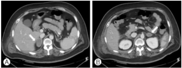
Fig. 1. Initial abdominal CT scan performed directly after blunt abdominal trauma. (A) 4 cm and (B) 2.3 cm adrenal masses wit- hout contrast enhancement are seen.
INTRODUCTION
Adrenal hemorrhage caused by blunt abdominal trauma is not unusual and is mostly unilateral and right-sided.
1,2In the autopsy series, it occurred about 7-26% after blunt abdomi- nal trauma.
1,3However, bilateral involvement of traumatic adrenal hemorrhage has been known to be rare and usually associated with predisposing conditions such as anticoagula- tion therapy, sepsis, and thromboembolism.
4Bilateral adrenal hemorrhage caused by blunt abdominal trauma is clinically important as it can cause acute adrenal insufficiency leading to death, if it is unsuspected and untreated immediately.
5In a literature search, there was no case report on the trau- matic bilateral adrenal hemorrhage in Korea. Therefore, we report a case of bilateral traumatic adrenal hemorrhage that developed in a man without any predisposing conditions.
CASE
A 40-year-old man was admitted to our department for the
evaluation of bilateral adrenal masses. He had a car accident
and admitted to a local clinic three months ago. Abdominal
CT taken directly after the car accident revealed splenic rupt-
ure, liver laceration, multiple rib fractures, hemothorax, and
bilateral adrenal masses (Fig. 1). Further diagnostic work-up
on the bilateral adrenal masses did not undergo as CT scan
findings suggested bilateral adrenal masses to be post-trauma-
Min Jung Lee, et al.
36
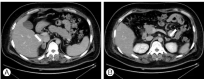
YUJM VOLUME 29, NUMBER 1, JUNE 2012Fig. 2. Follow-up abdominal CT scan obtained 2 months later.
Significant reduction of the bilateral adrenal masses is noted.
tic adrenal hemorrhage. However, follow-up CT scan taken two weeks later showed no significant interval change in the bila- teral adrenal masses. Moreover, on a careful physical examina- tion, he had abdominal obesity, buffalo humps, and abdominal striae suggesting Cushing’s syndrome. The hormonal evaluation for Cushing’s syndrome was performed: Urine free cortisol level for 24 hours was 133 ㎍/day and serum concentrations of free cortisol and adrenocorticotropic hormone (ACTH) were 16.3 ㎍/dL and 124 pg/mL, respectively. Thus the patient was referred to our hospital for the further evaluation of both adrenal masses and possible Cushing’s syndrome.
The past medical history of the patient was unremarkable except type 2 diabetes mellitus and hypertension diagnosed 6 months ago. He had been obese since he was young. There was no remarkable finding in his family history. On admi- ssion, blood pressure was 127/86 mmHg and pulse rate was 84 beats per minute. His height was 176.8 cm, body weight was 109 kg, and thus calculated body mass index was 35.13
kg/m
2. Physical examination revealed that he had cushingoid features such as buffalo hump, white abdominal striae, and abdominal obesity. He complained no specific symptom except pain around his ribs. A complete blood count showed hemo- globin 15.1 g/dL, hematocrit 44.0%, white blood cell count 6,100/mm³ and a platelet count 181,000/mm³. The serum sodium, potassium and chloride levels were 135, 3.8 and 101
mEq/L respectively. Outside initial contrast-enhanced abdo- minal CT scan displayed both adrenal masses with decreased attenuation (Fig. 1). Both ovoid masses were replacing normal adrenal structures. The longest diameter of the right and left mass was 4 cm and 2.3 cm, respectively (Fig. 1). We re-perfor- med hormonal evaluation for Cushing’s syndrome because of a rare incidence of bilateral involvement of traumatic adrenal hemorrhage and cushingoid features. Early morning (8 A.M.) blood cortisol and ACTH was 8.6㎍/dL and 46.2 pg/mL, both of which were within normal range. Urine free cortisol level for 24 hours was 50.4 ㎍/day. In the overnight dexamethasone suppression test, morning cortisol level was suppressed to be 1 ㎍/dL. These hormonal data did not satisfy the criteria for Cushing syndrome. Therefore, we decided to repeat abdomi- nal CT scan to differentiate adrenal adenomas from hema- tomas. On the follow-up abdominal CT scan which was done about 2 months after the initial work-up, the diameter of right adrenal mass decreased to 2 cm whereas that of left one was reduced to 1 cm (Fig. 2). A significant reduction in the size of both adrenal mass led us to diagnose bilateral adrenal masses as adrenal hematomas caused by blunt abdominal trauma. Elevated urinary cortisol excretion in initial work-up was thought to be due to post-trauma stress. Fortunately, he
had no clinical or biochemical evidence of acute adrenal insuffi- ciency during post-traumatic periods.
DISCUSSION
The frequency of post traumatic adrenal hemorrhage repor- ted to be between 7 and 26% in the autopsy series and app- roximately 2% in CT scan.
1,3,6Post traumatic adrenal injuries occur usually unilateral. Sevitt
3firstly reported in 1955 that 14 had an adrenal hemorrhage among 50 autopsies after severe torso injuries. Among them, bilateral involvement was observed only in 3 subjects while unilateral and right adrenal gland involvement was common. In 1991, Burk
6et al. repor- ted that 20 patients with adrenal hemorrhage were detected among the 1,120 subjects with blunt abdominal trauma using abdominal CT scans. Alike the study by Sevitt,
3injuries to adrenal gland were mostly unilateral (17/20, 85%) and right sided (12/17, 71%). Bilateral injuries were observed only in 3 patients (3/20, 15%).
6Several mechanisms for post-traumatic adrenal hemorrhage have been suggested although the exact mechanism is largely unknown. Sevitt
3reported that adrenal hemorrhage usually occurred in the adrenal medulla and loosely textured juxta- medullary cortex because these regions were vulnerable to damage. Therefore, after sudden compression and decompre- ssion, the vessels in those regions may be prone to rupture and thereby lead to hemorrhage.
3Regarding the mechanism for right-side preference, right adrenal gland appears to be more easily damaged because of its close opposition to the liver than the left one.
3Another explanation may come from the difference in drainage course of the adrenal veins between right and left adrenal gland. Severe compression of inferior vena cava may cause acute elevation of intra-adrenal venous pressure, leading to the adrenal hemorrhage. Since the right adrenal vein drains directly into the inferior vena cava, right adrenal gland is more susceptible to damage.
3Although traumatic adrenal injuries are usually unilateral, bila-
Traumatic Bilateral Adrenal Hemorrhage
YUJM VOLUME 29, NUMBER 1, JUNE 2012