CLAH, the most severe genetic disorder of steroid hor- mone biosynthesis (1), is a rare autosomal recessive dis- order characterized by the marked accumulation of lipids and cholesterol in the adrenal cortex and the fail- ure of adrenal steroids to synthesise (2). The disorder is characterized clinically by profound salt loss, complete feminization of male external genitalia, and hyperpig- mentation. Most patients have died in early infancy due to adrenal crisis (3). Early diagnosis and timely replace- ment treatment can, however, enable normal mental and physical development.
Prior to 1995 only one report describing a case of CLAH had been published in Korea (Lee et al.;4), though primarily a gene study of four Korean patients with CLAH, its findings based on clinical findings, has been published more recently (5). We describe the US, CT, and MR imaging findings of CLAH in a four-day-old female neonate.
Case report
A four-day-old female neonate was referred to our hospital because of frequent vomiting, lethargy, and dif- fuse pigmentation. The baby, whose parents were healthy, was the 3.5-kg product of an uncomplicated 41- week pregnancy and delivery, and their second child.
Their first, also a female, had died aged 3 days due to the same manifestation as this patient. Physical exami- nation disclosed a moderately lethargic patient with dif- fuse hyperpigmentation, including the oral mucosa. The external genitalia were normal. The body temperature was 37.0, pulse rate 160, and respiratory rate 60.
Laboratory studies indicated that blood urea nitrogen was 11 mg/dl, creatinine 0.4 (normal range, 0.7-1.5) mg/dl, sodium 123 (135-150) mEq/L, potassium 5.7 mEq/L, and chloride 111 (95-105) mEq/L. Serum aldos- terone was 91.3 pg/ml, testosterone 0.04 (0.09-0.9) ng/ml, plasma adrenocorticotropic hormone 799.1 (12- 76) pg/ml, and plasma renin activity 44 (0.2-2.5) ng/ml/h. The karyotype was normal.
Radiological studies
At 5 days of age, US examination revealed significant
J Korean Radiol Soc 2001;44:637-640
─ 637 ─
Radiological Findings of Congenital Lipoid Adrenal Hyperplasia: A Case Report1
Mi Jeong Kim, M.D., Joo Yong Shin, M.D., Hee Jung Lee, M.D., Jin Hee Lee, M.D., Cheol Ho Sohn, M.D., Sung Moon Lee, M.D., Hong Kim, M.D.,
Seong Ku Woo, M.D., Soo Ji Suh, M.D.
Congenital lipoid adrenal hyperplasia (CLAH) is a rare autosomal recessive disorder characterized by the marked accumulation of lipids and cholesterol in the adrenal cor- tex, and the failure of adrenal steroids to synthesise. We report the ultrasound (US), computed tomographic (CT), and magnetic resonance (MR) imaging findings in a four- day-old female neonate with CLAH.
Index words : Adrenal gland, hyperplasia Adrenal gland, CT
Adrenal gland, insufficiency Adrenal gland, MR
1Department of Diagnostic Radiology, Keimyung University School of Medicine
Received July 19, 2000; Accepted April 12, 2001
Address reprint requests to : Hee Jung Lee, M.D., Department of Diagnostic Radiology, Dongsan Medical Center, Keimyung University School of Medicine, 194 Dongsan-Dong, Jung-Ku, Taegu 700-712, Korea.
Tel. 82-53-250-7767 Fax. 82-53-250-7766 E-mail : [email protected]
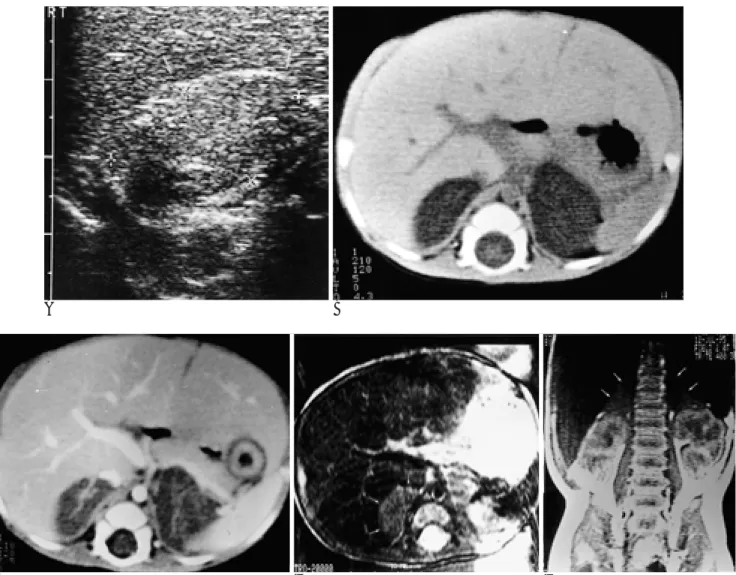
enlargement of both adrenal glands, which were iso- to slightly hyperechoic compared to the liver (Fig. 1A).
Follow-up US examinations at 18 and 32 days showed persistent adrenal enlargement with no change in echo- geneity (not shown). CT scanning of the abdomen was performed at 9 days of age; the precontrast scan showed massively enlarged adrenal glands, with fat-tissue atten- uation (H.U., -28) (Fig. 1B), enhanced scan demonstrat- ed that blood supply to the lesion was much reduced (Fig. 1C). MR imaging using the spin-echo technique was performed at 10 days of age; compared with hepatic parenchyma, the enlarged adrenal glands were hyperin- tense on T2-weighted images (2000/60) (Fig. 1D) and iso-
to slightly hyperintense on T1-weighted images (400/30) (Fig. 1E).
Clinical course
On the basis of the radiologic findings and laboratory data, CLAH was diagnosed and using the standard doses of glucocorticoid and sodium chloride, replacement ther- apy was initiated. This led to decreased pigmentation, and the subsequent clinical course was uneventful. At 20 days of age, the patient developed fever; antibiotic thera- py was initiated under the impression of sepsis but the parents refused further management. The patient was discharged at 30 days of age and was lost to follow-up.
Mi Jeong Kim, et al: Radiological Findings of Congenital Lipoid Adrenal Hyperplasia
─ 638 ─
A B
C D E
Fig. 1. Imaging features of CLAH in a 4-day-old female neonate.
A. A transverse scan of the upper abdomen with 7 MHz linear transducer demonstrates iso- to hyperechoic lesion in the right adrenal gland (arrows) with globular enlargement.
B. Precontrast CT scan shows massively enlarged both adrenal glands of fat-tissue attenuation (H.U., -28).
C. Enhanced CT scan demonstrates poor enhancement of the lesions (H.U., -20).
D. Axial T2-weighted image of the abdomen reveals diffusely enlarged right adrenal gland with slightly hyperintensity comparing to the hepatic parenchyma (arrows).
E. Coronal T1-weighted image shows slightly hyperintensity of the both adrenal glands compared to the liver (arrows).
Discussion
Congenital lipoid adrenal hyperplasia (CLAH) is the most severe form of congenital adrenal hyperplasia and is the most severe genetic disorder of steroid hormone biosynthesis (1). This failure to convert cholesterol to pregnenolon is due to a defect in 20, 22 desmolase.
There is a marked accumulation of lipids and choles- terol in the adrenal cortex, and adrenal steroids includ- ing all glucocorticoid, mineralocorticoid and the sex hor- mones fail to synthesise (2). The same enzyme defect is present in the testis, preventing the synthesis of testicu- lar hormones. As a consequence, males are phenotypi- cally female while females exhibit no genital abnormali- ty (3).
Prior to 1985, CLAH had been reported in 33 patients, 56% of whom were Japanese (3), recent studies have suggested that CLAH is more common in both Koreans and Japanese than in other ethnic populations (4-7).
The mutation Q258X, which is the leading defect in the key protein, has been found in 80% of affected alleles from Japanese and Korean patients (7), and a recent study reported four Korean patients with CLAH and an estimated incidence of the Q258X mutation of approxi- mately 1 in 250,000 among the Korean population (5).
This figure seems relatively high and CLAH is thus not a particularly rare disease in Korea.
The clinical findings of CLAH are remarkably con- stant : neonatal hyponatremia, hyperkalemia, hyperpig- mentation, and complete female external genitalia irre- spective of the gonadal sex (3-7). Hyperpigmentation, a sign of corticotropin hypersecretion, occurs in two thirds of newborns with CLAH (7), and because adrenal steroid levels are not elevated, affected infants are apt to be confused with those with other forms of adrenal hy- perplasia. Furthermore, the disease does not show am- biguity of the external genitalia, and early diagnosis and correct treatment tend to be delayed, resulting in early death from adrenal crisis in the majority of patients. If appropriate mineralocorticoid and glucocorticoid re- placement therapy is initiated, however, patients can survive to adulthood (3, 7).
To our knowledge, only two reports have described the role played by imaging in the diagnosis of CLAH (8, 9). Ogata et al. first described the CT findings of CLAH in a eight-day-old female infant in whom massively en- larged adrenal glands, with fat-tissue attenuation, were seen, as in our case (8). US also can provide a useful
method of evaluating the size and appearance of adrenal glands in neonates and young infants, and US has un- equivocally demonstrated enlarged adrenal glands (4, 5, 9). Both CT and US findings have correlated closely with known pathologic changes in adrenal glands char- acterized by the accumulation of lipid and cholesterol ester (2). In the case we describe, CLAH was diagnosed on the basis of the CT findings, and through a clinical approach the differential diagnosis of diseases causing adrenal crisis in neonates and young infants was nar- rowed. According to Ogata et al., a CT scan obtained af- ter 9 months of replacement therapy demonstrated in- tra-adrenal calcification and reduced adrenal size (8).
Takaya et al. described the serial changes in the adrenal glands depicted by US: after 24 days of replacement therapy the glands became smaller and their cortical echogenic pattern changed from hyperechoic to isoe- choic (9). In our case, the size and echogenicity of imag- ing showed that adrenal glands did not change within one month of replacement therapy. MR imaging, both adrenal glands were diffusely enlarged and isointense on T1-weighted images and iso- to slightly hyperintense on T2-weighted images. Viable fat may be isointense to subcutaneous fat on T1-weighted images and exhibit low signal intensity with fat suppression on T2-weight- ed images (10). Due to the failure of sedation we were unable to apply the fat saturation technique. The iso- to slight hyperintensity seen on T1-weighted images in our case may be due to field inhomogeneity or a technical problem. Takaya et al. (9) stated that whereas the adren- al cortex was hypointense, the adrenal medulla was markedly hyperintense, but did not attempt to explain why this was so.
The differential diagnosis includes adrenal hypoplasia, bilateral adrenal hemorrhage, congenital neuroblastoma and Wolman disease, all of which cause adrenal crisis in newborn infants. In an appropriate clinical setting, how- ever a diagnosis of CLAH would be suggested by find- ings of unequivocally delineated enlarged adrenal glands due to fat accumulation, as revealed by CT or MR imaging.
In summary, we have presented the US, CT and MR imaging findings of CLAH. A familarity with these facil- itates the diagnosis of CLAH, and appropriate manage- ment of this disorder is thus possible.
References
1. Degroot LJ. Endocrinology, 2nd ed, WB saunders Co, 1989:1693- J Korean Radiol Soc 2001;44:637-640
─ 639 ─
1694
2. Tsutsui Y, Hirabayashi N, Ito G. An autopsy case of congenital lipoid hyperplasia of the adrenal cortex. Acta Pathol Jpn 1970;20:227-237
3. Hauffa BP, Miller WL, Grumbach MM, Conte FA, Kaplan SL.
Congenital adrenal hyperplasia due to deficient cholesterol side- chain cleavage activity (20,22-desmolase) in a patient treated for 18 years. J Clin Endocrinol Metab 1985;23:481-493
4. Lee HK, Lee DH, Lee SJ. A case of congenital lipoid adrenal hyper- plasia. Pediatrics 1995;28:567-573
5. Yoo HW, Kim GH. Molecular and Clinical charac terization of Korean patients with congenital lipoid adrenal hyperliasia. J Pediatr Endocrinol Metab 1998;11:707-711
6. Lin D, Sugawara T, Strauss JF III, et al. Role of steroidogenic acute
regulatory protein in adrenal and gonadal steroidogenesis. Science 1995;267:1828-1831
7. Bose HS,Sugawara T, Strauss JF, Miller WL. The pathophysiology and genetics of congenital lipoid adrenal hyperplasia. N Engl J Med 1996;335:1870-1878
8. Ogata T, Ishikawa K, Kohda D, Matsuo N. Computed tomography in the early detection of congenital lipoid adrenal hyperplasia.
Pediatr Radiol 1988;18:360-361
9. Takaya J, Ishihara R, Kino M, Higashino H, Kobayashi Y. A pa- tient with congenital lipoid adrenal hyperplasia evaluated by serial abdominal ultrasonography. Eur J Pediatr 1998;157:544-546 10. Propeck T, Bullard MA, Lin J, Doi K, Martel W. Radiologic-patho-
logic correlation of intraosseous lipomas. AJR Am J Roentgenol 2000;175:673-678
Mi Jeong Kim, et al: Radiological Findings of Congenital Lipoid Adrenal Hyperplasia
─ 640 ─
대한방사선의학회지 2001;44:637-640
선천성 유지질 부신 증식증의 방사선학적 소견: 1예 보고1
1계명대학교 의과대학 진단방사선과학교실
김미정・신주용・이희정・이진희・손철호・이성문・김 홍・우성구・서수지
선천성 유지질 부신증식증은 체염색체 열성유전으로 부신의 모든 스테로이드 생합성의 장애로 인해 부신 피질에 유지질이 축적되는 매우 드문 질환이다. 저자들은 생후 4일 된 여자 신생아에 발생한 선천성 유지질 부신증식증 1 예를 경험하여 초음파 (US), 전산화단층촬영 (CT), 및 자기공명영상 (MR) 소견을 보고하고자 한다.