Received: 19 June 2010, Accepted: 28 August 2010 Corresponding author: Sung-Kil Lim
Department of Internal Medicine, Yonsei University College of Medicine, 250 Seongsan-ro, Seodaemun-gu, Seoul 120-752, Korea
Tel: +82-2-2228-1948, Fax: +82-2-393-6884, E-mail: [email protected]
Primary Bilateral Adrenal Non-Hodgkin’s Lymphoma Presented with Adrenal Insufficiency: A Case Report
Eun Young Lee, Kyoung Min Kim, Kwang Joon Kim, Songmi Noh1, Jin Seok Kim, Woo Ik Yang1, Sung-Kil Lim Departments of Internal Medicine and Pathology1, Yonsei University College of Medicine, Seoul, Korea
Primary adrenal lymphoma is a very rare disease and it is known to have a poor prognosis. We report here on a case of primary adre- nal insufficiency that was secondary to primary bilateral adrenal lymphoma. A 54-year old man was hospitalized because of easy fa- tigability, weight loss and consistent malaise for 6 months. The physical examination revealed hyperpigmentation on the anterior chest and hypotension. According these findings and symptoms, we did a rapid ACTH stimulation test with a clinical suspicion of adrenal insufficiency. He showed an inadequate adrenal response and so he was diagnosed with adrenal insufficiency. The abdom- inal CT images showed bilateral huge adrenal masses and increased uptake of the adrenal glands on PET. The pathologic diagnosis by ultrasound-guided gun biopsy of the right adrenal gland was diffuse large B cell lymphoma. The patient was administered combi- nation chemotherapy with the R-CHOP regimen, and after 8-cycles of chemotherapy, he achieved complete remission of tumor ac- cording to the image studies and he recovered his adrenal function. Primary adrenal lymphoma, although a rare disease, should be considered in patients with bilateral enlargement of the adrenal glands and when the adrenal glands show increased uptake on a PET scan, and especially there is adrenal insufficiency. (Endocrinol Metab 26:101-105, 2011)
Key Words: Adrenal insufficiency, Non-Hodgkin’s lymphoma
INTRODUCTION
Non-Hodgkin’s lymphoma (NHL) refers to all malignancies of the lymphoid system with the exception of Hodgkin’s lymphoma and is around 90% of all malignant lymphomas [1]. Most patients with NHL present with painless lymphadenopathy, commonly in the cervical or supraclavicular regions [2]. However, extranodal presentation of NHL occurs in 15% to 40% of patients at presenta- tion and varies depending on immune status and geographic differ- ences. The most common extranodal sites are the gastrointestinal tract and nasopharynx. Other common sites include skin, bone, brain, lung, thyroid, salivary glands, and testis [2-6]. Secondary in- volvement of the adrenal gland with NHL has been reported to oc- cur in up to 25% of patients during the course of disease [2,7].
However, primary adrenal lymphoma (PAL) is a very rare disease and is often accompanied by adrenal insufficiency. The first case of PAL with adrenal insufficiency was reported in 1970 and only ten cases of PAL had been reported until the year 1989 [8]. The
prognosis of PAL is known as poor [8-10], but early diagnosis and combination chemotherapy may improve not only the prognosis of lymphoma, but also adrenal function [8].
PAL should be considered in patients with bilateral enlargement of adrenal glands, especially with adrenal insufficiency [8]. We re- port a case of PAL with primary adrenal insufficiency, which showed early diagnosis and treatment could improve the prognosis.
CASE REPORT
A 54-year old man was hospitalized because of easy fatigability, weight loss of 10 kg and consistent malaise for 6 months. On ad- mission, his vital signs were as follows: body temperature, 36.7℃; pulse rate 113 beats per minute; blood pressure 98/77 mmHg.
Physical examination revealed some pigmented spots on his upper chest and face (Fig. 1). His tongue was dry and skin turgor was de- creased. Neither lymphadenopathy or hepatosplenomegaly was noted. Ascites and edema were not present.
A complete blood count showed hemoglobin 12.4 g/dL, hemato- crit 35.2%, white blood cell count 8320/mL with 58.7% segmented neutrophils, 24% lymphocytes, and 8.8% monocytes, and a platelet count 234,000/mL. The serum sodium, potassium and chloride
fatigability, weight loss, consistent malaise and hypotension. Clini- cal suspicion of adrenal insufficiency was confirmed by the finding of a low serum cortisol level of 47.85 ng/mL at 8 AM, a high plasma ACTH level of 147.29 pg/mL at 8 AM, and low cortisol level of
sion of right adrenal mass into the right hepatic lobe (Fig. 3). These findings are consistent with malignancy. In the rest of the body, there was no increased FDG uptake which suggests malignancy.
An ultrasound-guided percutaneous gun biopsy of the right adre- nal mass was performed. The gun biopsied specimen showed dif- fuse proliferation of large atypical cells without forming organoid structures. The tumor cells showed folded nuclei with one or mul- tiple prominent nucleoli and a variable amount of cytoplasm (Fig.
4A). They showed positive reactions with CD20 on immunohisto- chemical stain (Fig. 4B), but negative reactions with CD3, CD10, and cytokeratin. The final diagnosis was bilateral adrenal diffuse large B cell lymphoma (DLBCL) with adrenal insufficiency, and the clinical staging by the Ann Arbor system was ⅣB, and interna- tional prognostic index was low intermediate risk group.
The patient received adrenal hormone replacement with Cor- tico® (hydrocortisone) 30 mg daily. He also was administered com- bination chemotherapy with R-CHOP regimen; rituximab, cyclo- phosphamide, doxorubicin, vincristine, prednisolone. After 3 cy- cles of chemotherapy, left adrenal mass was not visible and right adrenal mass decreased in size from 6.4 cm to 3.7 cm by CT scan.
He received total 8 cycles of chemotherapy, because PET scan showed mild hypermetabolism on right adrenal gland after sixth
Fig. 3. FDG-PET scan at diagno- sis showed huge bilateral adrenal masses with intense FDG uptake.
Right adrenal mass invaded into the right hepatic lobe.
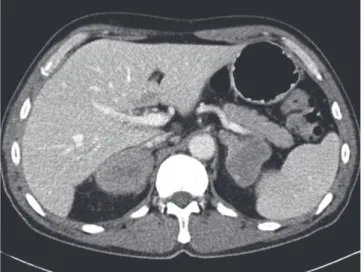
Fig. 2. Abdomen CT scan at diagnosis showed about 6.4 cm sized adrenal gland masses.
Fig. 1. Appearance of the patient: Hyperpigmentation on skin (A) face and the upper chest (B) back. Arrows: hyperpigmented spots.
A B
cycle of chemotherapy. After 8 cycles of chemotherapy, right adre- nal mass further decreased in size from 3.7 cm to 3.5 cm by CT scan and there were no increased FDG uptake by PET scan (Fig.
5). He had taken a prednisolone 10 mg per day for 6 months. Clini- cally adrenal insufficiency was also improved with disease remis- sion, so the dose of steroid replacement was tapered off. This pa- tient is continued to be followed up without any evidence of recur- rence. Furthermore, he did not have any signs or symptoms of ad- renal insufficiency for 1 year.
DISCUSSION
Adrenal glands are common metastatic site of primary cancers, such as breast, lung, lymphoma, melanoma, leukemia, renal cell and ovarian cancers [11]. In contrast to primary tumors, many me-
tastases tend to invade both adrenal glands. Primary malignant lymphomas arising in endocrine organs are rare with representing only 3% of extranodal lymphomas [8]. They are almost confined to the thyroid gland [5,12] and primary lymphoma originated from adrenal is extremely rare. The first case of PAL with adrenal insuf- ficiency was reported in 1970 and only ten cases of PAL had been reported until the year 1989. The development of imaging tech- niques and immunostaining procedures makes the diagnosis of PAL easier and so, about 120 cases of PAL have been reported in the literature [9].
Adrenal insufficiency is rare because it becomes apparent when approximately 90% of adrenal cortex is destructed [13]. And most of adrenal insufficiency are believed to be associated with atrophy of adrenal glands, secondary to autoimmune mechanisms [14]. Ap- proximately 50% of patients with primary adrenal lymphoma pres- Fig. 4. High-power view of the adrenal gland tumor (both, x 200). A. The gun biopsied specimen showed diffuse proliferation of large atypical cells, which had fold- ed nuclei with one or multiple prominent nucleoli (H&E staining). B. The tumor cells were positive to CD20 immunohistochemical staining.
A B
Fig. 5. Imaging studies after 8 cycle R-CHOP. A. Abdominal CT scan. B. FDG-PET scan.
A B
Because DLBCL is aggressive disease, most PAL patients with adre- nal insufficiency are not present with hyperpigmentation. How- ever, our patient had some pigmented spots on his upper chest and face (Fig. 1), which mean chronic progression of the disease.
Primary adrenal insufficiency can be confirmed by low basal se- rum cortisol level, high ACTH level, and inadequate adrenal re- sponse to the rapid ACTH stimulations [14].
In PAL, men are more frequently affected than women, with a ratio of 3:1. The mean age at the time of diagnosis is 68 years (range 39-89). More than two-third of PAL are bilateral. Most common histopathology is diffuse large cell lymphoma of B cell lineage [9].
These malignant cells express CD45, and antigens specific to B lymphocytes, such as CD19, CD20, and CD79a [15]. About 10% of de novo DLBCL cases express CD5 and CD5-positive DLBCL is more common in older women, with extranodal involvement and associated with a shorter survival [15]. T cell lymphoma, expressing antigens specific to T lymphocytes, such as CD3, CD5, and CD7 [16] occupies less than 10% of PAL. In our case, the tumor cells showed positive reactions with CD20 on immunohistochemical stain, but negative reactions with CD3 and CD10. Less common histopathology includes small-cell lymphoma, mixed small- and large-cell lymphoma, undifferentiated lymphoma, anaplastic large- cell and follicular lymphoma [9]. On CT scan, lymphomas usually show complex masses with variable density and sometimes ac- companied by necrosis and hemorrhage, but calcification is un- common. The contour of the involved adrenal gland may be pre- served. On MRI scan, lymphomas show low signal intensity on T1- weighted images and heterogeneous high signal intensity on T2- weighted images. However, CT and MRI finding of adrenal lym- phoma are not specific [17]. In our case, CT scan showed large bi- lateral adrenal gland masses with poor enhancement, and lym- phoma, metastasis or adrenal carcinoma were included in the dif-
expression of CD5 [18,19]. The cause of death is severe infection, pulmonary embolism, and tumor recurrence [20]. One study re- ported that mean overall-survival rate is 15.3 months; 34 months of the patients who achieve a partial or complete remission after che- motherapy and 3.6 months of the patients who have no response to chemotherapy [8]. The longest survival was 50 months in cases of treating with CHOP chemotherapy [13]. The treatment of PAL is not established, but recent studies show that chemotherapy includ- ing CHOP can extend survival. The regimens of chemotherapy in- clude CHOP (cyclophosphamide, doxorubicin, vincristine, and prednisone), R-CHOP (adding rituximab to CHOP), CHO (cyclo- phosphamide, doxorubicin, vincristine), CVP (cyclophosphamide, vincristine, and prednisone) or MACOP (cyclophosphamide, doxo- rubicin, prednisone, methotrexate, bleomycin) [9]. Surgical resec- tion only is not recommended for PAL treatment because of a poor outcome. The role of radiation therapy on PAL is unknown. But in some cases, the patients with PAL had radiation therapy with che- motherapy [9]. CNS prophylaxis with intrathecal methotrexate is also considered in patients with high risk of CNS involvement. Risk factors for CNS involvement are elevated serum LDH level, high/
intermediate or high International Prognostic Index and involve- ment of more than one extranodal site including bone marrow [9].
In this case, the patient had taken abdomen ultrasonography and CT at first visit of our hospital, and diagnosed of NHL by percuta- neous gun biopsy. After pathologic diagnosis, he received combi- nation chemotherapy without delay. After 8-cycle of chemotherapy, he achieved complete remission clinically. Primary adrenal lym- phoma, although a rare disease, should be considered in patients with bilateral enlargement of adrenal glands, especially with adre- nal insufficiency. And early diagnosis can lead to better prognosis by early treatment.
REFERENCES
1. Ekstrom-Smedby K: Epidemiology and etiology of non-Hodgkin lym- phoma-a review. Acta Oncol 45:258-271, 2006
2. Rosenberg SA, Diamond HD, Jaslowitz B, Craver LF: Lymphosarcoma: a review of 1269 cases. Medicine (Baltimore) 40:31-84, 1961
3. Jones SE, Fuks Z, Bull M, Kadin ME, Dorfman RF, Kaplan HS, Rosen- berg SA, Kim H: Non-Hodgkin’s lymphomas. IV. Clinicopathologic cor- relation in 405 cases. Cancer 31:806-823, 1973
4. Vega F, Lin P, Medeiros LJ: Extranodal lymphomas of the head and neck.
Ann Diagn Pathol 9:340-350, 2005
5. AlShemmari SH, Ameen RM, Sajnani KP: Extranodal lymphoma: a com- parative study. Hematology 13:163-169, 2008
6. Said J: Diffuse aggressive B-cell lymphomas. Adv Anat Pathol 16:216-235, 2009
7. Paling MR, Williamson BR: Adrenal involvement in non-Hodgkin lym- phoma. AJR Am J Roentgenol 141:303-305, 1983
8. Kumar R, Xiu Y, Mavi A, El-Haddad G, Zhuang H, Alavi A: FDG-PET imaging in primary bilateral adrenal lymphoma: a case report and review of the literature. Clin Nucl Med 30:222-230, 2005
9. Ozimek A, Diebold J, Linke R, Heyn J, Hallfeldt K, Mussack T: Bilateral primary adrenal non-Hodgkin’s lymphoma and primary adrenocortical carcinoma-review of the literature preoperative differentiation of adrenal tumors. Endocr J 55:625-638, 2008
10. Wang J, Sun NC, Renslo R, Chuang CC, Tabbarah HJ, Barajas L, French SW: Clinically silent primary adrenal lymphoma: a case report and review of the literature. Am J Hematol 58:130-136, 1998
11. Mansmann G, Lau J, Balk E, Rothberg M, Miyachi Y, Bornstein SR: The clinically inapparent adrenal mass: update in diagnosis and management.
Endocr Rev 25:309-340, 2004
12. Krol AD, le Cessie S, Snijder S, Kluin-Nelemans JC, Kluin PM, Noordijk EM: Primary extranodal non-Hodgkin’s lymphoma (NHL): the impact of alternative definitions tested in the Comprehensive Cancer Centre West population-based NHL registry. Ann Oncol 14:131-139, 2003
13. Kuyama A, Takeuchi M, Munemasa M, Tsutsui K, Aga N, Goda Y, Kane- tada K: Successful treatment of primary adrenal non-Hodgkin’s lymphoma associated with adrenal insufficiency. Leuk Lymphoma 38:203-205, 2000 14. Oelkers W: Adrenal insufficiency. N Engl J Med 335:1206-1212, 1996 15. de Leval L, Hasserjian RP: Diffuse large B-cell lymphomas and burkitt
lymphoma. Hematol Oncol Clin North Am 23:791-827, 2009
16. Libe R, Giavoli C, Barbetta L, Dall’Asta C, Passini E, Buffa R, Beck-Pec- coz P, Ambrosi B: A primary adrenal non-Hodgkin’s lymphoma present- ing as an incidental adrenal mass. Exp Clin Endocrinol Diabetes 114:140- 144, 2006
17. Zhang LJ, Yang GF, Shen W, Qi J: Imaging of primary adrenal lympho- ma: case report and literature review. Acta Radiol 47:993-997, 2006 18. Yamaguchi M, Seto M, Okamoto M, Ichinohasama R, Nakamura N, Yo-
shino T, Suzumiya J, Murase T, Miura I, Akasaka T, Tamaru J, Suzuki R, Kagami Y, Hirano M, Morishima Y, Ueda R, Shiku H, Nakamura S: De novo CD5+ diffuse large B-cell lymphoma: a clinicopathologic study of 109 patients. Blood 99:815-821, 2002
19. Schreiber CS, Sakon JR, Simiao FP, Tomarchio MP, Huayllas M, Pereira LC, Stella LC, Santomauro AC Jr, Bueno SS, Fraige FF: Primary adrenal lymphoma: a case series study. Ann Hematol 87:859-861, 2008
20. Hsu CW, Ho CL, Sheu WH, Harn HJ, Chao TY: Adrenal insufficiency caused by primary aggressive non-Hodgkin’s lymphoma of bilateral adre- nal glands: report of a case and literature review. Ann Hematol 78:151- 154, 1999