Received February 9, 2019 / Revised February 19, 2019 / Accepted February 25, 2019
Correspondence: In Ho Choi, MD, Department of Pathology, Soonchunhyang University Seoul Hospital, 59 Daesagwan-ro, Yongsan-gu, Seoul 04401, Korea
Tel: 82-2-709-9429, Fax: 82-2-709-9441, E-mail: [email protected]
Copyright ⓒ 2019, the Korean Thyroid Association. All rights reserved.
This is an open-access article distributed under the terms of the Creative Commons Attribution Non-Commercial License (http://creative- commons.org/licenses/by-nc/4.0/), which permits unrestricted non-commercial use, distribution, and reproduction in any medium, provided the
Yeeun Han
1, Yon Hee Kim
1, Hye Jeong Kim
2and In Ho Choi
1Departments of Pathology1 and Internal Medicine2, Soonchunhyang University Seoul Hospital, Seoul, Korea
Cases of simultaneously occurring medullary thyroid carcinoma (MTC) and lymphoma are extremely rare. An 84-year-old woman visited the hospital due to dyspnea, resulting from rapidly aggravated enlarged neck mass.
Ultrasonography revealed two lesions in the thyroid and they were diagnosed as concurrent medullary thyroid carcinoma and diffuse large B cell lymphoma after total thyroidectomy. A few cases simultaneously diagnosed with MTC and systemic lymphoma have been reported. However, the coexistence of MTC and primary thyroid lymphoma is extremely rare.
Key Words: Primary thyroid lymphoma, Diffuse large B cell lymphoma, Medullary carcinoma
Introduction
Medullary thyroid carcinoma (MTC) and primary thyroid lymphoma (PTL) occupy small proportions of thyroid malignancies (MTC <2 to 3%, PTL <5%).1-4) MTC can exhibit extensive local tumor growth, result- ing in upper airway obstruction and dysphagia.1) PTL also can exhibit rapidly enlarging neck mass accom- panied by hoarseness, dyspnea.1) The most common type of PTL is diffuse large B-cell lymphoma (DLBCL), followed by extranodal marginal zone B-cell lympho- ma of mucosa-associated lymphoid tissue (MALT lymphoma).1) As mentioned above, PTL is rare and a few cases with concurrent papillary thyroid carcinoma (PTC) have been reported.5-8) To the best of our knowledge, cases of simultaneously occurring MTC and lymphoma are extremely rare. Herein, we report a case of concurrent MTC and DLBCL in the thyroid.
Case Report
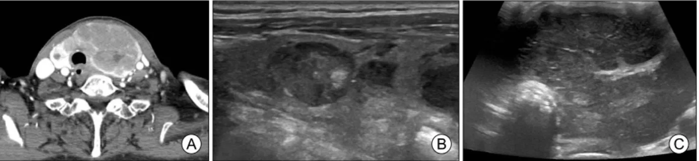
An 84-year-old female visited the hospital due to dyspnea that developed 10 days previously, and a rapidly aggravated enlarged neck mass. She had a history of diabetes mellitus, hypertension, coronary 2 vessel disease, and severe dementia. Neck CT re- vealed about a 4.3×6.6×8.3 cm thyroid mass in- volving the left lobe, isthmus and medial portion of the right lobe and resulting in tracheal deviation to the right side without tracheal stenosis (Fig. 1A) Ultrasono- graphy showed two lesions. The first lesion was a 1.64 cm sized, hypoechoic mass at the right upper pole (Fig. 1B), and the second lesion was a 6.85 cm sized hypoechogenic thyroid mass involving the left thyroid lobe, isthmus and medial portion of the right lobe with intrathoracic extension (Fig. 1C). Several en- larged lymph nodes were noted in the left neck level
Fig. 1. (A) Neck CT reveals about 4.3×6.6×8.3 cm thyroid mass involving the left lobe, isthmus and medial portion of the right lobe and resulting in tracheal deviation. (B, C) Thyroid ultrasonography reveals 1.64 cm sized, hypoechoic mass at the right upper pole (B) and on the left lobe, there is a 6.85 cm sized hypoechogenic mass (C), which shows extension to the isthmus, medial portion of the right lobe and intrathoracic area.
Fig. 2. (A) Specimen from total thyroidectomy. The right lobe measures 3×4.5×1.5 cm and the left lobe is markedly enlarged by the mass, mea- sures 7×9×3.5 cm. (B) On cut section of right lobe, there is a well-circumscribed ovoid, tan, yellowish solid mass, measuring 1.2×1.1 cm. (C) Cut sections of left lobe of thyroid gland reveals multiple iirregular, patchy or nodular, gray lesions occupying almost entire left lobe and extending to the right lobe with focal necrosis.
III, and level IV, but the lymph node image findings fa- vored benign reactive hyperplasia. Laboratory findings showed normal thyroid hormone levels (T3: <0.40 ng/mL, fT4: 0.85 ng/dL, TSH: 1.02 uIU/mL), in- creased levels of procalcitonin (1.52 ng/mL) and slightly increased serum CEA levels (5.4 ng/mL).
Ultrasound-guided needle biopsies were done on the lesions of both thyroid lobes. Based on the findings of the needle biopsy, the MTC and atypical lymphoid proliferation were suspected. Total thyroidectomy was done.
On the operative field, there was gross extension of the lesion to the esophagus, recurrent laryngeal nerve, tracheal, and sternothyroid muscle. The sur- geon thought complete resection was unattainable, and he decided to perform a debulking total thyroi- dectomy, instead.
On the gross examination, the resected specimen (Fig. 2A) revealed 2 lesions: (A), a well-demarcated ovoid, tan, yellowish solid mass at the right mid pole (1.2×1.1 cm) (Fig. 2B), and (B) a large solid mass with patchy or multinodular cut surface with ill define margin and focal necrosis (9×7 cm) involving almost the entire left lobe and extending to the right lobe (Fig.
2C). Microscopically, lesion of right mid pole was a relatively well-defined solid mass composed of plas- macytoid cells, and the tumor was transected by dense fibrous tissue (Fig. 3A [left] and 3B). Lesion of left lobe exhibited a relatively well-demarcated mass showing diffuse growth of large atypical cells with fre- quent mitoses and apoptotic bodies (Fig. 3A [right]
and 3C, D). The diagnoses of MTC for mass of right lobe and high-grade lymphoma for lesion of left lobe were made. Further immunohistochemical studies
Fig. 3. (A) Border of medullary thyroid carcinoma (left side) abutting with primary thyroid lymphoma (right side). The left side reveals a well-defined solid mass (detailed view is presented in (B)), and the right side lesion shows diffuse growth pattern of lymphoid cells (detailed view is presented in (C)) (H&E, ×40). (B) The mass of right lobe consists of plasmacytoid cells on the fibrous background, suggesting medullary thyroid carcinoma (H&E, ×400). (C) The large lesion of left lobe shows diffuse growth of large atypical cells with frequent mitoses and apoptotic bodies (H&E, ×400). (D) At the peripheral portion of lesion in left lobe, destruction of thyroid follicles with infiltrate of large atypical lymphoid cells is noted (H&E, ×200).
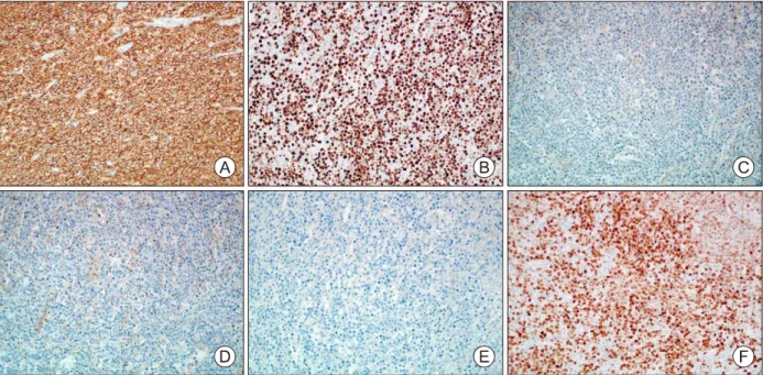
Fig. 4. Immunohistochemical staining of medullary thyroid carcinoma. Right mid pole mass shows positivity for calcitonin (Calcitonin, ×200).
were performed on both lesions. The mass of right lobe showed strong positivity for calcitonin (Fig. 4), and the mass of left lobe revealed strong reactivity for CD20 and MUM-1 with high proliferative index (90%) and are negative for Bcl-2, CD10, and Bcl-6 (Fig.
5A-F). Finally, diagnosis of concurrent medullary thy- roid carcinoma and DLBCL was made. Besides the thyroid specimens, bilateral paratracheal lymph nodes were sent separately and shows partial involvement of malignant lymphoma in one paratracheal node (Fig.
6A, B).
After the surgery, the patient was discharged, ac- cording to the legal guardian’s request. About three months later, patient was readmitted to the hospital due to general weakness and increased sputum.
Chest CT showed tumor recurrence at the operative bed with direct invasion of the mediastinal structures and tracheo-esophageal fistula at the T2 level. Due to the tracheo-esophageal fistula, bronchoscopy was performed, and bronchial washing cytology revealed
only necrotic inflammatory cells without malignant cells.
Despite administration of antibiotics accompanied by conservative treatment, aspiration pneumonia devel- oped, and the patient expired four months after the surgery.
Fig. 5. Immunohistochemical staining of primary thyroid lymphoma. Mass shows diffuse strong positivity for CD20 (A, ×200).
It shows 90% proliferative rate on Ki-67 (B, ×200). It is negative for Bcl-2 (C, ×200), Bcl-6 (D, ×200) and CD-10 (E, ×200).
It is positive for MUM-1 (F, ×200).
Fig. 6. (A) Metastatic lymph node by diffuse large B-cell lymphoma. Left paratracheal lymph node is partly involved with diffuse large B-cell lymphoma (right side) (H&E,
×40). (B) Detailed view of (A) (H&E, ×100).
Discussion
MTC comprises less than 2 to 3% of thyroid malig- nancies, and PTL is less than 5 percent.1-4) The most common histological type of PTL is non-Hodgkin’s lymphoma. Considering the low incidences of both diseases in the thyroid, a combination of both malig- nancies in the thyroid would be very rare, unlike the combination of PTC and lymphoma.
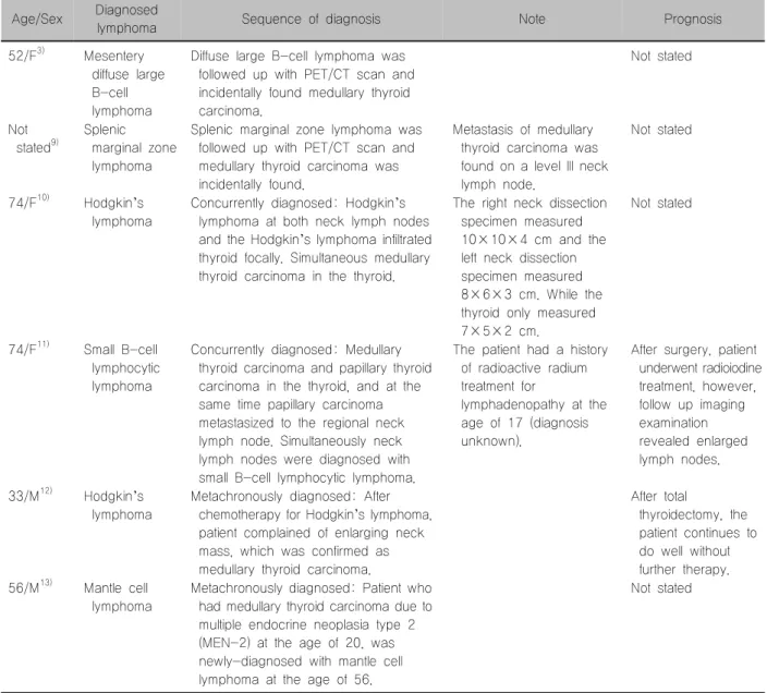
According to the literature, six cases of co-occur- ring MTC and lymphoma have been described. Four cases of MTC coexisting with systemic lymphoma have been reported, including cases with Hodgkin’s lymphoma in the bilateral neck nodes, splenic margin-
al zone lymphoma (SMZL), and mesenteric DLBCL and small lymphocytic lymphoma (SLL).3,9-11) Two cases of SMZL and mesenteric DLBCL included MTC incidentally found on the work-up for lymphoma by 18F-fluorodeoxyglucose-positron emission tomog- raphy (F-18 FDG PET).3,9) The case with SLL (B-cell type) had a simultaneous occurrence of both medul- lary and papillary carcinomas of the thyroid gland with metastases of a papillary carcinoma.11) Two cases with MTC and lymphoma that developed metachronously also were reported; one case was diagnosed with MTC after treatment for Hodgkin’s lymphoma, and the other case with MTC due to multiple endocrine neo- plasia (MEN) type II, later developed mantle cell
Table 1. Summary of cases with medullary thyroid carcinoma and lymphoma Age/Sex Diagnosed
lymphoma Sequence of diagnosis Note Prognosis
52/F3) Mesentery diffuse large B-cell lymphoma
Diffuse large B-cell lymphoma was followed up with PET/CT scan and incidentally found medullary thyroid carcinoma.
Not stated
Not stated9)
Splenic marginal zone lymphoma
Splenic marginal zone lymphoma was followed up with PET/CT scan and medullary thyroid carcinoma was incidentally found.
Metastasis of medullary thyroid carcinoma was found on a level III neck lymph node.
Not stated
74/F10) Hodgkin’s lymphoma
Concurrently diagnosed: Hodgkin’s lymphoma at both neck lymph nodes and the Hodgkin’s lymphoma infiltrated thyroid focally. Simultaneous medullary thyroid carcinoma in the thyroid.
The right neck dissection specimen measured 10×10×4 cm and the left neck dissection specimen measured 8×6×3 cm. While the thyroid only measured 7×5×2 cm.
Not stated
74/F11) Small B-cell lymphocytic lymphoma
Concurrently diagnosed: Medullary thyroid carcinoma and papillary thyroid carcinoma in the thyroid, and at the same time papillary carcinoma metastasized to the regional neck lymph node. Simultaneously neck lymph nodes were diagnosed with small B-cell lymphocytic lymphoma.
The patient had a history of radioactive radium treatment for
lymphadenopathy at the age of 17 (diagnosis unknown).
After surgery, patient underwent radioiodine treatment, however, follow up imaging examination revealed enlarged lymph nodes.
33/M12) Hodgkin’s lymphoma
Metachronously diagnosed: After chemotherapy for Hodgkin’s lymphoma, patient complained of enlarging neck mass, which was confirmed as medullary thyroid carcinoma.
After total
thyroidectomy, the patient continues to do well without further therapy.
56/M13) Mantle cell lymphoma
Metachronously diagnosed: Patient who had medullary thyroid carcinoma due to multiple endocrine neoplasia type 2 (MEN-2) at the age of 20, was newly-diagnosed with mantle cell lymphoma at the age of 56.
Not stated There should be some consideration whether
DLBCL in this case could be of primary thyroid origin.
It is reasonable that it is PTL with involvement of re- gional nodes, rather than systemic lymphoma involv- ing the thyroid gland because the size of the DLBCL
neck dissection specimens were overwhelming (10×10×4 cm on the right side and 8×6×3 cm on the left side). In contrast, in our case, the size of the thyroid mass was overwhelming (9×7 cm), and the involved lymph node was small, which supports our
opinion concerning the primary thyroid lymphoma.
One more point needs to be considered in this case. Considering the association of Hashimoto’s thy- roiditis (HT) with thyroid malignancy,14) it is possible that the patient in our case developed lymphoma from HT.
However, the evidence for HT was not definitive, as almost entire thyroid was replaced by DLBCL. An as- sociation between HT and lymphoma, and between HT and PTC have been documented by previous studies. Specifically, Resende de Paiva et al.14) gen- erated their association by a systematic review, and no association was found between HT and follicular, medullary, or anaplastic thyroid cancers.14) Nevertheless, a few cases of MTC developed in the background of HT have been reported, and some of them occurred on the background of MEN type II.15-17)
In conclusion, a few cases have been reported of patients diagnosed with both MTC and lymphoma, however the coexistence of MTC and primary thyroid lymphoma is extremely rare. We present one such case here.
References
1) Lloyd RV, Osamura RY, Kloppel G, Rosai J. WHO classification of tumours of endocrine organs. WHO classification of tumors, 4th ed. International Agency for Research on Cancer (IARC); 2017.
2) Ahmed M, Al-Saihati B, Greer W, Al-Nuaim A, Bakheet S, Abdulkareem AM, et al. A study of 875 cases of thyroid cancer observed over a fifteen-year period (1975-1989) at the King Faisal Specialist Hospital and Research Centre. Ann Saudi Med 1995;15(6):579-84.
3) Papajik T, Myslivecek M, Sedova Z, Buriankova E, Prochazka V, Raida L, et al. Synchronous second primary neoplasms detected by initial staging F-18 FDG PET/CT examination in patients with non-Hodgkin lymphoma. Clin Nucl Med 2011;36(7):509-12.
4) Catana R, Boila A, Borda A. Thyroid cancer profile in Mures County (Romania): a 20 years study. Rom J Morphol Embryol 2012;53(4):1007-12.
5) Lan XB, Cao J, Zhu XH, Han Z, Huang YQ, Ge MH, et al. Concomitant papillary thyroid carcinoma and mucosa- associated lymphoid tissue thyroid lymphoma in the setting of Hashimoto thyroiditis. Int J Clin Exp Pathol 2018;11(6):3076- 83.
6) Cheng V, Brainard J, Nasr C. Co-occurrence of papillary thyroid carcinoma and primary lymphoma of the thyroid in a patient with long-standing Hashimoto’s thyroiditis. Thyroid 2012;22(6):647-50.
7) Shen G, Ji T, Hu S, Liu B, Kuang A. Coexistence of papillary thyroid carcinoma with thyroid MALT lymphoma in a patient with Hashimoto’s thyroiditis: A clinical case report. Medicine (Baltimore) 2015;94(52):e2403.
8) Mathai C, Ruby E. Case Report: Coexistence of papillary thyroid cancer and thyroid lymhoma. The Medicine Forum 2018;19:
15-6.
9) Paschali A, Moonim M, Hubbard J, Mohan H. 18F-FDG PET detection of a medullary thyroid carcinoma in a patient with SMZL. Clin Nucl Med 2016;41(10):e447-8.
10) Acosta-Ortega J, Montalban-Romero S, Garcia-Solano J, Sanchez-Sanchez C, Perez-Guillermo M. Simultaneous medul- lary carcinoma of the thyroid gland and Hodgkin’s lymphoma in bilateral lymph nodes of the neck: a potential pitfall in fine-needle aspiration cytology. Diagn Cytopathol 2004;31(4):
255-8.
11) Bocian A, Kopczynski J, Rieske P, Piaskowski S, Sluszniak J, Kupnicka D, et al. Simultaneous occurrence of medullary and papillary carcinomas of the thyroid gland with metastases of papillary carcinoma to the cervical lymph nodes and the coinciding small B-cell lymphocytic lymphoma of the lymph nodes--a case report. Pol J Pathol 2004;55(3):23-30.
12) Willard EM. Medullary carcinoma of the thyroid in a patient treated for Hodgkin’s disease. Am J Med 1990;89(5):690.
13) Machaczka M. Mantle cell lymphoma successfully treated in a patient with multiple endocrine neoplasia type 2: a rare com- bination of two malignancies. Med Oncol 2012;29(3):2174-5.
14) Resende de Paiva C, Gronhoj C, Feldt-Rasmussen U, von Buchwald C. Association between Hashimoto’s thyroiditis and thyroid cancer in 64,628 patients. Front Oncol 2017;7:53.
15) Dasgupta S, Chakrabarti S, Mandal PK, Das S. Hashimoto’s thyroiditis and medullary carcinoma of thyroid. JNMA J Nepal Med Assoc 2014;52(194):831-3.
16) Gaskin D, Parai SK, Parai MR. Hashimoto’s thyroiditis with medullary carcinoma. Can J Surg 1992;35(5):528-30.
17) Patel BK, Roy A, Badhe BA, Siddaraju N. Cytologic aspects of an interesting case of medullary thyroid carcinoma coexisting with Hashimoto’s thyroiditis. J Cytol 2016;33(2):100-2.