비골두부위에서의 비골신경의 내부국소해부와 전기진단결과의 연관성
서울대학교 의과대학 재활의학교실
정태두∙김재호∙오병모∙김정환∙정선근∙한태륜
– Abstract –
The Relationship of Internal Topography of Peroneal Nerve at the Fibular Head and Electrodiagnositc Findings
Tae Du Jung, M.D., Je Ho Kim, M.D., Byung Mo Oh, M.D., Jung Hwan Kim, M.D., Sun Gun Chung, M.D., Tai Ryoon Han, M.D.
Department of Rehabilitation Medicine, Seoul National University College of Medicine
Objectives: To find the relationship of internal topography of peroneal nerve at the fibular head and electrodiagnositc findings
Method: Six limbs of 3 cadavers are evaluated to know how the peroneal nerve is innervating the mus- cles of lower extremity. We observed the fibular tunnel and extracted the peroneal nerve at that point. We stained it with hematoxillin-and-eosin and observed internal topography of peroneal nerve with microscopy. Additionally we compared it with electrodiagnostic findings of 24 patients with peroneal neuropathy.
Results: In cadaver dissection, we observed that peroneal nerve has many nerve fascicles and is inner- vating the muscles of lower extremity with a rule. Superficial peroneal nerve (SPN) is located most later- ally. Deep peroneal nerves (DPN) to extensor digitorum longus (EDL), extensor hallucis longus (EHL), extensor digitorum brevis (EDB), tibialis longus (TA) are in order from laterally to medially. In electrodi- agnostic studies of 24 patients, 3 nerve fascicles of SPN and DPN to EDB, TA are evaluated. Among them, 16 patients are common peroneal neuropathy and 8 patients are deep peroneal neuropathy. In 22 limbs of the former, the abnormalities of SPN, DPN to EDB, TA are 22, 22, 8 limbs in order at nerve con- duction study (NCS) and 22, 21, 17 limbs in order at electromyographic study (EMG) respectively. In 11 limbs of the latter, the abnormalities of DPN to EDB, TA are 10, 2 limbs in order at NCS and 11, 11 limbs in order at EMG respectively.
Conclusion: We observed SPN, DPN to EDL, EHL, EDB, TA are in order from laterally to medially.
We can find there is the relationship of internal topography at the fibular head and electrodiagnositc find- ings in peroneal nerve
Key Words: Peroneal nerve, Internal topography, Electrodiagnosis
Address reprint requests to Tae Du Jung, M.D.
Department of Rehabilitation Medicine, Seoul National University College of Medicine 28, Yongon-dong, Chongno-gu, Seoul, 110-744, Korea
TEL : 82-2-760-2619, FAX : 82-2-743-7473, E-mail : [email protected]
서 론
비골신경의 비골두에서의 신경병증은 하지에서 가장 흔한 신경병증이나 이는 비골굴(fibular tunnel)과 같 은 내부구조물에 의한 포착성 신경병증인 경우보다 외 부 압력에 의한 외상성 신경병증인 경우가 많은 것으로 알려져 있다.1 비골신경은 피부와 인접하여 외부 압력에 상당히 취약하며 비골굴이라고 불리는 장비골근과 비골 이 만나는 끝지점에서 긴장에 상당히 취약한 것으로 알 려져 있다. 상지에서는 1980년대부터 포착신경증후군을 잘 일으키는 말초신경의 내부국소해부(internal topography) 대한 연구가 많은 반면에2,3 하지에서의 말초신경의 내부국소해부에 대한 연구가 부족하였고 내 부국소해부와 전기진단의 소견을 동시에 본 경우는 더 욱 드물어 본 연구진은 사체 해부를 통하여 비골두부위 에서의 비골신경의 분포를 육안과 현미경으로 확인하였 고 이를 근전도 검사결과와 비교하여 연관성이 있는지 를 알아보기로 하였다.
연구대상 및 방법
3구의 사체, 6개의 하지를 해부하여 비골두부위에서 총비골신경을 노출시키고 이 신경이 지배하고 있는 근 육까지 추적하여 신경분지들을 육안으로 확인하였다.
비골신경손상환자에서 압박이 잘 일어나는 곳으로 알려 진 비골굴을 확인하고 그 위치에서 신경을 가로 절단하 여 포르말린으로 고정한후 검은 잉크로 외측을 표시한 후 hematoxillin-eosin 염색을 실시한 후 현미경으로 내부국소해부를 확인하였다.
그리고 1998년부터 2004년까지 본원에서 전기진단검 사상 비골신경손상으로 진단된 환자 24명을 조사하여 전기진단결과가 비골신경의 내부국소해부와 연관성이 있는지를 비교하였다. 신경전도검사에서 발가락단신전 근(extensor digitorum brevis, EDB)과 전경골근 (tibialis anterior, TA) 모두에서 비골신경검사가 이 루어지지 않은 환자들을 제외하였고 예전에 비골신경이 나 다른 말초신경의 손상이 있었던 환자는 제외하였으 며 말초신경손상의 재분포를 대변하는 다상성 운동단위 활동전위가 두드러지게 나타나는 기간이 손상시점에서 2개월 이후라는 Dumitru4의 주장을 근거로 2개월 이 후에 전기진단이 이루어진 검사도 제외하였다. 신경전 도검사는 얕은비골신경(superficial peroneal nerve, SPN), EDB와 TA로 가는 깊은 비골신경(deep per- oneal nerve, DPN)의 잠시와 진폭을 측정하였다, 비 정상기준은 본원에서 사용하고 있는 정상치를 기준으로 했으며 TA에서 기록한 DPN의 비정상은 반대쪽과 비 교하여 50%이하인경우로 하였다. 근전도검사는 장비골
근(peroneus longus, PL), EDB, TA에서 실시하여 자발성활동전위와 운동단위활동전위를 관찰하였는데 비 정상기준은 탈분근전위나 운동단위활동전위의 이상을 가지고 판단하였고 다상성만 보이는 경우는 비정상으로 간주하지 않았다. 또한 비골두 부위에서의 병변임을 확 진을 위해 단머리넙적다리두갈래근(Bicpes femoris short head)에서 이상소견이 보인 경우는 제외하였다.
결 과
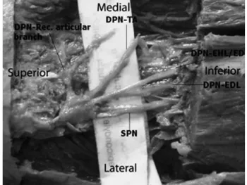
사체 해부를 실시하여 비골두부위에서 총비골신경 (common peroneal nerve, CPN)을 노출시키고 이 신경이 지배하고있는 근육까지 추적하여 신경분지들을 육안적으로 확인하였고(Fig. 1) 비골굴에서 적출한 비 골신경을 횡절단하여 현미경으로 내부국소해부를 확인 하였다.(Fig. 2) 가장 외측에 위치하는 것이 SPN이 고 다음보이는 4개의 신경다발이 DPN인데 외측에서 내측으로 가면서 EDL, EHL, EDB, TA의 순서로 내 부 국소해부가 형성되어 있음을 확인하였고 육안사진에 서 가장위쪽에 보이는 분지는 DPN의 회귀관절분지 (recurrent articular branch)인데 비골굴 위치에서 는 외측에 있다가 주행하면서 내측으로 교차됨을 확인 하였다.
또한 6년간 본원에서 비골신경손상으로 진단된 24명 의 환자 33개의 하지의 근전도 검사를 조사한 결과 남 자 18명, 여자 6명이었고 총비골신경손상이 16명, 깊은 비골신경손상이 8명이었다. 평균나이는 45세이었고 발 병 후 평균 1개월 정도 지나서 전기진단을 실시한 환자
Fig. 1. Peroneal nerve after dissection of the fibular cannel SPN: superficial peroneal nerve, DPN: deep peroneal nerve, TA: tibialis anterior, EDB: extensor digitorum brevis, EDL: extensor digitorum longus, EHL: extensor hallucis longus
들이었다. 총비골신경손상으로 진단된 16명, 22개의 하 지를 검토한 결과(Fig. 3) 신경전도검사에서 SPN의 이상이 22개중 22개 모두에서 보였고 EDB로 가는 DPN의 이상도 22개중 22개 모두에서 보였고 TA로 가는 DPN의 이상은 22개중 8개에서만 보였다. TA로 가는 DPN은 SPN이나 EDB로 가는 DPN에 비해 통 계적 의미 있게 적게 침범되었다.(p<0.05) 근전도 검사 소견에서는 PL의 이상이 22개중 22개 모두에서 보였고 EDB의 이상은 22개중 21개에서 보였고 TA의 이상은 22개중 17개에서 보였다. PL에 비해 EDB나 TA가 모 두 적게 침범됨을 알 수 있고 PL을 제외하고 EDB와 TA만을 비교해본 결과역시 통계적으로 의미 있게 적게 침범되었다.(p<0.05)
SPN을 침범하지 않는 깊은비골신경손상으로 진단된 환자는 8명이었고 11개의 하지를 검토한 결과(Fig. 4) 신경전도검사에서 EDB로 가는 DPN의 이상이 11개중 10개 모두에서 보였고 TA로 가는 DPN의 이상은 11 개중 2개에서만 보였다. TA로 가는 DPN이 EDB로 가는 DPN에 비해 통계적 의미있게 적게 침범되었 다.(p<0.05) 그러나 근전도 검사소견에서는 EDB, TA 에서 11개중 11개 모두에서 보였다.
고 찰
비골두부위에서의 비골신경포착증후군은 선천적인 원 인이든지 후천적 원인이든지 하지에서 가장 흔한 것으
Fig. 2. Peroneal nerve internal topography in microscopy (cross section view at the fibular tunnel)
SPN: superficial peroneal nerve, DPN: deep peroneal nerve, TA: tibialis anterior, EDB: extensor digitorum brevis, EDL: extensor digitorum longus, EHL: extensor hallucis longus, RAB: recurrent articular branch
Fig. 3. Electrodiagnostic findings of patienis with common peroneal neuropathy (N=22)
SPN: superficial peroneal nerve, DPN: deep peroneal nerve, TA: tibialis anterior, EDB: extensor digitorum brevis
* p<0.05 (chi-square test)
로 알려져 있다.5,6 비골두부위에서의 비골신경의 해부학 적 위치관계에 대한 연구를 보면 Ryan 등1은 비골신경 이 비골두부위에서 어떻게 주행하는지를 알기위해 비골 두의 꼭지점에서 비골굴까지의 거리, CPN의 갈림 (bifurcation)이 생기는 위치까지의 거리와 DPN이 외 측구획으로부터 분리되는 지점까지의 거리등을 사체해 부를 통해 밝혀 비골두부위에서 비골신경이 다른 주변 구조물과의 관계를 밝혔고 Ihunwo와 Dimitrov7는 사 체해부를 통해 족하수의 원인으로 생각되는 비골두부위 에서의 신경손상이 PL과 주변의 근막, 외측비골인대등 의 복합체에 의해 생기는 비탄력성 띠에 의한 것임을 확인하였다. 수술이나 조직검사를 실시할 때 비골신경 손상을 일으키지 않기 위한 외과분야에서의 연구가 이 루어져 왔으나8,9 비골신경의 내부국소해부, 특히 횡단면 에 대한 연구는 이루어져 있지 않았다. 이에 저자들은 비골두부위 특히 비골굴에서 비골신경의 내부국소해부 가 신경손상의 차이를 나타낼 수 있을 것으로 생각하고 사체해부를 실시하여 비골신경을 적출 한 후 현미경으 로 횡단면을 확인한 결과 비골신경에서 가장 외측에 위 치하는 신경다발은 SPN이고 다음보이는 4개의 신경다 발이 DPN인데 외측에서 내측으로 가면서 EDL, EHL, EDB, TA의 순서로 내부국소해부가 형성되어 있음을 확인하였고 육안에서 가장위쪽에 보이는 분지는 DPN의 회귀관절분지인데 비골굴 위치에서는 외측에 있다가 주행하면서 내측으로 교차됨을 확인하였다.
이에 저자들은 비골두부위에서의 비골신경손상은 일 정한 내부국소해부를 가지므로 전기진단학적 소견과도 연관성이 있을 것으로 생각하여 비골신경손상으로 진단 된 24명의 환자들의 기록을 조사해 보았다. 총비골신경 손상으로 진단된 16명, 22개의 하지를 검토한 결과 신 경전도검사에서는 TA로 가는 DPN이 SPN이나 EDB 로 가는 DPN에 비해 통계적 의미있게 적게 침범됨을 알수 있었고 근전도 검사소견에서는 PL에 비해 EDB 나 TA가 모두 적게 침범됨을 알 수 있고 PL을 제외하 고 EDB와 TA만을 비교해본 결과역시 통계적으로 의 미있게 적게 침범됨을 알 수 있었다.(p<0.05) 또한 SPN을 침범하지 않는 깊은비골신경손상으로 진단된 8 명의 11개 하지를 검토한 결과 신경전도검사에서는 TA 로 가는 DPN이 EDB로 가는 DPN에 비해 통계적 의 미있게 적게 침범됨을 알 수 있었고(p<0.05) 근전도 검 사소견에서는 EDB, TA에서 11개중 11개 모두 이상소 견을 보여 침범의 차이가 어느정도인지 알 수는 없었 다. 이 결과는 Raudino10의 비골두부위에서의 비골신 경손상 환자의 전기진단소견과 일치한다. 그는 EDB와 TA에서의 근전도소견을 비교하여 두 근육이 서로 다르 게 침범 받을 수 있음을 주장하며 두 근육 모두 근전도 시 검사의 가치가 있다고 주장하였다. 물론 EDB가 TA보다 외측에 위치하는 내부구조를 확인함으로써 근 전도 검사시 다르게 침범될 수 있음을 알 수는 있었지 만 이는 내부구조상 매우 인접하여 있어 경미한 손상시
Fig. 4. Electrodiagnostic findings of patienis with deep peroneal neuropathy (N=11) DPN: deep peroneal nerve, TA: tibialis anterior, EDB: extensor digitorum brevis
* p<0.05 (chi-square test)
근전도 검사에서 적절하지 않은 선택으로 생각된다.
EDB는 신경전도검사시 일반적으로 운동신경을 검사시 기록전극을 붙이는 근육이라 근전도검사시 선택하는것 에는 동의하지만 경미한 얕은 비골신경손상에서는 발가 락장신전근(Extensor digitorum longus, EDL)이 큰 진단적 가치가 있을것으로 생각된다. 본 연구에서는 거의 모든 환자가 심한 축삭손상이었고 근전도검사시 비골신경이 지배하는 모든 근육을 모두 검사하지 않아 경한 압박으로 인한 탈수초화에 의해 생길 수 있는 전 기진단학적 소견을 얻을 수 없었다는 제한점이 있다.
그러나 비록 신경전도검사와 일부 근육에서의 근전도검 사를 통해 신경손상이 해부학적 내부 국소해부를 따를 것이라고 추측할 수 있었고 비골신경의 내부국소해부를 밝힘으로서 이를 확인 할 수 있었다. 본 결과는 향후 전 기진단검사에 더욱 도움이 될 수 있을 것으로 생각된다.
결 론
비골두부위에서의 비골신경의 내부국소해부는 외측에 서 내측으로 SPN, DPN to EDL, EHL, EDB, TA순서로 위치하고 있으며 전기진단검사결과가 내부국 소해부를 잘 반영하여 준다.
참고문헌
01. Campbell WW: Diagnosis and management of common compression and entrapment neuropathies. Neurol Clin
1997: 15: 549-567.
02. Jabaley ME, Wallace WH, Heckler FR: Internal topogra- phy of major nerves of the forearm and hand: a current view. J Hand Surg 1980: 5: 1-18.
03. Chow JA, Van Beek AL, Meyer DL, Johnson MC: Surgi- cal significance of the motor fascicular group of the ulnar nerve in the forearm. J Hand Surg 1985: 10: 867-872.
04. Dumitru D, Amato AA, Zwarts MJ: Electrodiagnostic medicine, 2nd edition, Philadelphia: Hanley & Belfus, 2002, pp281-283.
05. Hong YH, Kim M, Kim HJ, Sung JJ, Kim SH, Lee KW:
Clinical and electrophysiologic features of HNPP patients with 17p11.2 deletion. Acta Neurol Scand 2003: 108: 352- 358.
06. Cruz-Martinez A, Arpa J, Palau F: Peroneal neuropathy after weight loss. J Peripher Nerv Syst 2000: 5: 101-105.
07. Ihunwo AO, Dimitrov ND: Anatomical basis for pressure on the common peroneal nerve. Cent Afr J Med 1999: 45:
77-79.
08. Dellon AL, Ebmer J, Swier P: Anatomic variations related to decompression of the common peroneal nerve at the fibular head. Ann Plast Surg 2002: 48: 30-34.
09. Takeda A, Tsuchiya H, Mori Y, Tanaka S, Kikuchi S, Tomita K: Anatomical aspects of biopsy of the proximal fibula. Int Orthop 2001: 24: 335-337.
10. Raudino F: Electrophysiological study of peroneal nerve palsy at the fibular head. Electromyogr Clin Neurophysiol 1996: 36: 439-440.